Revision 1

#43547Store at -20C

1 Kit

(10 slides)

UniProt ID:

#Q86VB7

Entrez-Gene Id:

#9332

Cell Signaling Technology

Orders: 877-616-CELL (2355) [email protected]

Support: 877-678-TECH (8324)

Web: [email protected] cellsignal.com

3 Trask LaneDanversMassachusetts01923USA
For Research Use Only. Not for Use in Diagnostic Procedures.
Product Includes Item # Volume Reactivity Isotype
CD163 (D6U1J) Rabbit mAb (SignalStar Conjugate 0022) 44568 50 µl H Rabbit IgG
Complementary Oligo (CO-0022-594) 93466 22 µl

Storage

SignalStar conjugates are supplied in PBS (pH 7.2), less than 0.1% sodium azide, 2 mM EDTA, 0.05% Triton X-100, 2 mg/mL BSA, and 50% glycerol. Complementary oligos are supplied in nuclease-free water. Store at -20°C. Do not aliquot the antibody. All components in this kit are stable for at least 12 months when stored at the recommended temperature.

Description

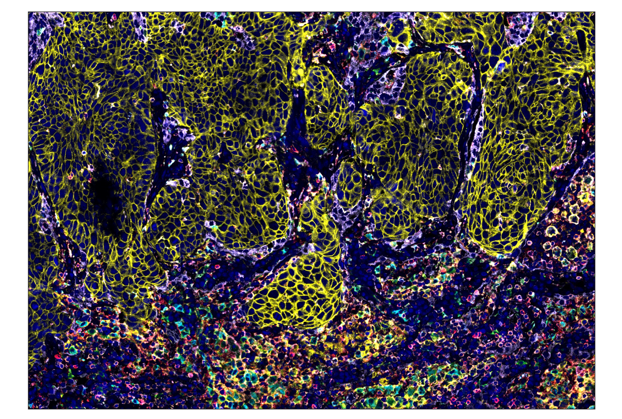
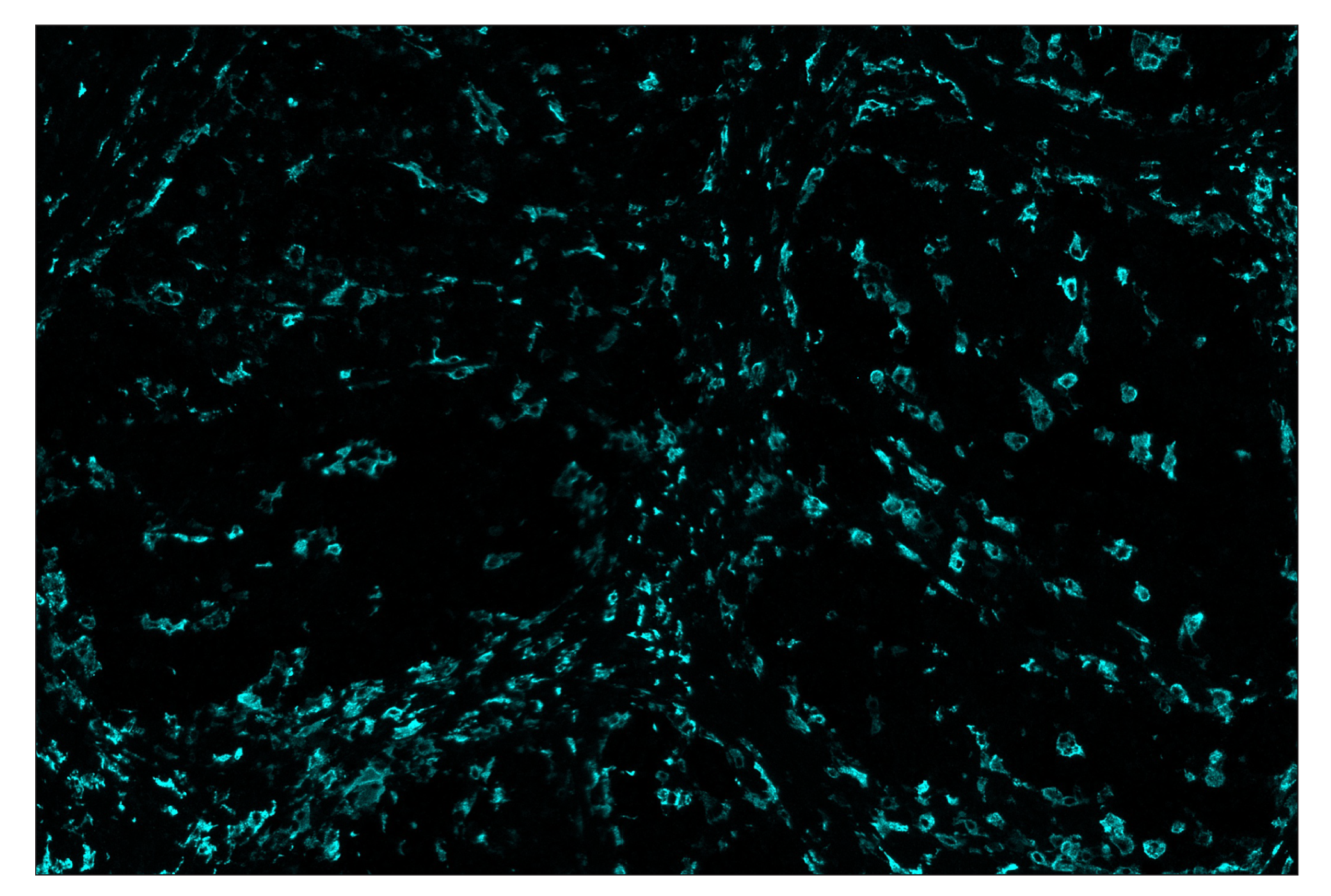
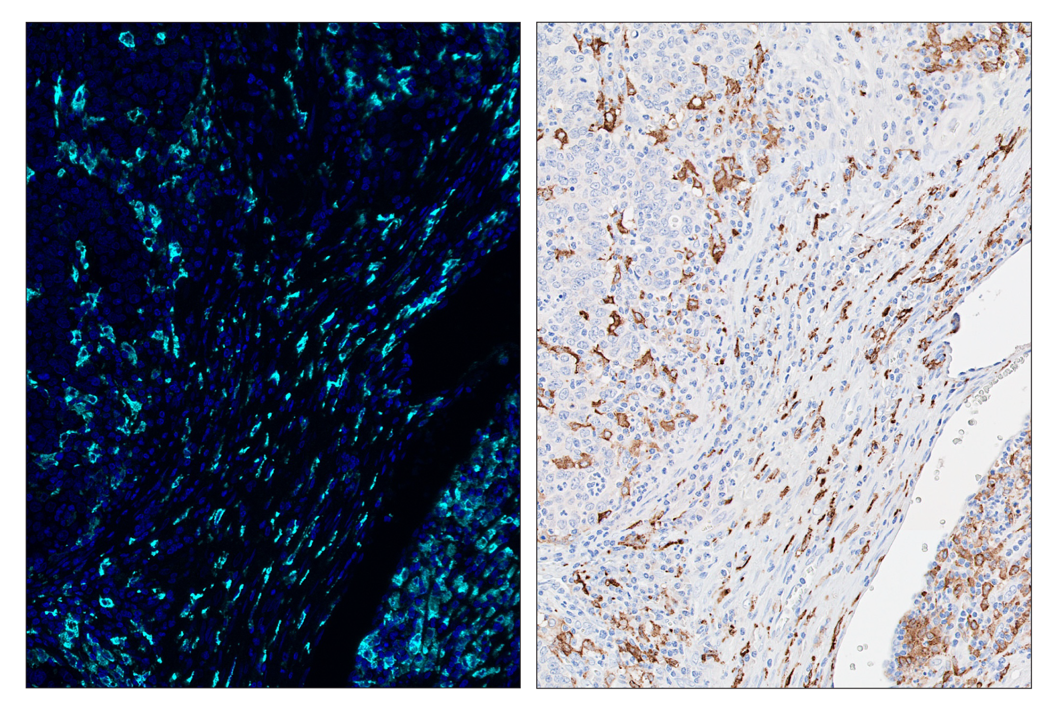
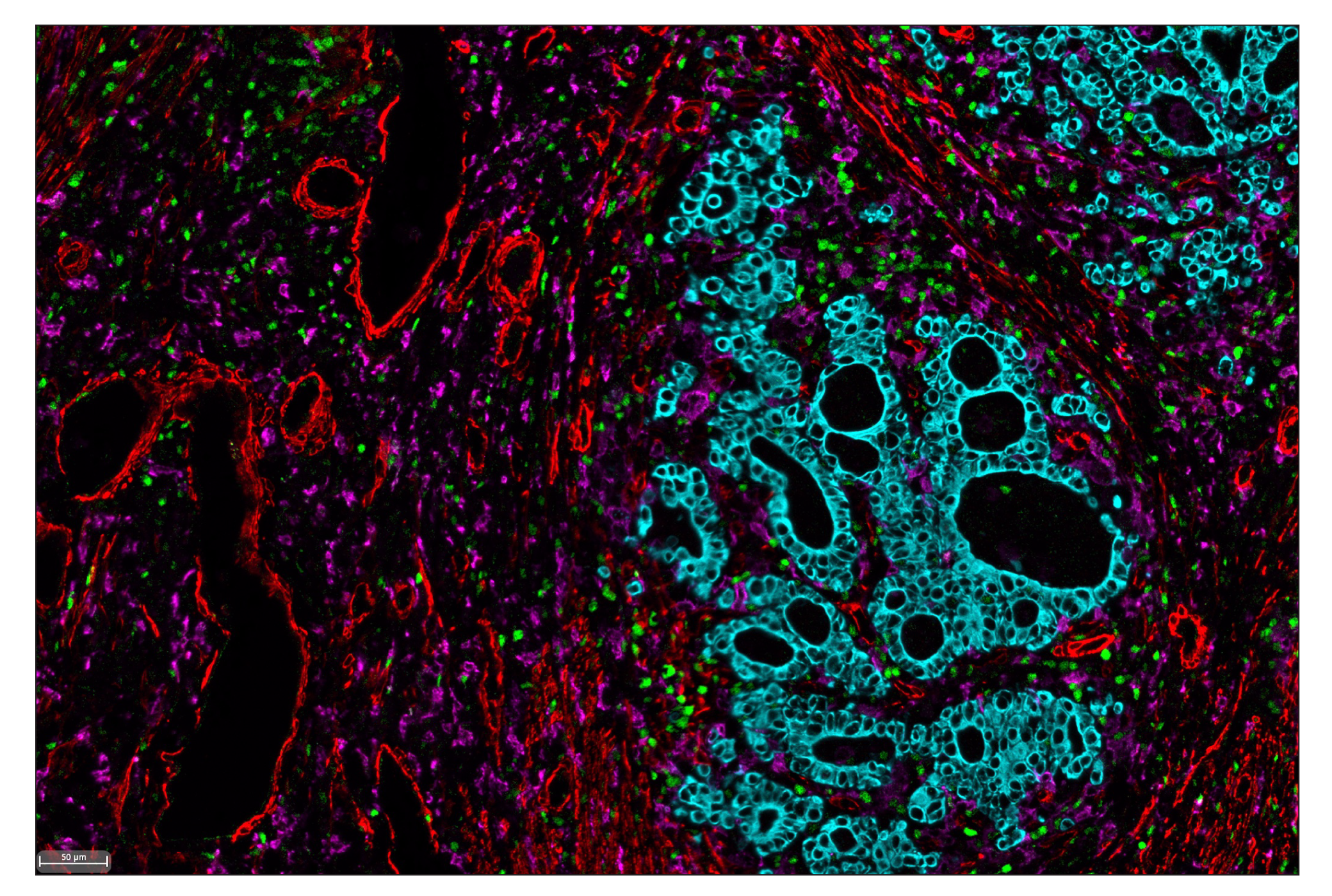
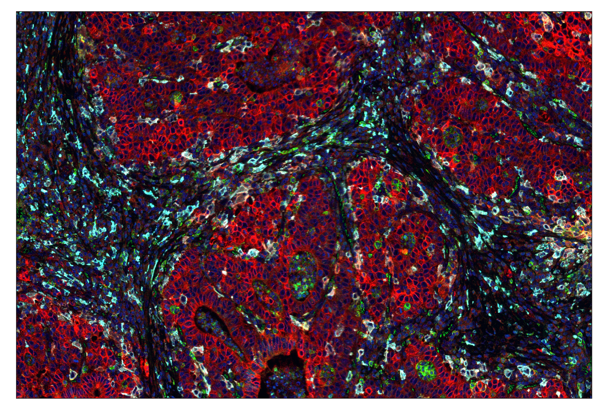
SignalStar multiplex immunohistochemistry (IHC) is an advanced technology for labeling multiple proteins simultaneously in tissue samples using specific primary antibodies and fluorescent detection reagents. This technology offers accuracy and reliability in visualizing and analyzing protein expression while maintaining spatial context and tissue architecture.

SignalStar Oligo-Antibody Pairs are compatible with the SignalStar Multiplex IHC Buffer Kits for use in fluorescent multiplex imaging experiments. This product includes the oligo-conjugated antibodies and complementary oligos required for labeling your target protein on up to 10 slides. SignalStar Multiplex IHC Buffer Kits are required to amplify and image the target signal. Multiple oligo-antibody pairs can be conveniently combined into a multiplex panel using the SignalStar Multiplex IHC Panel Builder. SignalStar Multiplex IHC Kits & Reagents are not compatible with all of Cell Signaling Technology® products and protocols that are recommended for use in immunohistochemical assays.

Specificity/Sensitivity

CD163 (D6U1J) Rabbit mAb (SignalStar Conjugate 0022) recognizes endogenous levels of total CD163 protein.

Source / Purification

Monoclonal antibody is produced by immunizing animals with recombinant protein specific to human CD163 protein.

Background

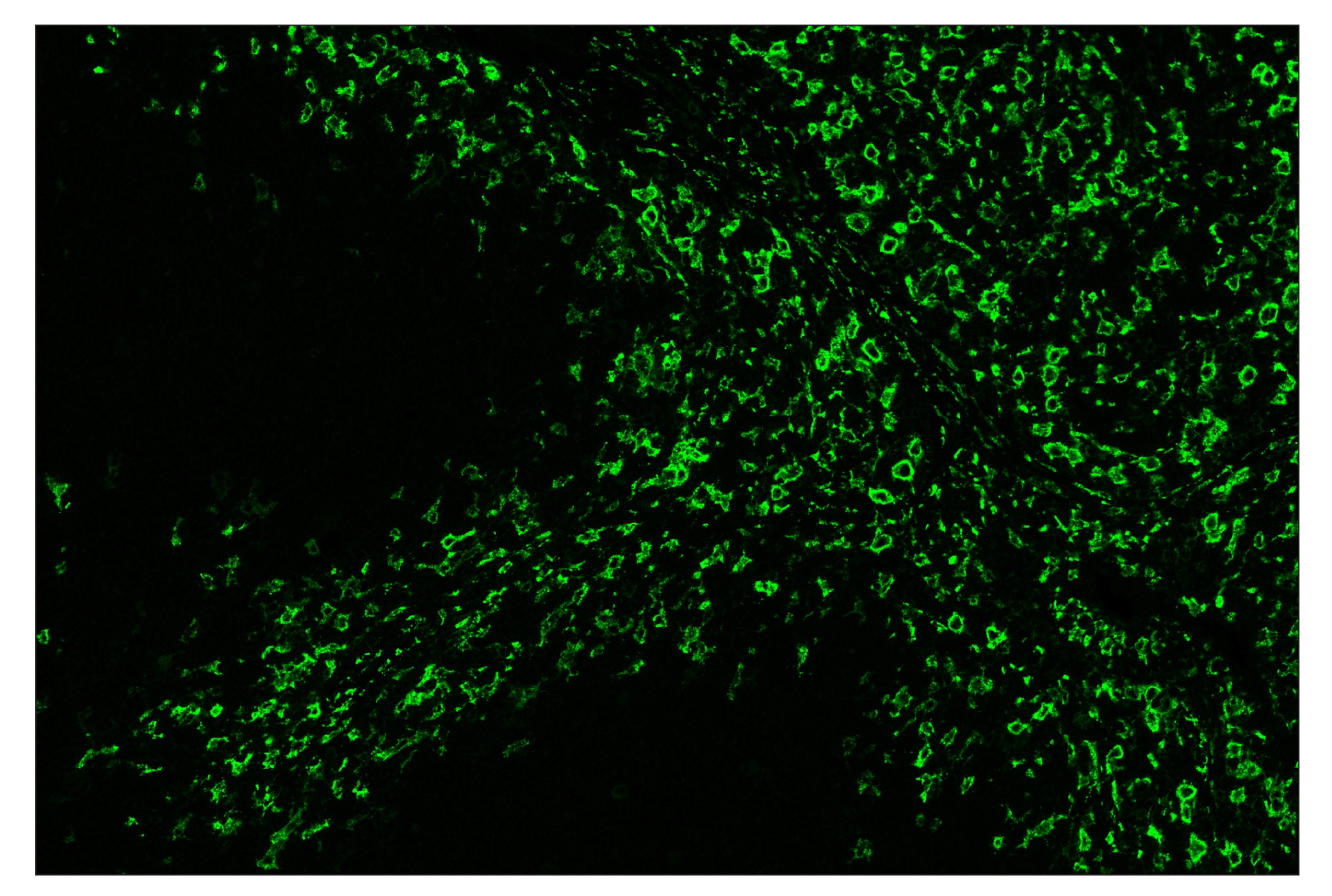
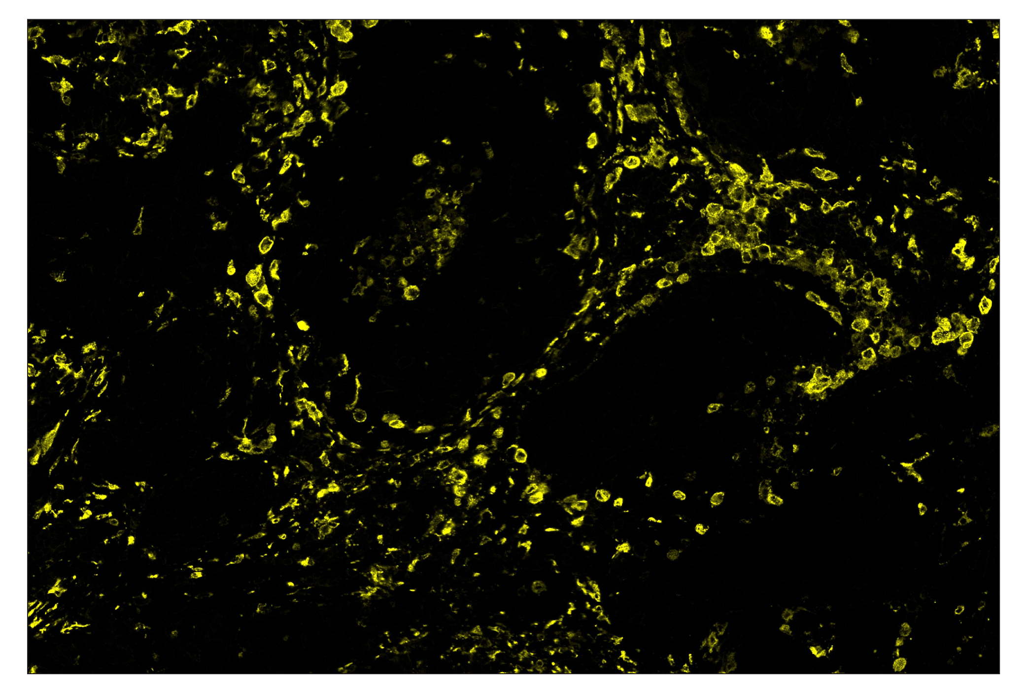
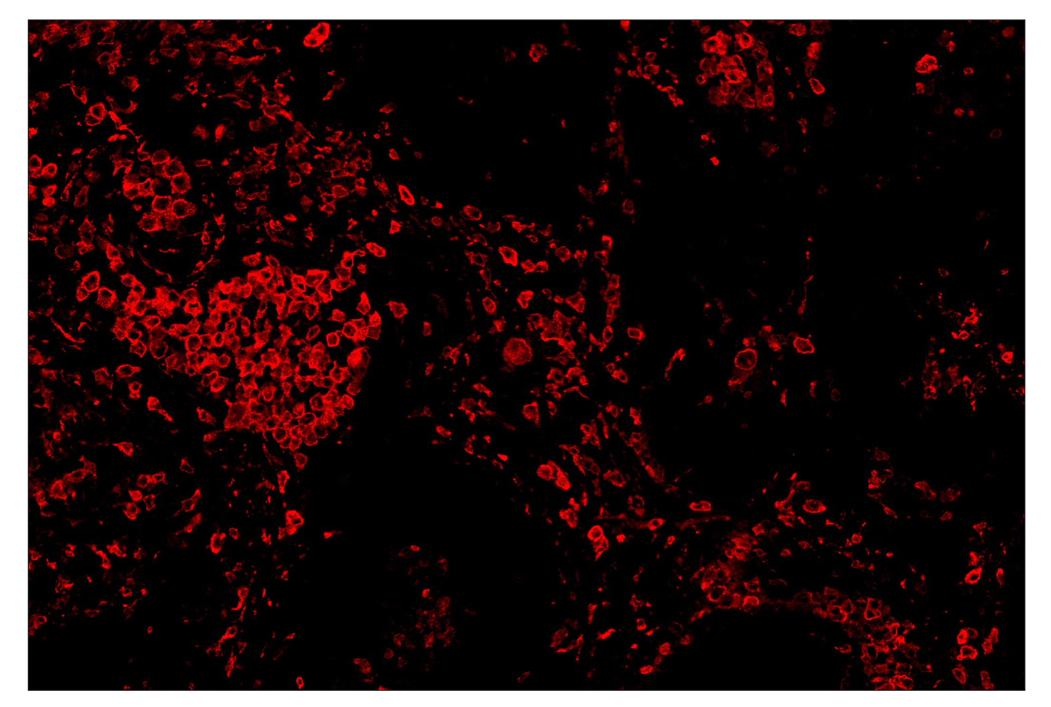
CD163 is a transmembrane scavenger receptor expressed on the macrophage surface. It has 9 B-type Scavenger Receptor Cysteine-Rich (SRCR) extracellular domains mediating serum haptoglobin clearing/endocytosis, pathogen binding and signal transduction, and calcium binding (1,2). CD163 is used as a surface marker of M2 type macrophages, including M2 type tumor-associated macrophages (TAMs), which facilitate cancer progression by secreting cytokines to promote angiogenesis, immunosuppression, and metastasis (3). Inflammatory stimulation and stress signals can induce extracellular domain shedding of CD163 to generate soluble CD163 (sCD163). The increased sCD163 level in serum is associated with low-grade inflammation in disease conditions (4-7).

  1. Graversen, J.H. and Moestrup, S.K. (2015) Membranes (Basel) 5, 228-52.
  2. Etzerodt, A. and Moestrup, S.K. (2013) Antioxid Redox Signal 18, 2352-63.
  3. Komohara, Y. et al. (2014) Cancer Sci 105, 1-8.
  4. Zhi, Y. et al. (2017) Mol Med Rep 15, 2931-2939.
  5. Møller, H.J. et al. (2011) Clin Chem 57, 291-7.
  6. Grønbaek, H. et al. (2012) Aliment Pharmacol Ther 36, 173-80.
  7. Aristoteli, L.P. et al. (2006) Atherosclerosis 184, 342-7.

Species Reactivity

Species reactivity is determined by testing in at least one approved application (e.g., western blot).

Cross-Reactivity Key

H: human M: mouse R: rat Hm: hamster Mk: monkey Vir: virus Mi: mink C: chicken Dm: D. melanogaster X: Xenopus Z: zebrafish B: bovine Dg: dog Pg: pig Sc: S. cerevisiae Ce: C. elegans Hr: horse GP: Guinea Pig Rab: rabbit All: all species expected

Trademarks and Patents

Cell Signaling Technology is a trademark of Cell Signaling Technology, Inc.
SignalStar is a trademark of Cell Signaling Technology, Inc.
All other trademarks are the property of their respective owners. Visit cellsignal.com/trademarks for more information.

Limited Uses

Except as otherwise expressly agreed in a writing signed by a legally authorized representative of CST, the following terms apply to Products provided by CST, its affiliates or its distributors. Any Customer's terms and conditions that are in addition to, or different from, those contained herein, unless separately accepted in writing by a legally authorized representative of CST, are rejected and are of no force or effect.

Products are labeled with For Research Use Only or a similar labeling statement and have not been approved, cleared, or licensed by the FDA or other regulatory foreign or domestic entity, for any purpose. Customer shall not use any Product for any diagnostic or therapeutic purpose, or otherwise in any manner that conflicts with its labeling statement. Products sold or licensed by CST are provided for Customer as the end-user and solely for research and development uses. Any use of Product for diagnostic, prophylactic or therapeutic purposes, or any purchase of Product for resale (alone or as a component) or other commercial purpose, requires a separate license from CST. Customer shall (a) not sell, license, loan, donate or otherwise transfer or make available any Product to any third party, whether alone or in combination with other materials, or use the Products to manufacture any commercial products, (b) not copy, modify, reverse engineer, decompile, disassemble or otherwise attempt to discover the underlying structure or technology of the Products, or use the Products for the purpose of developing any products or services that would compete with CST products or services, (c) not alter or remove from the Products any trademarks, trade names, logos, patent or copyright notices or markings, (d) use the Products solely in accordance with CST Product Terms of Sale and any applicable documentation, and (e) comply with any license, terms of service or similar agreement with respect to any third party products or services used by Customer in connection with the Products.

Revision 1
#43547

CD163 (D6U1J) & CO-0022-594 SignalStar Oligo-Antibody Pair

Immunohistochemistry Image 1: CD163 (D6U1J) & CO-0022-594 SignalStar™ Oligo-Antibody Pair Expand Image
SignalStar multiplex immunohistochemical analysis of paraffin-embedded human squamous cell lung carcinoma using CD163 (D6U1J) & CO-0022-488 SignalStar Oligo-Antibody Pair #35238 (green), PD-L1 (E1L3N®) & CO-0005-594 SignalStar Oligo-Antibody Pair #28249 (yellow), PD-1 (Intracellular Domain) (D4W2J) & CO-0008-647 SignalStar Oligo-Antibody Pair #56837 (red), CD11c (D3V1E) & CO-0017-647 SignalStar Oligo-Antibody Pair #96411 (magenta), CD68 (D4B9C) & CO-0007-750 SignalStar Oligo-Antibody Pair #91044 (cyan), HLA-DRA (E9R2Q) & CO-0023-488 SignalStar Oligo-Antibody Pair #92941 (pink), CD3ε (D7A6E) & CO-0001-594 SignalStar Oligo-Antibody Pair #84634 (orange), Pan-Keratin (C11) & CO-0003-750 SignalStar Oligo-Antibody Pair #97227 (white), and ProLong Gold Antifade Reagent with DAPI #8961 (blue). All fluorophores have been assigned a pseudocolor, as indicated. Staining was performed on the BOND RX by Leica Biosystems.
Immunohistochemistry Image 2: CD163 (D6U1J) & CO-0022-594 SignalStar™ Oligo-Antibody Pair Expand Image
SignalStar immunohistochemical analysis of paraffin-embedded human B-cell non-Hodgkin lymphoma using CD163 (D6U1J) & CO-0022-488 SignalStar Oligo-Antibody Pair #35238 (green). All fluorophores have been assigned a pseudocolor, as indicated. Staining was performed on the BOND RX by Leica Biosystems.
Immunohistochemistry Image 3: CD163 (D6U1J) & CO-0022-594 SignalStar™ Oligo-Antibody Pair Expand Image
SignalStar immunohistochemical analysis of paraffin-embedded human squamous cell lung carcinoma using CD163 (D6U1J) & CO-0022-594 SignalStar Oligo-Antibody Pair #43547 (yellow). All fluorophores have been assigned a pseudocolor, as indicated. Staining was performed on the BOND RX by Leica Biosystems.
Immunohistochemistry Image 4: CD163 (D6U1J) & CO-0022-594 SignalStar™ Oligo-Antibody Pair Expand Image
SignalStar immunohistochemical analysis of paraffin-embedded human squamous cell lung carcinoma using CD163 (D6U1J) & CO-0022-647 SignalStar Oligo-Antibody Pair #57183 (red). All fluorophores have been assigned a pseudocolor, as indicated. Staining was performed on the BOND RX by Leica Biosystems.
Immunohistochemistry Image 5: CD163 (D6U1J) & CO-0022-594 SignalStar™ Oligo-Antibody Pair Expand Image
SignalStar immunohistochemical analysis of paraffin-embedded human gastric adenocarcinoma using CD163 (D6U1J) & CO-0022-750 SignalStar Oligo-Antibody Pair #71043 (cyan). All fluorophores have been assigned a pseudocolor, as indicated. Staining was performed on the BOND RX by Leica Biosystems.
Immunohistochemistry Image 6: CD163 (D6U1J) & CO-0022-594 SignalStar™ Oligo-Antibody Pair Expand Image
SignalStar multiplex immunohistochemical analysis of paraffin-embedded human gastric adenocarcinoma using CD163 (D6U1J) & CO-0022-750 SignalStar Oligo-Antibody Pair #71043 (left, cyan) and ProLong Gold Antifade Reagent with DAPI #8961 (left, blue) compared to chromogenic immunohistochemical analysis of a serial section of paraffin-embedded human gastric adenocarcinoma using CD163 (D6U1J) Rabbit mAb #93498 (right). All fluorophores have been assigned a pseudocolor, as indicated. Staining was performed on the BOND RX by Leica Biosystems.
Immunohistochemistry Image 7: CD163 (D6U1J) & CO-0022-594 SignalStar™ Oligo-Antibody Pair Expand Image
SignalStar multiplex immunohistochemical analysis of paraffin-embedded human gastric adenocarcinoma using CD163 (D6U1J) & CO-0022-594 SignalStar Oligo-Antibody Pair #43547 (magenta), Tox/Tox2 (E6I3Q) & CO-0016-488 SignalStar Oligo-Antibody Pair #31189 (green), α-Smooth Muscle Actin (D4K9N) & CO-0024-647 SignalStar Oligo-Antibody Pair #63902 (red), and Pan-Keratin (C11) & CO-0003-750 SignalStar Oligo-Antibody Pair #97227 (cyan). All fluorophores have been assigned a pseudocolor, as indicated. Staining was performed on the BOND RX by Leica Biosystems.
Immunohistochemistry Image 1: CD163 (D6U1J) & CO-0022-594 SignalStar™ Oligo-Antibody Pair Expand Image
SignalStar multiplex immunohistochemical analysis of paraffin-embedded human gastric adenocarcinoma using CD163 (D6U1J) & CO-0022-750 SignalStar Oligo-Antibody Pair #71043 (cyan), CD16 (D1N9L) & CO-0031-488 SignalStar Oligo-Antibody Pair #29967 (green), PD-L1 (E1L3N®) & CO-0005-647 SignalStar Oligo-Antibody Pair #52085 (red), and ProLong Gold Antifade Reagent with DAPI #8961. All fluorophores have been assigned a pseudocolor, as indicated.