Revision 1

#83271Store at -20C

1 Kit

(10 slides)

UniProt ID:

#Q13094

Entrez-Gene Id:

#3937

Cell Signaling Technology

Orders: 877-616-CELL (2355) [email protected]

Support: 877-678-TECH (8324)

Web: [email protected] cellsignal.com

3 Trask LaneDanversMassachusetts01923USA
For Research Use Only. Not for Use in Diagnostic Procedures.
Product Includes Item # Volume Reactivity Isotype
Phospho-SLP-76 (Ser376) (E3G9U) XP® Rabbit mAb (SignalStar Conjugate 0018) 67572 50 µl H M Rabbit IgG
Complementary Oligo (CO-0018-647) 88830 22 µl

Storage

SignalStar conjugates are supplied in PBS (pH 7.2), less than 0.1% sodium azide, 2 mM EDTA, 0.05% Triton X-100, 2 mg/mL BSA, and 50% glycerol. Complementary oligos are supplied in nuclease-free water. Store at -20°C. Do not aliquot the antibody. All components in this kit are stable for at least 12 months when stored at the recommended temperature.

Description

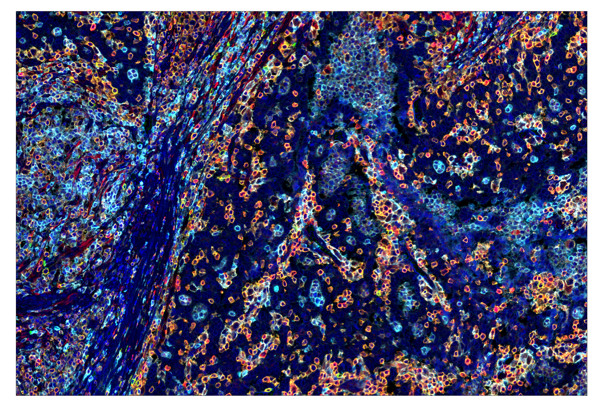
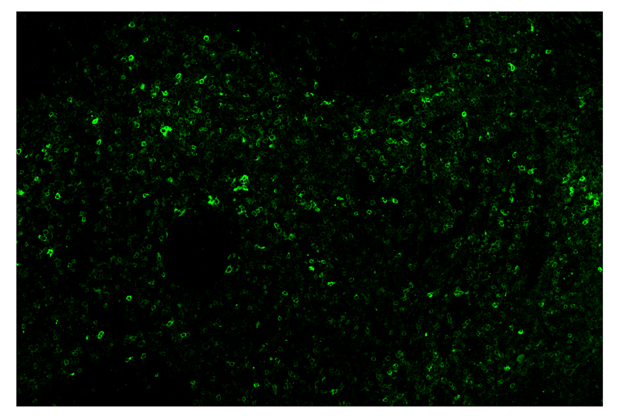
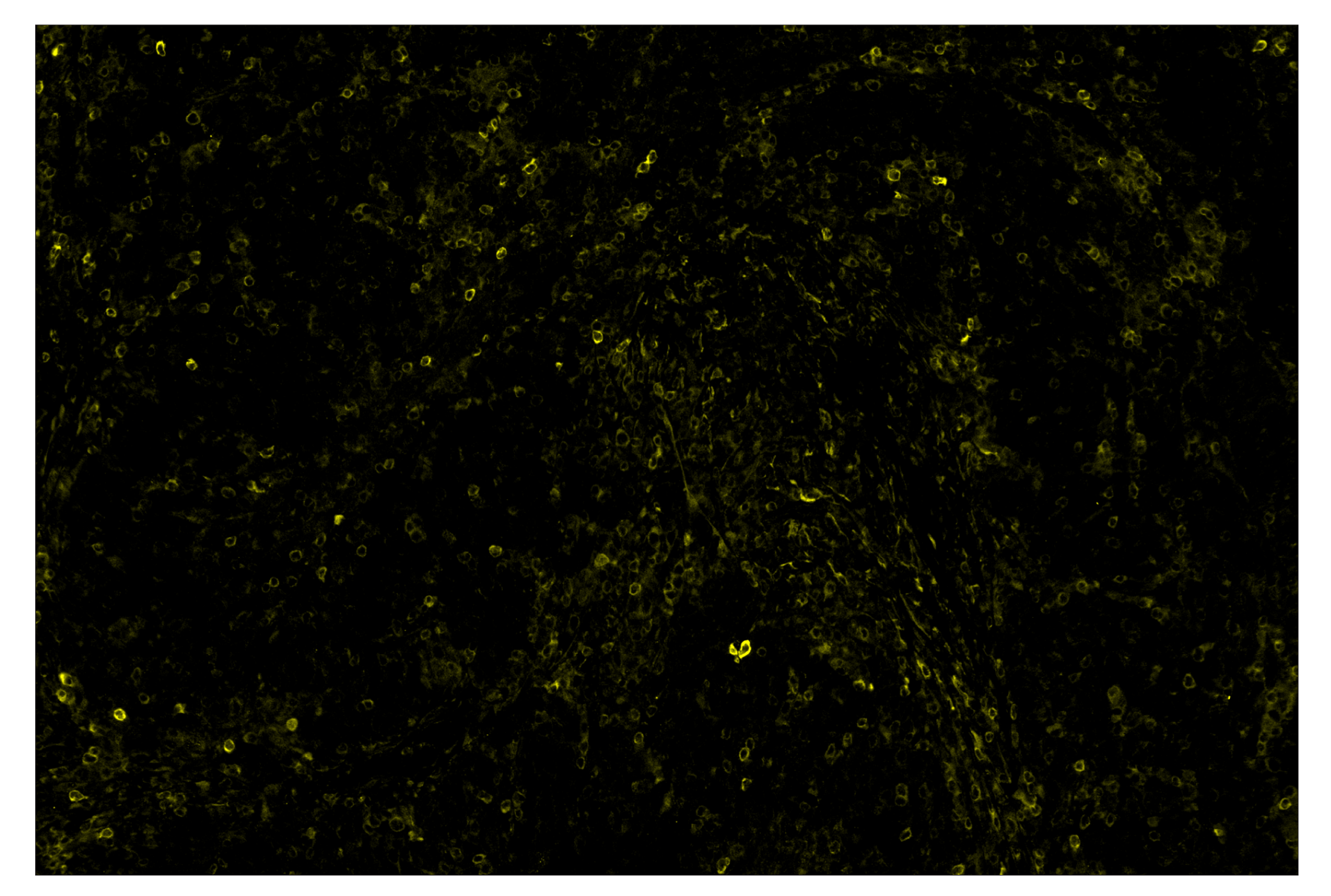
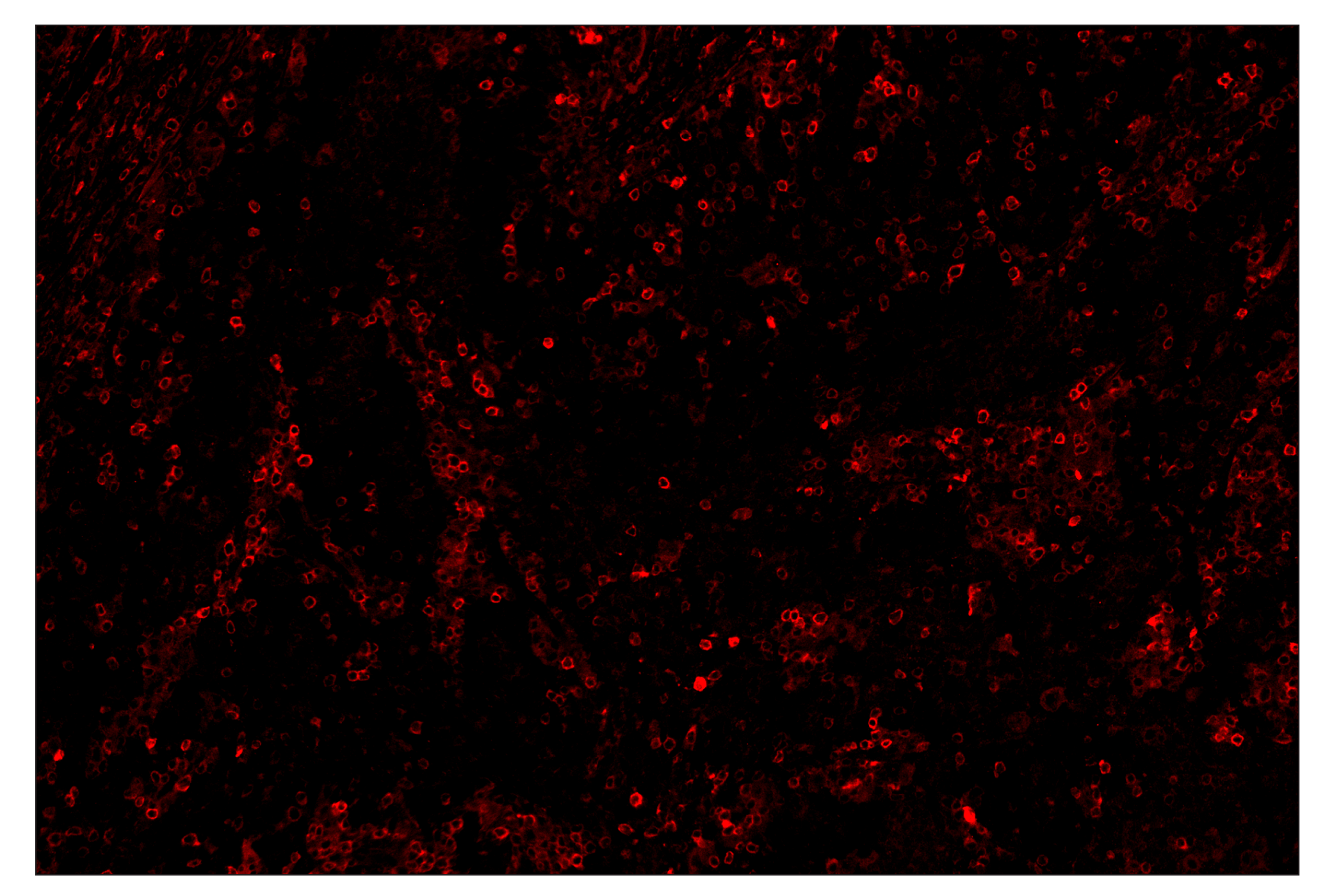
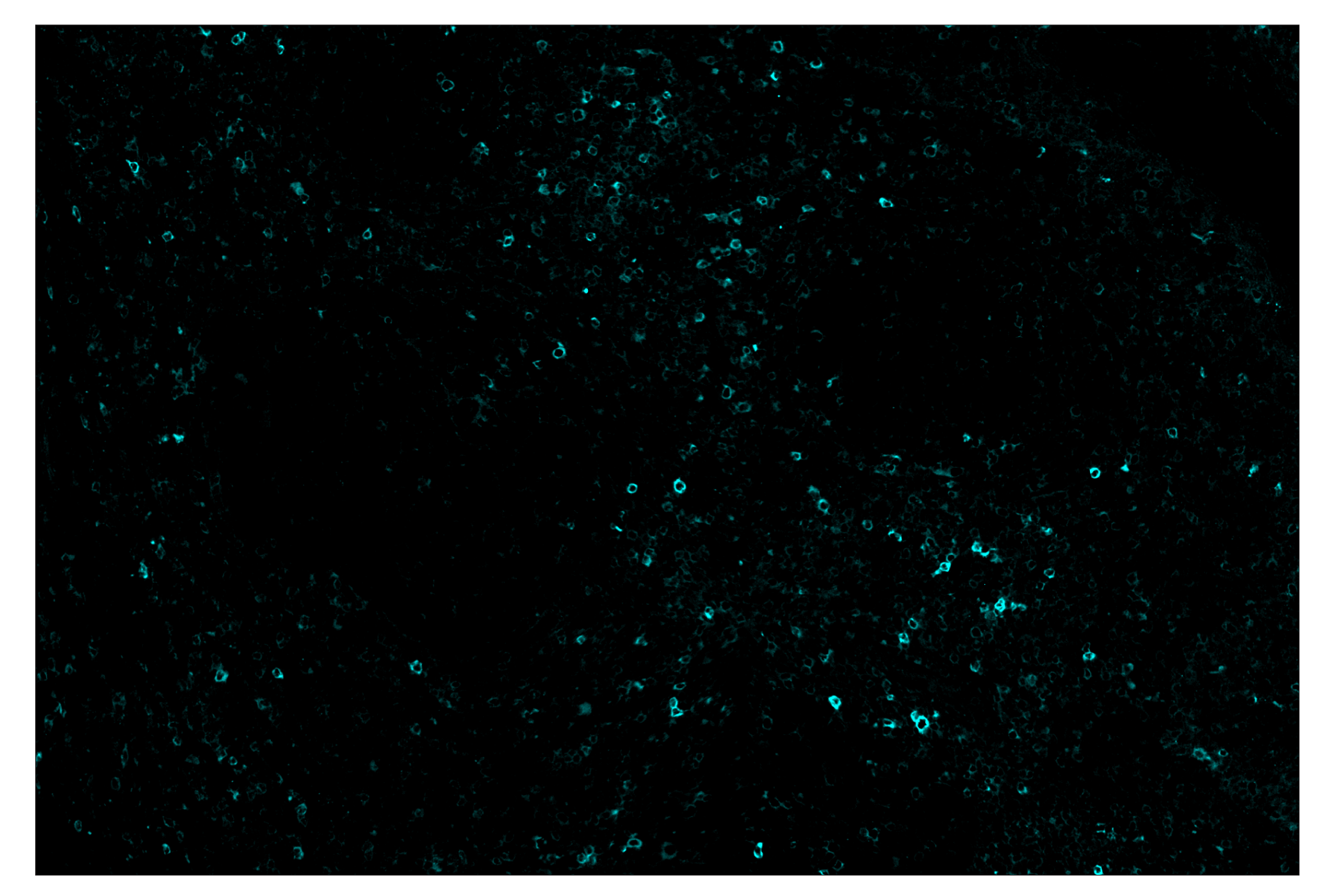
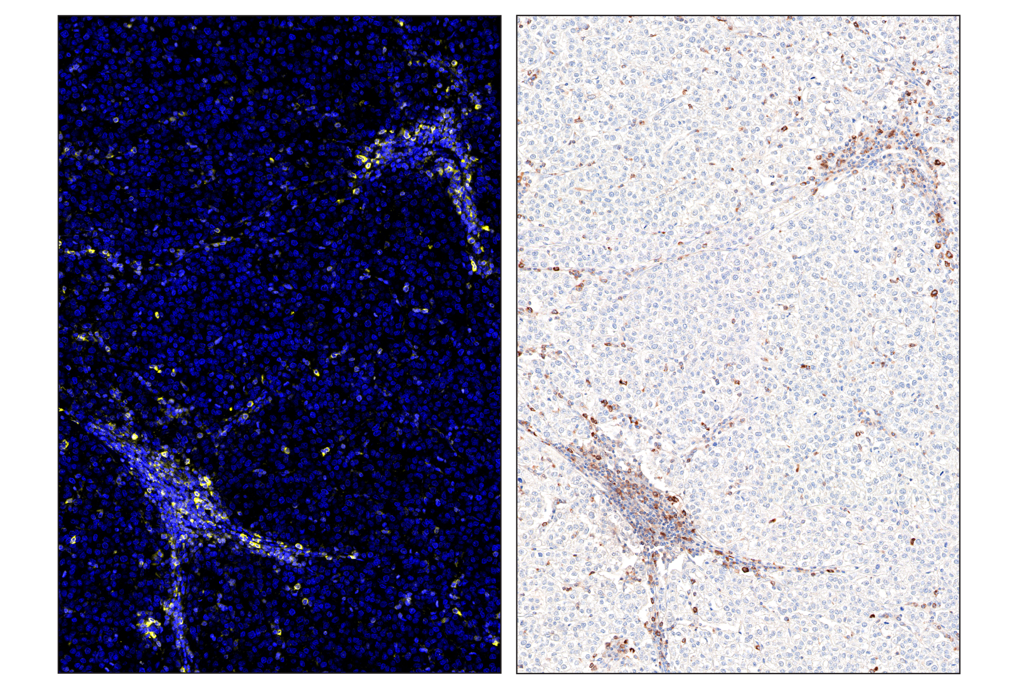
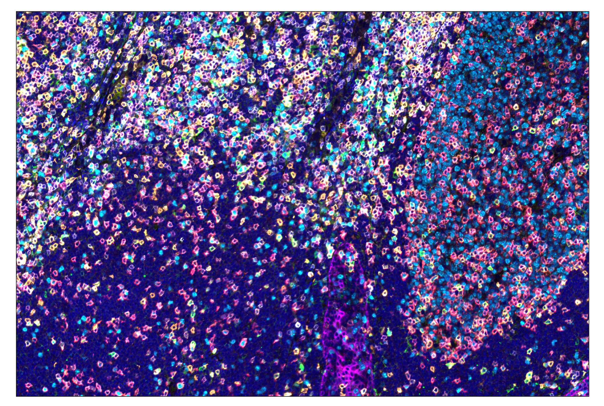
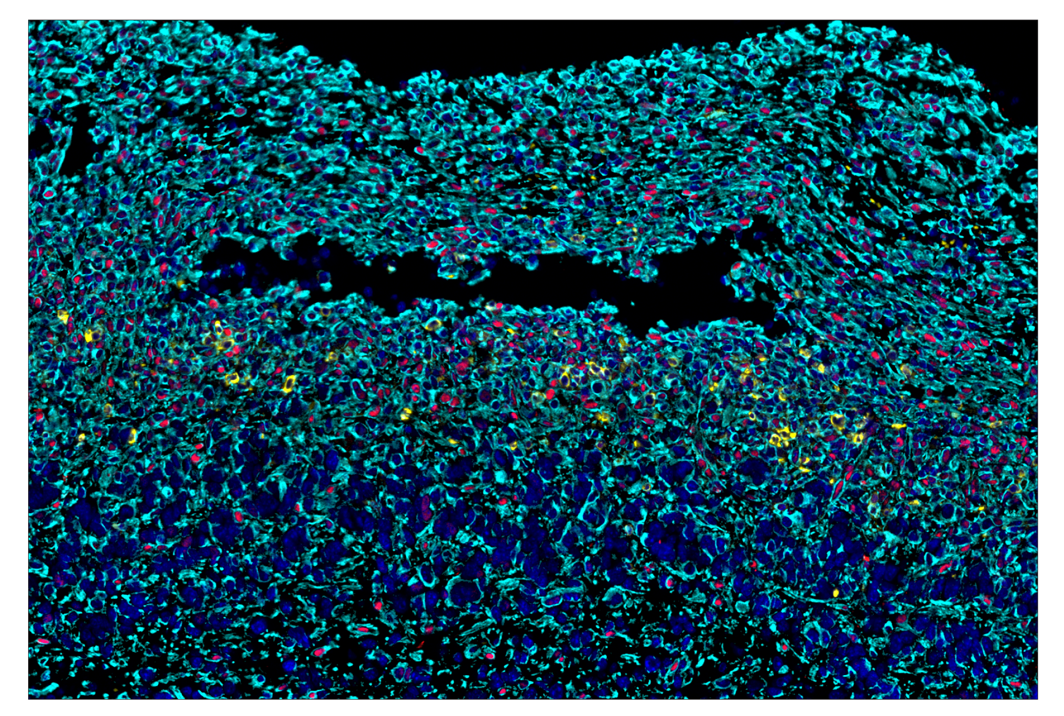
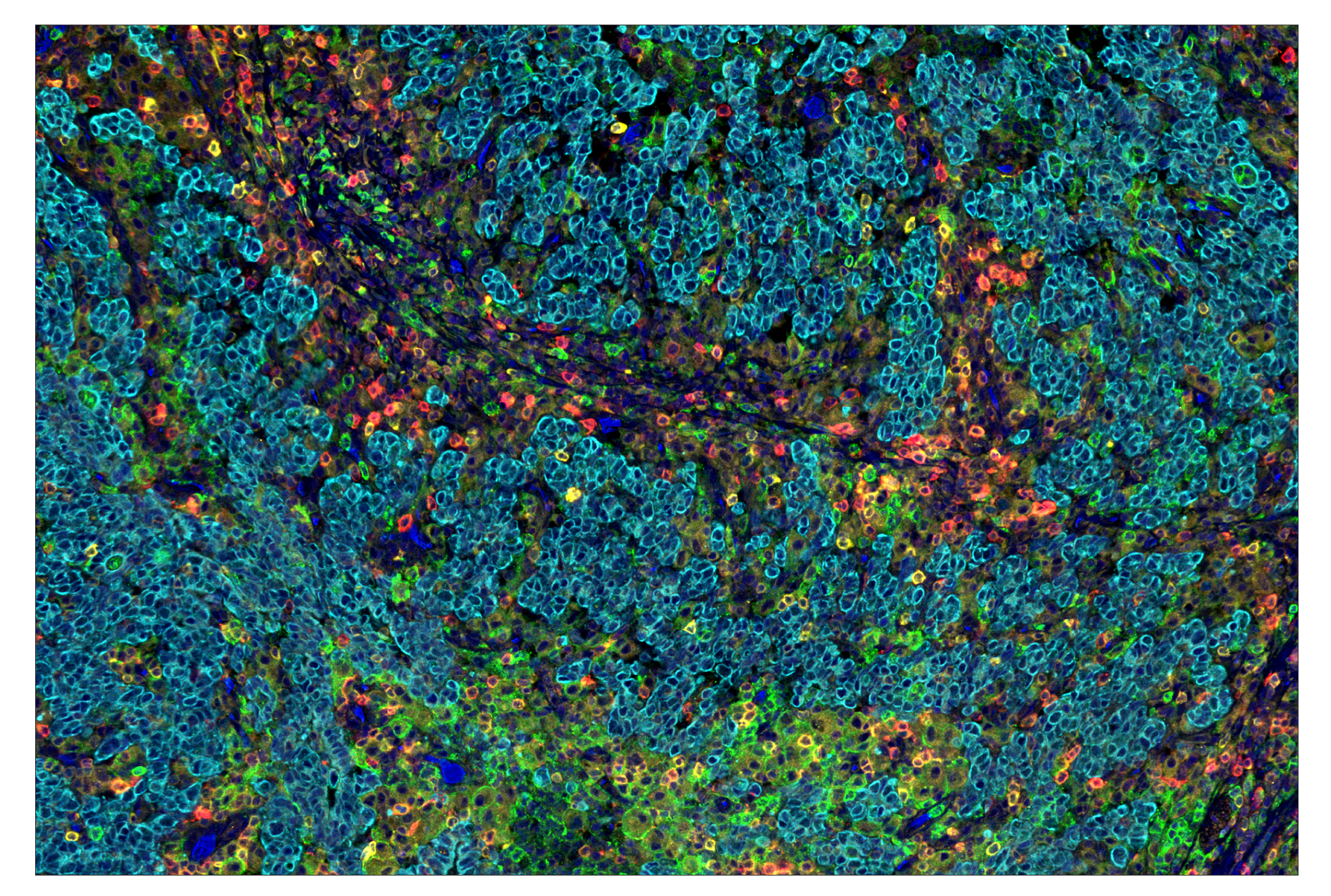
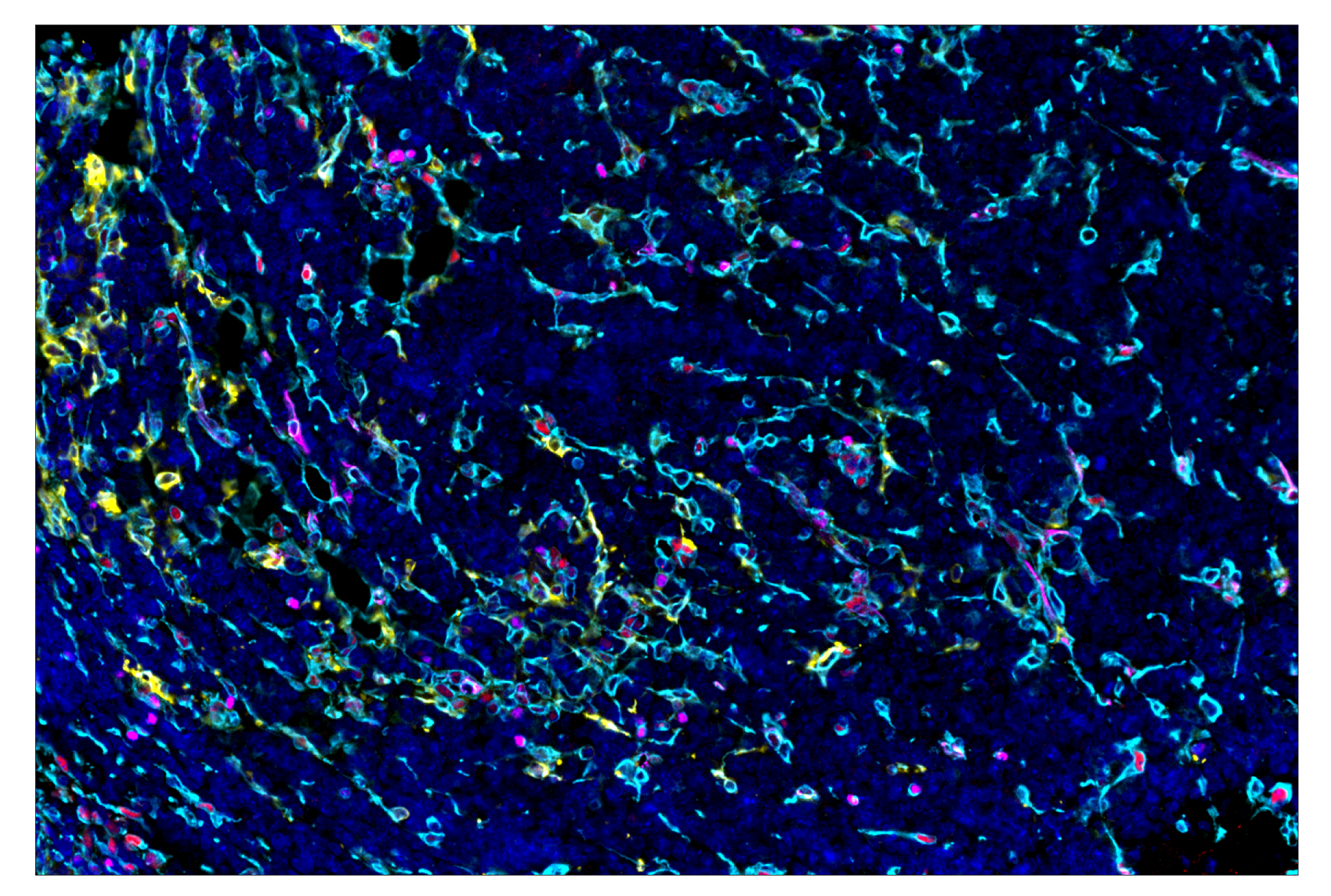
SignalStar multiplex immunohistochemistry (IHC) is an advanced technology for labeling multiple proteins simultaneously in tissue samples using specific primary antibodies and fluorescent detection reagents. This technology offers accuracy and reliability in visualizing and analyzing protein expression while maintaining spatial context and tissue architecture.

SignalStar Oligo-Antibody Pairs are compatible with the SignalStar Multiplex IHC Buffer Kits for use in fluorescent multiplex imaging experiments. This product includes the oligo-conjugated antibodies and complementary oligos required for labeling your target protein on up to 10 slides. SignalStar Multiplex IHC Buffer Kits are required to amplify and image the target signal. Multiple oligo-antibody pairs can be conveniently combined into a multiplex panel using the SignalStar Multiplex IHC Panel Builder. SignalStar Multiplex IHC Kits & Reagents are not compatible with all of Cell Signaling Technology® products and protocols that are recommended for use in immunohistochemical assays.

Specificity/Sensitivity

Phospho-SLP-76 (Ser376) (E3G9U) XP® Rabbit mAb (SignalStar Conjugate 0018) recognizes endogenous levels of SLP-76 protein only when phosphorylated at Ser376. Non-specific staining was observed in human stomach crypts by immunohistochemistry. Clone E3G9U is more sensitive by flow cytometry than clone D7S1K.

Source / Purification

Monoclonal antibody is produced by immunizing animals with a synthetic peptide corresponding to residues surrounding Ser376 of human SLP-76 protein.

Background

SH2 domain-containing leukocyte protein of 76 kDa (SLP-76) is a hematopoietic adaptor protein that is important in multiple biochemical signaling pathways and necessary for T cell development and activation (1). ZAP-70 phosphorylates SLP-76 and LAT as a result of TCR ligation. SLP-76 has amino-terminal tyrosine residues followed by a proline-rich domain and a carboxy-terminal SH2 domain. Phosphorylation of Tyr113 and Tyr128 result in recruitment of the GEF Vav and the adaptor protein Nck (2). TCR ligation also leads to phosphorylation of Tyr145, which mediates an association between SLP-76 and Itk, which is accomplished in part via the proline-rich domain of SLP-76 and the SH3 domain of Itk (3). Furthermore, the proline-rich domain of SLP-76 binds to the SH3 domains of Grb2-like adaptor Gads (3,4). In resting cells, SLP-76 is predominantly in the cytosol. Upon TCR ligation, SLP-76 translocates to the plasma membrane and promotes the assembly of a multi-protein signaling complex that includes Vav, Nck, Itk, and PLCγ1 (1). The expression of SLP-76 is tightly regulated; the protein is detected at very early stages of thymocyte development, increases as thymocyte maturation progresses, and is reduced as cells mature to CD4+ CD8+ double-positive thymocytes (5).

  1. Clements, J.L. (2003) Immunol Rev 191, 211-9.
  2. Bubeck Wardenburg, J. et al. (1998) Immunity 9, 607-16.
  3. Bunnell, S.C. et al. (2000) J Biol Chem 275, 2219-30.
  4. Liu, S.K. et al. (1999) Curr Biol 9, 67-75.
  5. Clements, J.L. et al. (1998) J Immunol 161, 3880-9.

Species Reactivity

Species reactivity is determined by testing in at least one approved application (e.g., western blot).

Cross-Reactivity Key

H: human M: mouse R: rat Hm: hamster Mk: monkey Vir: virus Mi: mink C: chicken Dm: D. melanogaster X: Xenopus Z: zebrafish B: bovine Dg: dog Pg: pig Sc: S. cerevisiae Ce: C. elegans Hr: horse GP: Guinea Pig Rab: rabbit All: all species expected

Trademarks and Patents

Cell Signaling Technology is a trademark of Cell Signaling Technology, Inc.
SignalStar is a trademark of Cell Signaling Technology, Inc.
U.S. Patent No. 10,781,477, foreign equivalents, and child patents deriving therefrom.
All other trademarks are the property of their respective owners. Visit cellsignal.com/trademarks for more information.

Limited Uses

Except as otherwise expressly agreed in a writing signed by a legally authorized representative of CST, the following terms apply to Products provided by CST, its affiliates or its distributors. Any Customer's terms and conditions that are in addition to, or different from, those contained herein, unless separately accepted in writing by a legally authorized representative of CST, are rejected and are of no force or effect.

Products are labeled with For Research Use Only or a similar labeling statement and have not been approved, cleared, or licensed by the FDA or other regulatory foreign or domestic entity, for any purpose. Customer shall not use any Product for any diagnostic or therapeutic purpose, or otherwise in any manner that conflicts with its labeling statement. Products sold or licensed by CST are provided for Customer as the end-user and solely for research and development uses. Any use of Product for diagnostic, prophylactic or therapeutic purposes, or any purchase of Product for resale (alone or as a component) or other commercial purpose, requires a separate license from CST. Customer shall (a) not sell, license, loan, donate or otherwise transfer or make available any Product to any third party, whether alone or in combination with other materials, or use the Products to manufacture any commercial products, (b) not copy, modify, reverse engineer, decompile, disassemble or otherwise attempt to discover the underlying structure or technology of the Products, or use the Products for the purpose of developing any products or services that would compete with CST products or services, (c) not alter or remove from the Products any trademarks, trade names, logos, patent or copyright notices or markings, (d) use the Products solely in accordance with CST Product Terms of Sale and any applicable documentation, and (e) comply with any license, terms of service or similar agreement with respect to any third party products or services used by Customer in connection with the Products.

Revision 1
#83271

Phospho-SLP-76 (Ser376) (E3G9U) & CO-0018-647 SignalStar Oligo-Antibody Pair

Immunohistochemistry Image 1: Phospho-SLP-76 (Ser376) (E3G9U) & CO-0018-647 SignalStar™ Oligo-Antibody Pair Expand Image
SignalStar multiplex immunohistochemical analysis of paraffin-embedded human gastric adenocarcinoma using ICOS (D1K2T) & CO-0027-488 SignalStar Oligo-Antibody Pair #92797 (green), CD45 (Intracellular Domain) (D9M8I) & CO-0013-594 SignalStar Oligo-Antibody Pair #15734 (yellow), Phospho-SLP-76 (Ser376) (E3G9U) & CO-0018-647 SignalStar Oligo-Antibody Pair #83271 (red), CD16 (D1N9L) & CO-0031-750 SignalStar Oligo-Antibody Pair #48056 (cyan), and ProLong Gold Antifade Reagent with DAPI #8961 (blue). All fluorophores have been assigned a pseudocolor, as indicated. Staining was performed on the BOND RX autostainer by Leica Biosystems.
Immunohistochemistry Image 2: Phospho-SLP-76 (Ser376) (E3G9U) & CO-0018-647 SignalStar™ Oligo-Antibody Pair Expand Image
SignalStar immunohistochemical analysis of paraffin-embedded human tonsil using Phospho-SLP-76 (Ser376) (E3G9U) & CO-0018-488 SignalStar Oligo-Antibody Pair #34567 (green). All fluorophores have been assigned a pseudocolor, as indicated. Staining was performed on the BOND RX autostainer by Leica Biosystems.
Immunohistochemistry Image 3: Phospho-SLP-76 (Ser376) (E3G9U) & CO-0018-647 SignalStar™ Oligo-Antibody Pair Expand Image
SignalStar immunohistochemical analysis of paraffin-embedded human gastric adenocarcinoma using Phospho-SLP-76 (Ser376) (E3G9U) & CO-0018-594 SignalStar Oligo-Antibody Pair #53299 (yellow). All fluorophores have been assigned a pseudocolor, as indicated. Staining was performed on the BOND RX autostainer by Leica Biosystems.
Immunohistochemistry Image 4: Phospho-SLP-76 (Ser376) (E3G9U) & CO-0018-647 SignalStar™ Oligo-Antibody Pair Expand Image
SignalStar immunohistochemical analysis of paraffin-embedded human gastric adenocarcinoma using Phospho-SLP-76 (Ser376) (E3G9U) & CO-0018-647 SignalStar Oligo-Antibody Pair #83271 (red). All fluorophores have been assigned a pseudocolor, as indicated. Staining was performed on the BOND RX autostainer by Leica Biosystems.
Immunohistochemistry Image 5: Phospho-SLP-76 (Ser376) (E3G9U) & CO-0018-647 SignalStar™ Oligo-Antibody Pair Expand Image
SignalStar immunohistochemical analysis of paraffin-embedded human tonsil using Phospho-SLP-76 (Ser376) (E3G9U) & CO-0018-750 SignalStar Oligo-Antibody Pair #94745 (cyan). All fluorophores have been assigned a pseudocolor, as indicated. Staining was performed on the BOND RX autostainer by Leica Biosystems.
Immunohistochemistry Image 6: Phospho-SLP-76 (Ser376) (E3G9U) & CO-0018-647 SignalStar™ Oligo-Antibody Pair Expand Image
SignalStar multiplex immunohistochemical analysis of paraffin-embedded human hepatocellular carcinoma using Phospho-SLP-76 (Ser376) (E3G9U) & CO-0018-594 SignalStar Oligo-Antibody Pair #53299 (left, yellow) and DAPI #4083 (left, blue) compared to chromogenic immunohistochemical analysis of a serial section of paraffin-embedded human hepatocellular carcinoma using Phospho-SLP-76 (Ser376) (E3G9U) XP® Rabbit mAb #76384 (right). All fluorophores have been assigned a pseudocolor, as indicated. Staining was performed on the BOND RX autostainer by Leica Biosystems.
Immunohistochemistry Image 7: Phospho-SLP-76 (Ser376) (E3G9U) & CO-0018-647 SignalStar™ Oligo-Antibody Pair Expand Image
SignalStar multiplex immunohistochemical analysis of paraffin-embedded human tonsil using Phospho-SLP-76 (Ser376) (E3G9U) & CO-0018-488 SignalStar Oligo-Antibody Pair #34567 (green), CD16 (D1N9L) & CO-0031-594 SignalStar Oligo-Antibody Pair #51630 (yellow), ICOS (D1K2T) & CO-0027-647 SignalStar Oligo-Antibody Pair #24147 (red), Ki-67 (8D5) & CO-0014-750 SignalStar Oligo-Antibody Pair #56398 (cyan), CD3ε (D7A6E) & CO-0001-488 SignalStar Oligo-Antibody Pair #92856 (pink), CD8α (D8A8Y) & CO-0004-594 SignalStar Oligo-Antibody Pair #19166 (orange), Granzyme B (D6E9W) & CO-0009-647 SignalStar Oligo-Antibody Pair #37369 (white), Pan-Keratin (C11) & CO-0003-750 SignalStar Oligo-Antibody Pair #97227 (magenta), and ProLong Gold Antifade Reagent with DAPI #8961 (blue). All fluorophores have been assigned a pseudocolor, as indicated. Staining was performed on the BOND RX autostainer by Leica Biosystems.
Immunohistochemistry Image 8: Phospho-SLP-76 (Ser376) (E3G9U) & CO-0018-647 SignalStar™ Oligo-Antibody Pair Expand Image
SignalStar multiplex immunohistochemical analysis of paraffin-embedded GL261 syngeneic tumor using Phospho-SLP-76 (Ser376) (E3G9U) & CO-0018-594 SignalStar Oligo-Antibody Pair #53299 (yellow), Phospho-Stat3 (Tyr705) (D3A7) & CO-0029-647 SignalStar Oligo-Antibody Pair #94459 (red), Vimentin (D21H3) & CO-0012-750 SignalStar Oligo-Antibody Pair #61002 (cyan), and DAPI #4083 (blue). All fluorophores have been assigned a pseudocolor, as indicated. Staining was performed on the BOND RX autostainer by Leica Biosystems.
Immunohistochemistry Image 1: Phospho-SLP-76 (Ser376) (E3G9U) & CO-0018-647 SignalStar™ Oligo-Antibody Pair Expand Image
SignalStar multiplex immunohistochemical analysis of paraffin-embedded human gastric adenocarcinoma using ICOS (D1K2T) & CO-0027-647 SignalStar Oligo-Antibody Pair #24147 (red), CD16 (D1N9L) & CO-0031-488 SignalStar Oligo-Antibody Pair #29967 (green), Phospho-SLP-76 (Ser376) (E3G9U) & CO-0018-594 SignalStar Oligo-Antibody Pair #53299 (yellow), Pan-Keratin (C11) & CO-0003-750 SignalStar Oligo-Antibody Pair #97227 (cyan), and ProLong Gold Antifade Reagent with DAPI #8961 (blue). All fluorophores have been assigned a pseudocolor, as indicated.
Immunohistochemistry Image 2: Phospho-SLP-76 (Ser376) (E3G9U) & CO-0018-647 SignalStar™ Oligo-Antibody Pair Expand Image
SignalStar multiplex immunohistochemical analysis of paraffin-embedded A20 syngeneic tumor using TCF1/TCF7 (C63D9) & CO-0006-488 SignalStar Oligo-Antibody Pair #83016 (magenta), Phospho-SLP-76 (Ser376) (E3G9U) & CO-0018-594 SignalStar Oligo-Antibody Pair #53299 (yellow), Phospho-Stat3 (Tyr705) (D3A7) & CO-0029-647 SignalStar Oligo-Antibody Pair #94459 (red), Vimentin (D21H3) & CO-0012-750 SignalStar Oligo-Antibody Pair #61002 (cyan), and DAPI #4083 (blue). All fluorophores have been assigned a pseudocolor, as indicated.