Revision 5
Cell Signaling Technology

Orders: 877-616-CELL (2355) [email protected]

Support: 877-678-TECH (8324)

Web: [email protected] cellsignal.com

3 Trask LaneDanversMassachusetts01923USA
For Research Use Only. Not for Use in Diagnostic Procedures.
Applications:

StarBond, StarManual

:

:

:

:

Product Information

Storage

All components in this kit are stable for at least 12 months when stored at the recommended temperature.

Product Description

SignalStar multiplex immunohistochemistry (IHC) is an advanced technology for labeling multiple proteins simultaneously in tissue samples using specific primary antibodies and fluorescent detection reagents. This technology offers accuracy and reliability in visualizing and analyzing protein expression while maintaining spatial context and tissue architecture.

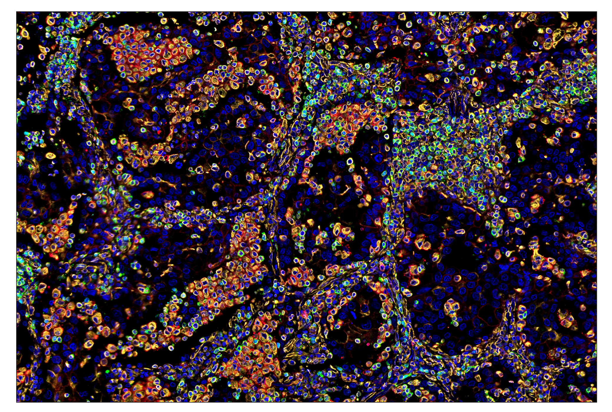
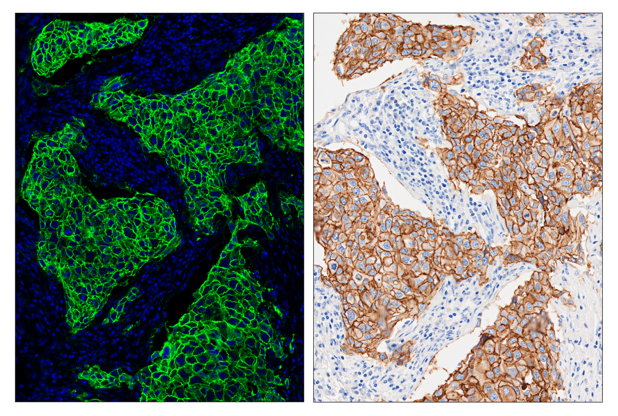
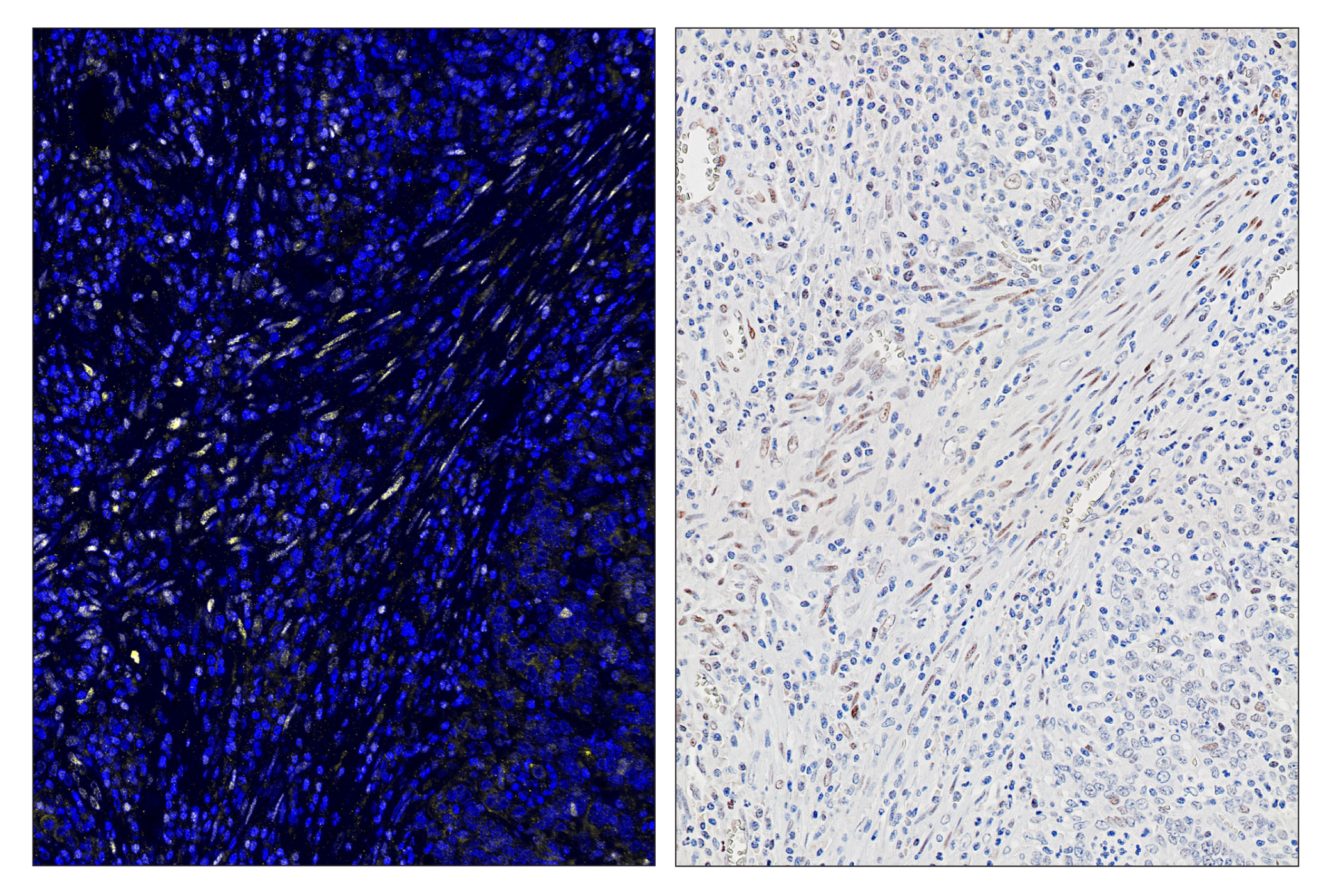
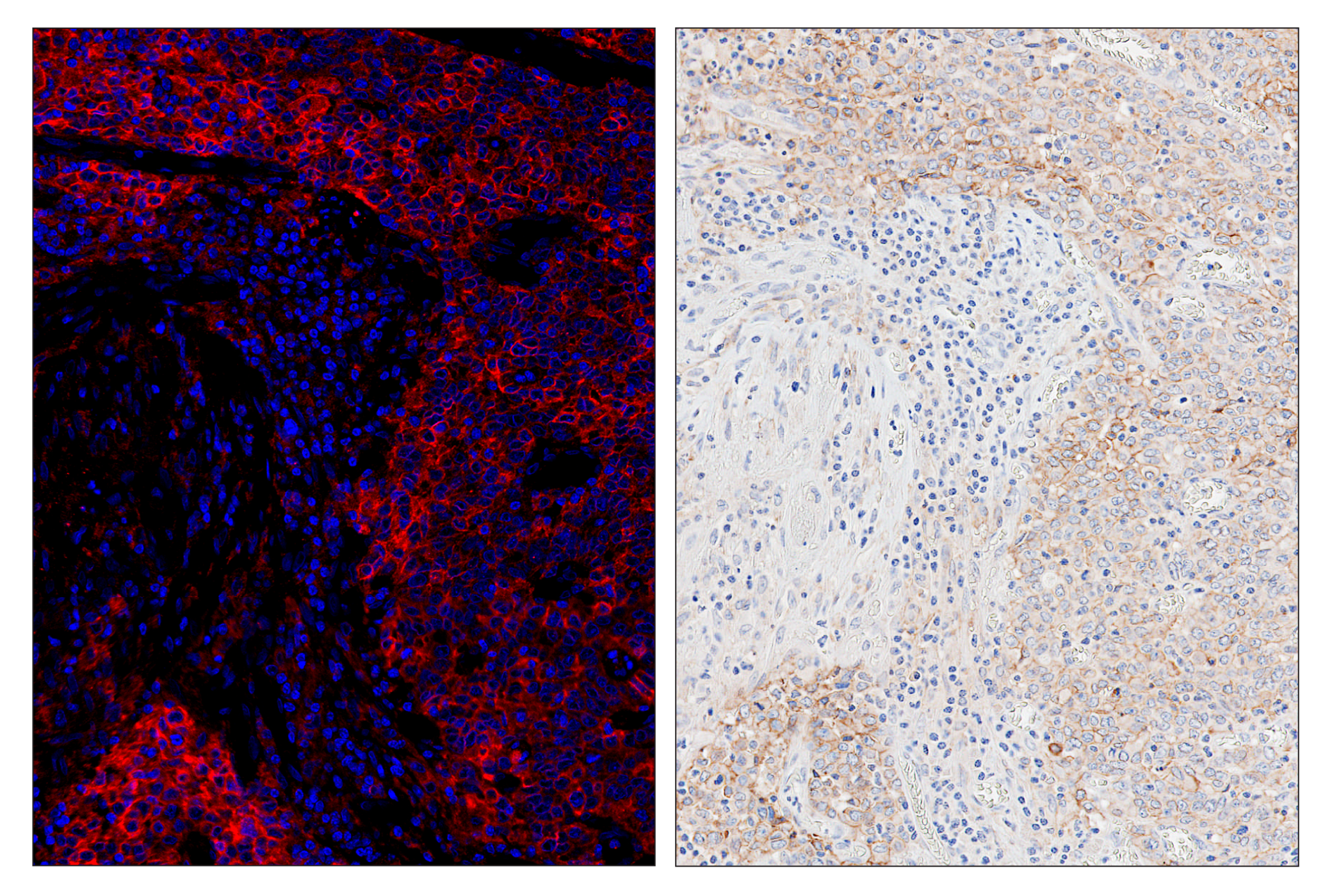
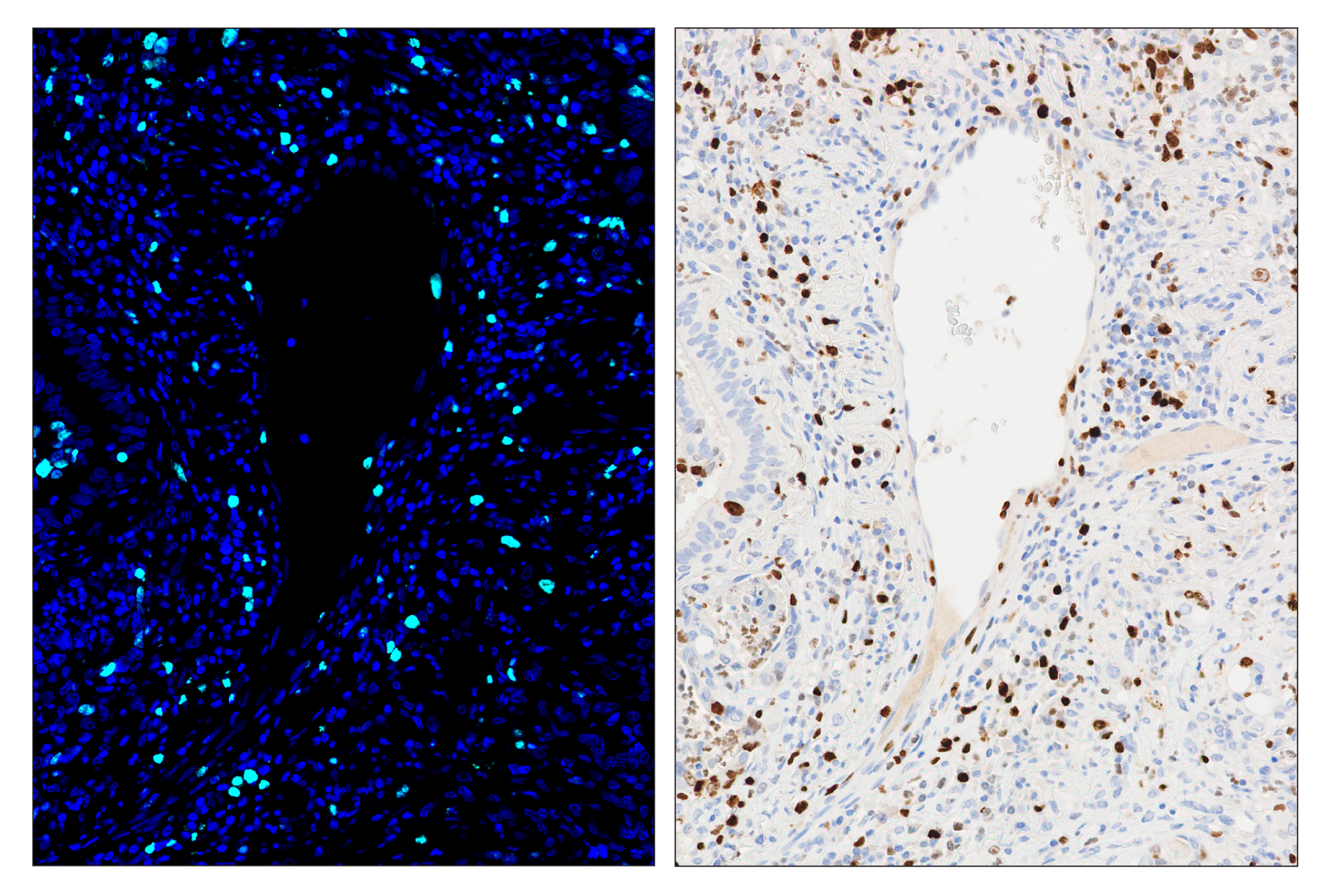
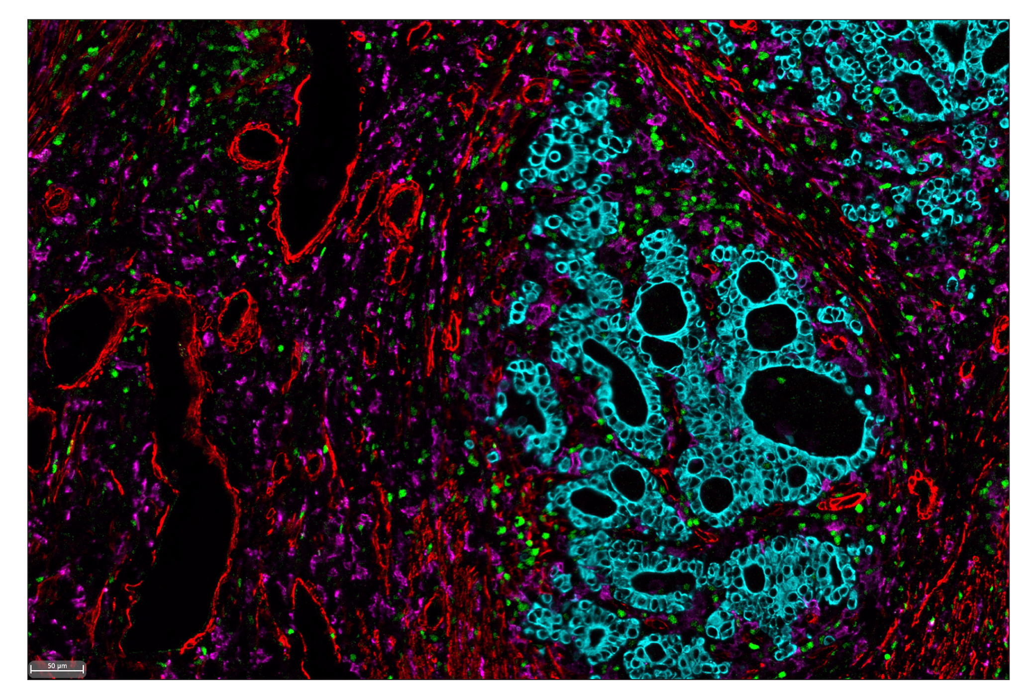
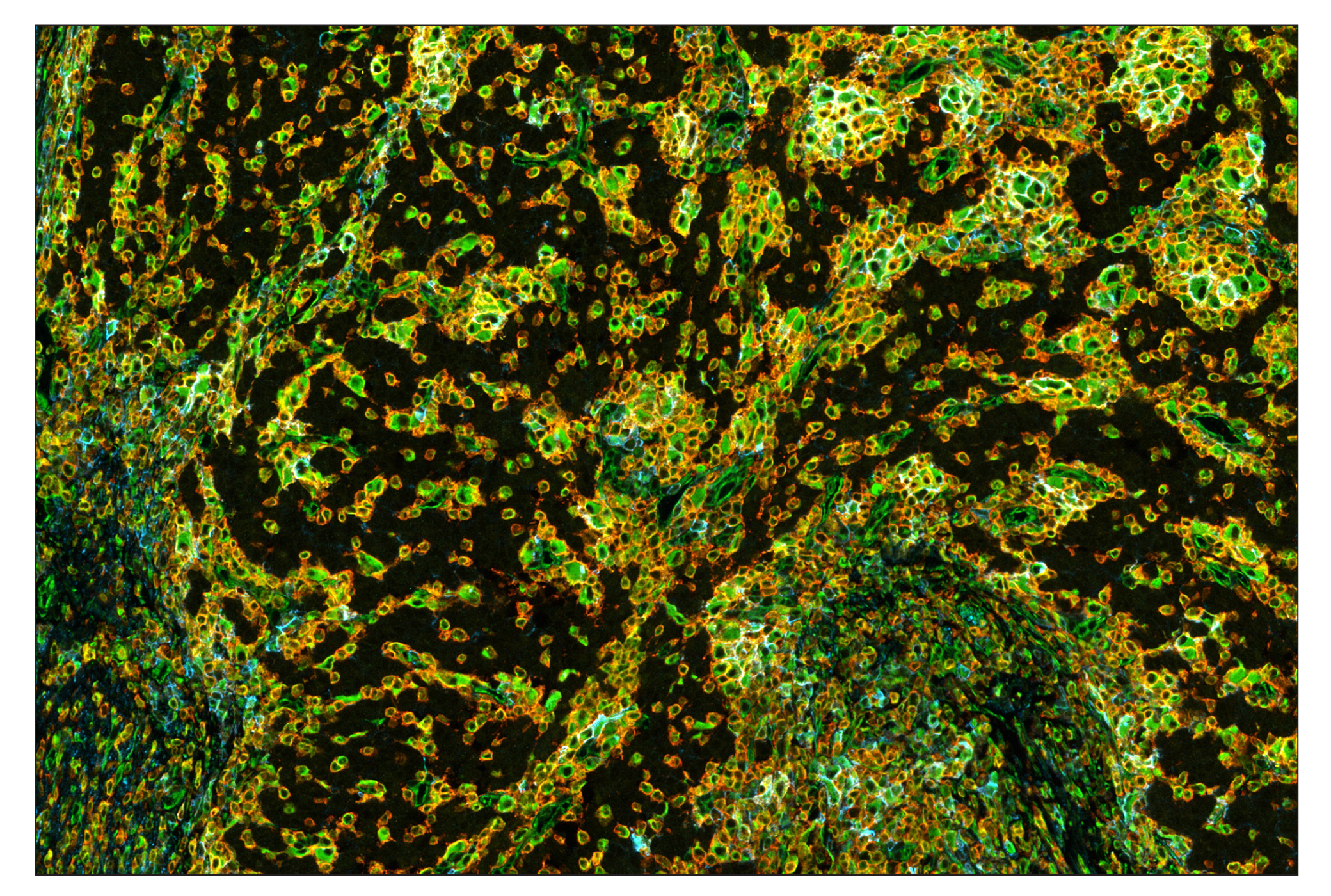
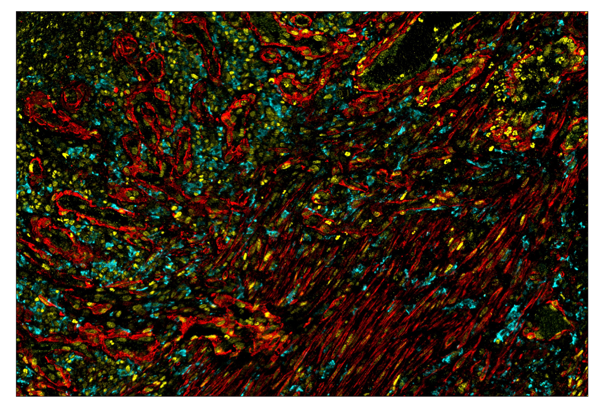
The SignalStar Miniplex IHC Buffer Kit is compatible with SignalStar Oligo-Antibody Pairs for use in fluorescent multiplex imaging experiments. This product is required to amplify and image the signal of 3-4 SignalStar Oligo-Antibody Pairs selected using the SignalStar Multiplex IHC Panel Builder. This buffer kit includes the reagents required for labeling 10 slides. SignalStar Multiplex IHC Kits & Reagents are not compatible with all of Cell Signaling Technology® products and protocols that are recommended for use in immunohistochemical assays.

Species Reactivity

Species reactivity is determined by testing in at least one approved application (e.g., western blot).

Applications Key

StarBond: SignalStar™ Leica Bond StarManual: SignalStar™ Manual

Cross-Reactivity Key

H: human M: mouse R: rat Hm: hamster Mk: monkey Vir: virus Mi: mink C: chicken Dm: D. melanogaster X: Xenopus Z: zebrafish B: bovine Dg: dog Pg: pig Sc: S. cerevisiae Ce: C. elegans Hr: horse GP: Guinea Pig Rab: rabbit All: all species expected

Trademarks and Patents

Cell Signaling Technology is a trademark of Cell Signaling Technology, Inc.
SignalStar is a trademark of Cell Signaling Technology, Inc.
U.S. Patent No. 10,781,477, foreign equivalents, and child patents deriving therefrom.
U.S. Patent No. 7,429,487, foreign equivalents, and child patents deriving therefrom.
All other trademarks are the property of their respective owners. Visit cellsignal.com/trademarks for more information.

Limited Uses

Except as otherwise expressly agreed in a writing signed by a legally authorized representative of CST, the following terms apply to Products provided by CST, its affiliates or its distributors. Any Customer's terms and conditions that are in addition to, or different from, those contained herein, unless separately accepted in writing by a legally authorized representative of CST, are rejected and are of no force or effect.

Products are labeled with For Research Use Only or a similar labeling statement and have not been approved, cleared, or licensed by the FDA or other regulatory foreign or domestic entity, for any purpose. Customer shall not use any Product for any diagnostic or therapeutic purpose, or otherwise in any manner that conflicts with its labeling statement. Products sold or licensed by CST are provided for Customer as the end-user and solely for research and development uses. Any use of Product for diagnostic, prophylactic or therapeutic purposes, or any purchase of Product for resale (alone or as a component) or other commercial purpose, requires a separate license from CST. Customer shall (a) not sell, license, loan, donate or otherwise transfer or make available any Product to any third party, whether alone or in combination with other materials, or use the Products to manufacture any commercial products, (b) not copy, modify, reverse engineer, decompile, disassemble or otherwise attempt to discover the underlying structure or technology of the Products, or use the Products for the purpose of developing any products or services that would compete with CST products or services, (c) not alter or remove from the Products any trademarks, trade names, logos, patent or copyright notices or markings, (d) use the Products solely in accordance with CST Product Terms of Sale and any applicable documentation, and (e) comply with any license, terms of service or similar agreement with respect to any third party products or services used by Customer in connection with the Products.

Revision 5
#63043

SignalStar Miniplex IHC Buffer Kit

Immunohistochemistry Image 1: SignalStar™ Miniplex IHC Buffer Kit Expand Image
SignalStar multiplex immunohistochemical analysis of paraffin-embedded human non-small cell lung carcinoma using Vimentin (D21H3) & CO-0012-594 SignalStar Oligo-Antibody Pair #16471 (yellow), CD3ε (D7A6E) & CO-0001-488 SignalStar Oligo-Antibody Pair #92856 (green), TIM-3 (D5D5R) & CO-0010-647 SignalStar Oligo-Antibody Pair #15231 (red), TCF1/TCF7 (C63D9) & CO-0006-750 SignalStar Oligo-Antibody Pair #53114 (cyan), and ProLong Gold Antifade Reagent with DAPI #8961 (blue). All fluorophores have been assigned a pseudocolor, as indicated. Staining was performed on the BOND RX autostainer by Leica Biosystems.
Immunohistochemistry Image 2: SignalStar™ Miniplex IHC Buffer Kit Expand Image
SignalStar multiplex immunohistochemical analysis of paraffin-embedded human infiltrating ductal carcinoma of the breast using HER2/ErbB2 (D8F12) & CO-0019-488 SignalStar Oligo-Antibody Pair #31149 (left, green) and ProLong Gold Antifade Reagent with DAPI #8961 (left, blue) compared to chromogenic immunohistochemical analysis of a serial section of paraffin-embedded human infiltrating ductal carcinoma of the breast using HER2/ErbB2 (D8F12) XP® Rabbit mAb #4290 (right). All fluorophores have been assigned a pseudocolor, as indicated. Staining was performed on the BOND RX autostainer by Leica Biosystems.
Immunohistochemistry Image 3: SignalStar™ Miniplex IHC Buffer Kit Expand Image
SignalStar multiplex immunohistochemical analysis of paraffin-embedded human gastric adenocarcinoma using Phospho-Stat3 (Tyr705) (D3A7) & CO-0029-594 SignalStar Oligo-Antibody Pair #86323 (left, yellow) and ProLong Gold Antifade Reagent with DAPI #8961 (left, blue) compared to chromogenic immunohistochemical analysis of a serial section of paraffin-embedded human gastric adenocarcinoma using Phospho-Stat3 (Tyr705) (D3A7) XP® Rabbit mAb #9145 (right). All fluorophores have been assigned a pseudocolor, as indicated. Staining was performed on the BOND RX autostainer by Leica Biosystems.
Immunohistochemistry Image 4: SignalStar™ Miniplex IHC Buffer Kit Expand Image
SignalStar multiplex immunohistochemical analysis of paraffin-embedded human gastric adenocarcinoma using PD-L1 (E1L3N®) &; CO-0005-647 SignalStar Oligo-Antibody Pair #52085 (left, red) and ProLong Gold Antifade Reagent with DAPI #8961 (left, blue) compared to chromogenic immunohistochemical analysis of a serial section of paraffin-embedded human gastric adenocarcinoma using PD-L1 (E1L3N®) XP® Rabbit mAb #13684 (right). All fluorophores have been assigned a pseudocolor, as indicated. Staining was performed on the BOND RX autostainer by Leica Biosystems.
Immunohistochemistry Image 5: SignalStar™ Miniplex IHC Buffer Kit Expand Image
SignalStar multiplex immunohistochemical analysis of paraffin-embedded human gastric adenocarcinoma using PD-L1 (E1L3N®) & CO-0005-647 SignalStar Oligo-Antibody Pair #52085 (left, red) and ProLong Gold Antifade Reagent with DAPI #8961 (left, blue) compared to chromogenic immunohistochemical analysis of a serial section of paraffin-embedded human gastric adenocarcinoma using PD-L1 (E1L3N®) XP® Rabbit mAb #13684 (right). All fluorophores have been assigned a pseudocolor, as indicated. Staining was performed on the BOND RX autostainer by Leica Biosystems.
Immunohistochemistry Image 6: SignalStar™ Miniplex IHC Buffer Kit Expand Image
SignalStar multiplex immunohistochemical analysis of paraffin-embedded human gastric adenocarcinoma using CD163 (D6U1J) & CO-0022-594 SignalStar Oligo-Antibody Pair #43547 (magenta), Tox/Tox2 (E6I3Q) & CO-0016-488 SignalStar Oligo-Antibody Pair #31189 (green), α-Smooth Muscle Actin (D4K9N) & CO-0024-647 SignalStar Oligo-Antibody Pair #63902 (red), and Pan-Keratin (C11) & CO-0003-750 SignalStar Oligo-Antibody Pair #97227 (cyan). All fluorophores have been assigned a pseudocolor, as indicated. Staining was performed on the BOND RX autostainer by Leica Biosystems.
Immunohistochemistry Image 1: SignalStar™ Miniplex IHC Buffer Kit Expand Image
SignalStar multiplex immunohistochemical analysis of paraffin-embedded human colorectal adenocarcinoma using Ki-67 (8D5) & CO-0014-750 SignalStar Oligo-Antibody Pair #56398 (left, cyan) and ProLong Gold Antifade Reagent with DAPI #8961 (left, blue) compared to chromogenic immunohistochemical analysis of a serial section of paraffin-embedded human colorectal adenocarcinoma using Ki-67 (8D5) Mouse mAb #9449 (right). All fluorophores have been assigned a pseudocolor, as indicated.
Immunohistochemistry Image 2: SignalStar™ Miniplex IHC Buffer Kit Expand Image
SignalStar multiplex immunohistochemical analysis of paraffin-embedded human gastric adenocarcinoma using Phospho-Stat3 (Tyr705) (D3A7) & CO-0029-594 SignalStar Oligo-Antibody Pair #86323 (yellow), α-Smooth Muscle Actin (D4K9N) & CO-0024-647 SignalStar Oligo-Antibody Pair #63902 (red), and CD163 (D6U1J) & CO-0022-750 SignalStar Oligo-Antibody Pair #71043 (cyan). All fluorophores have been assigned a pseudocolor, as indicated.