| Product Includes | Item # | Volume | Reactivity | Isotype |
|---|---|---|---|---|
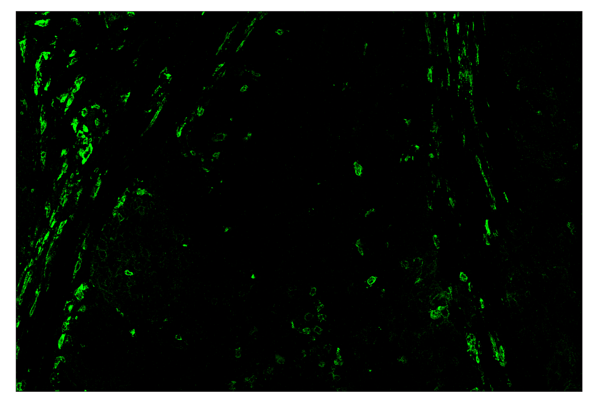
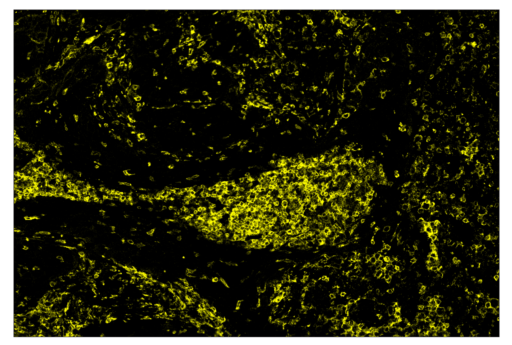
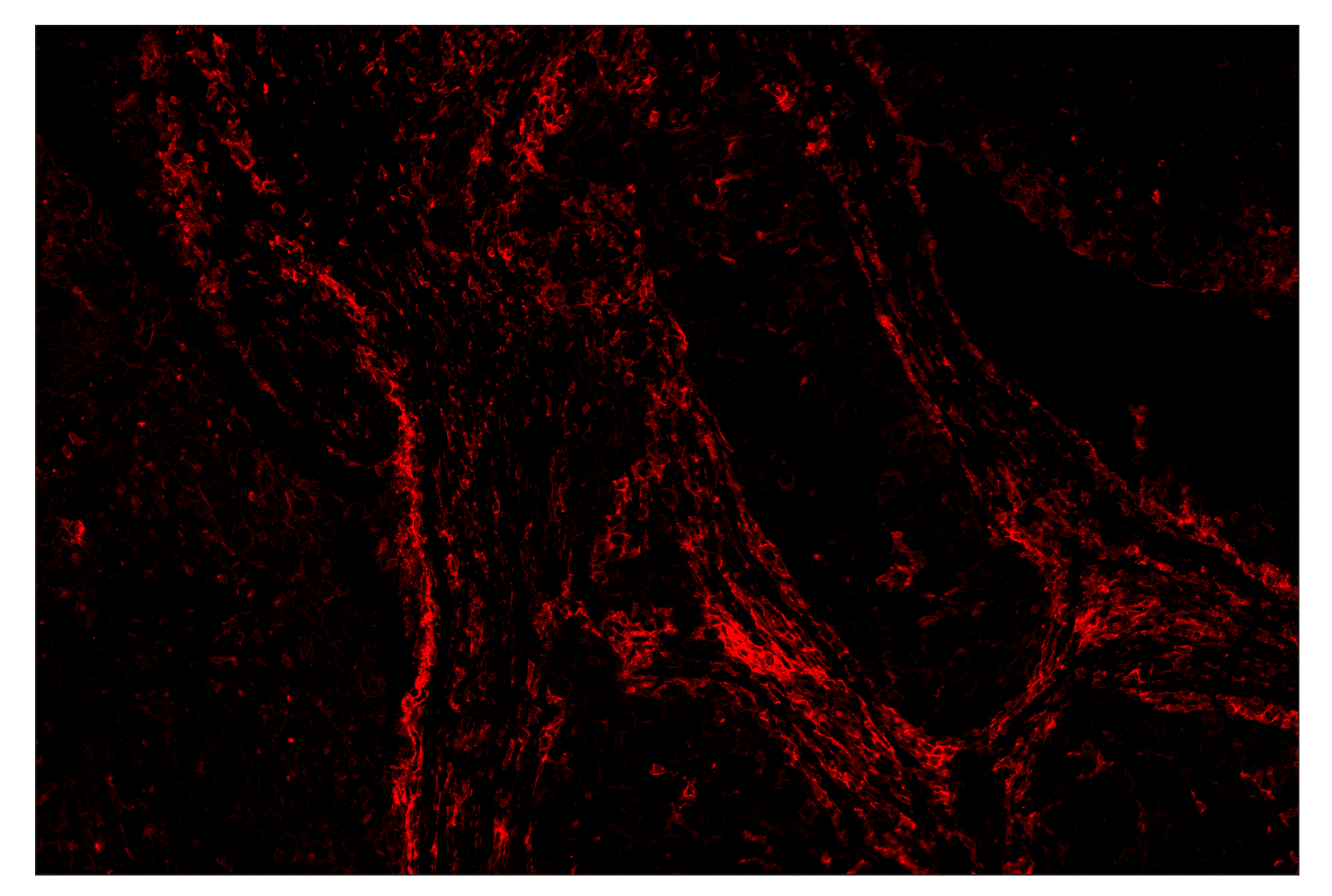
| SIRPα/SHPS1 (D6I3M) Rabbit mAb (SignalStar™ Conjugate 0034) | 69545 | 50 µl | H M | Rabbit IgG |
| Complementary Oligo (CO-0034-750) | 80048 | 22 µl |
Storage
Description
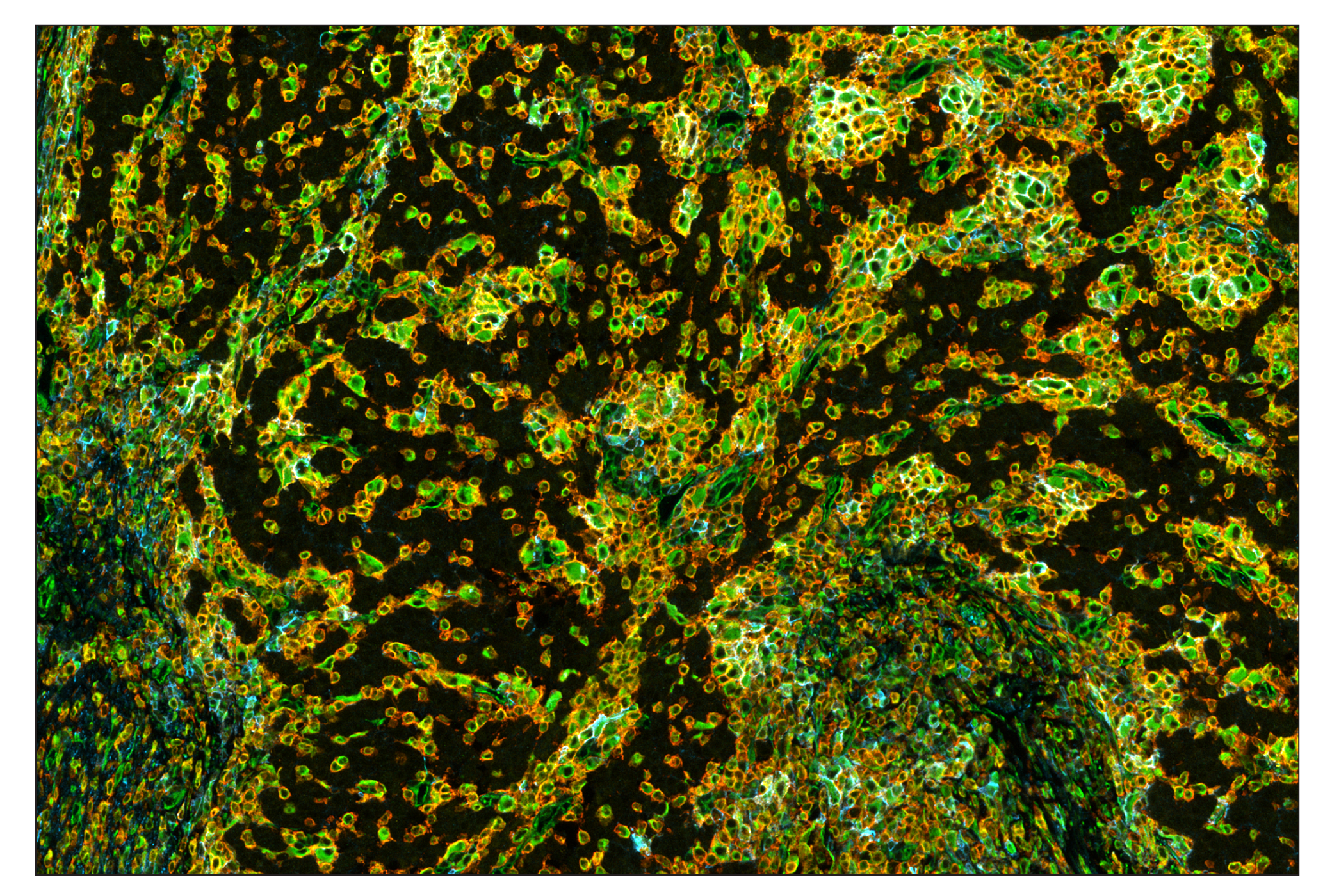
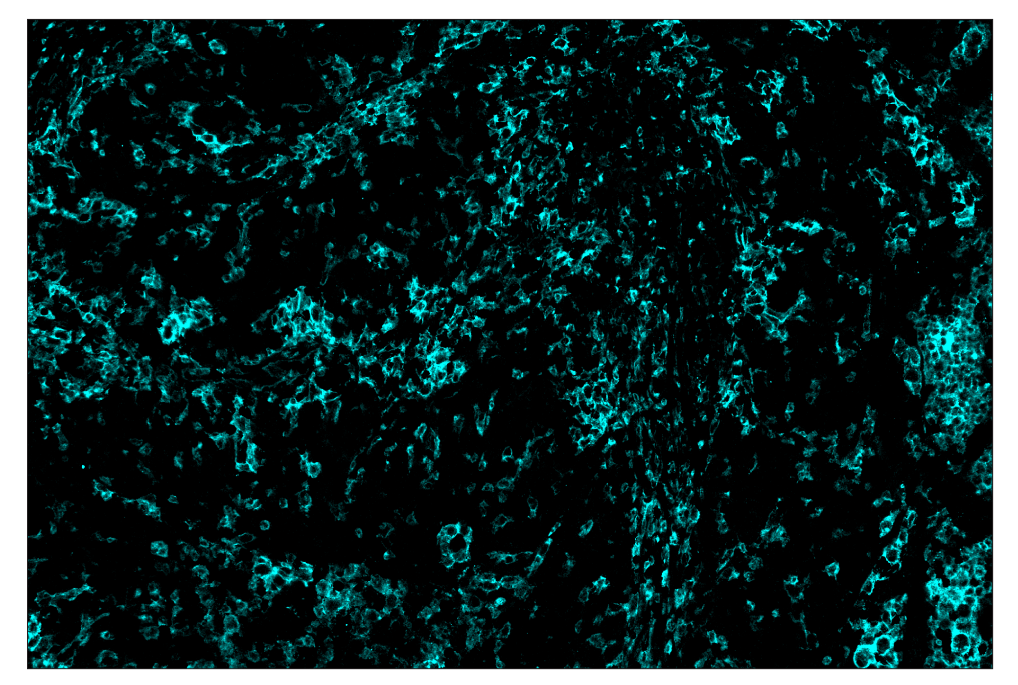
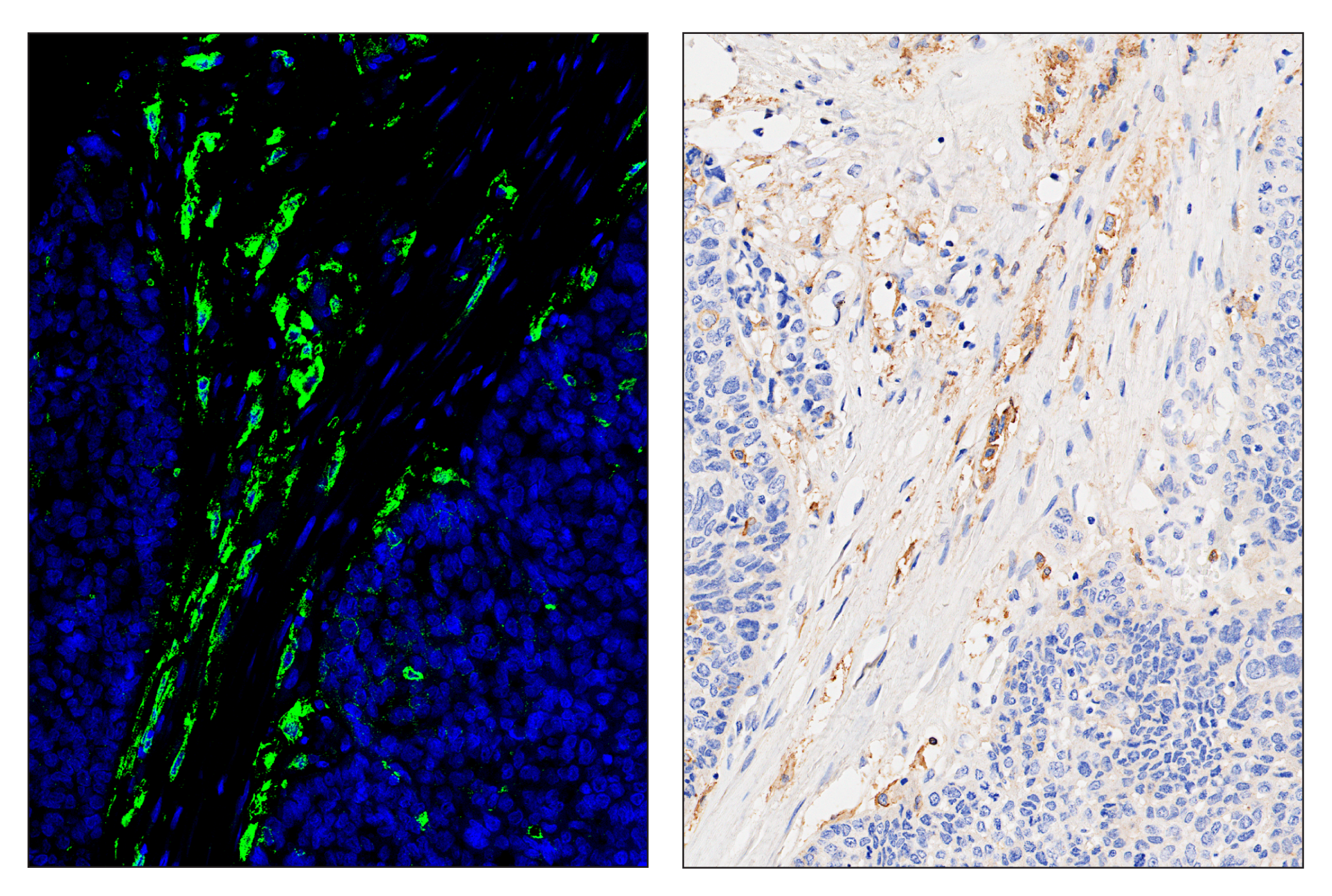
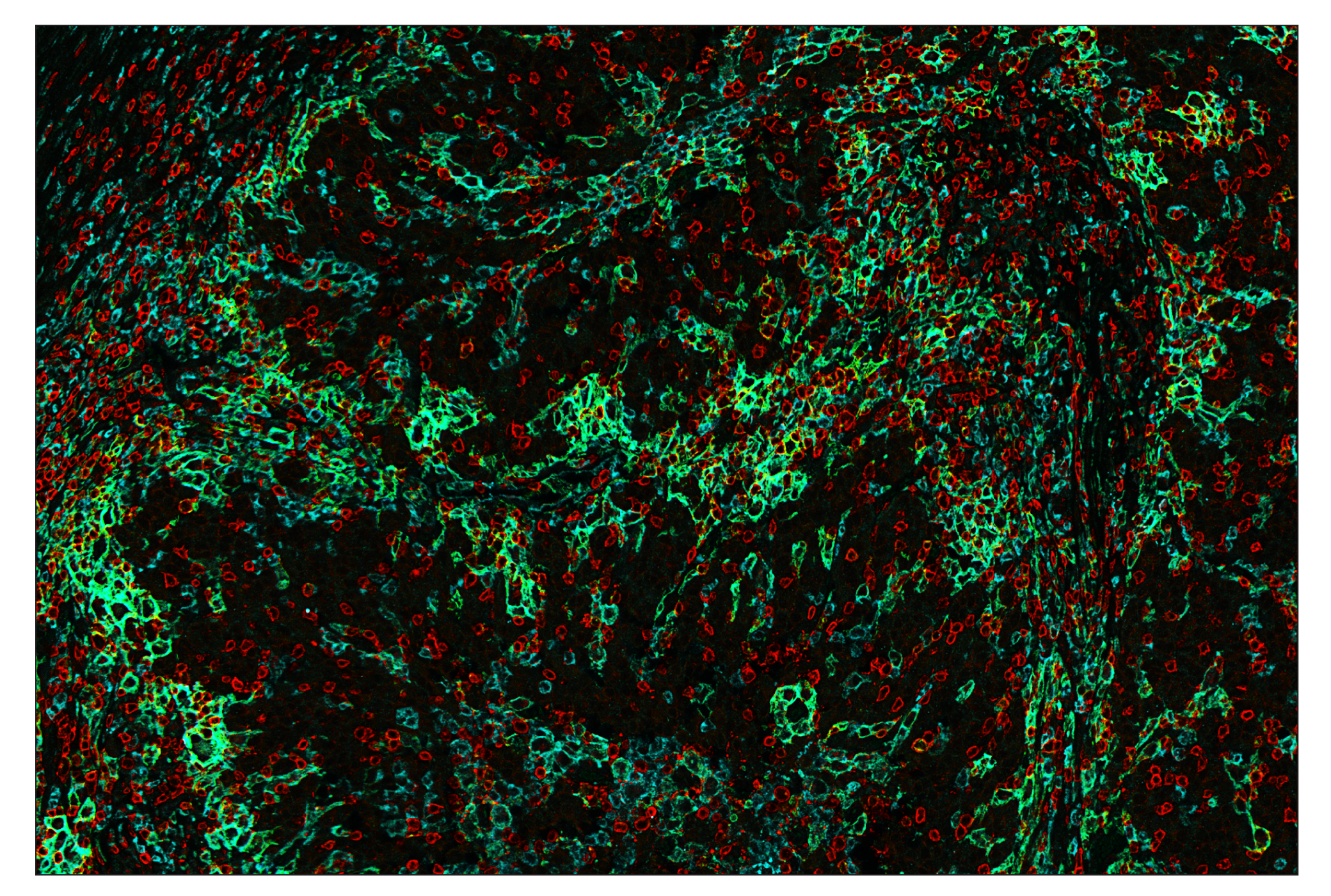
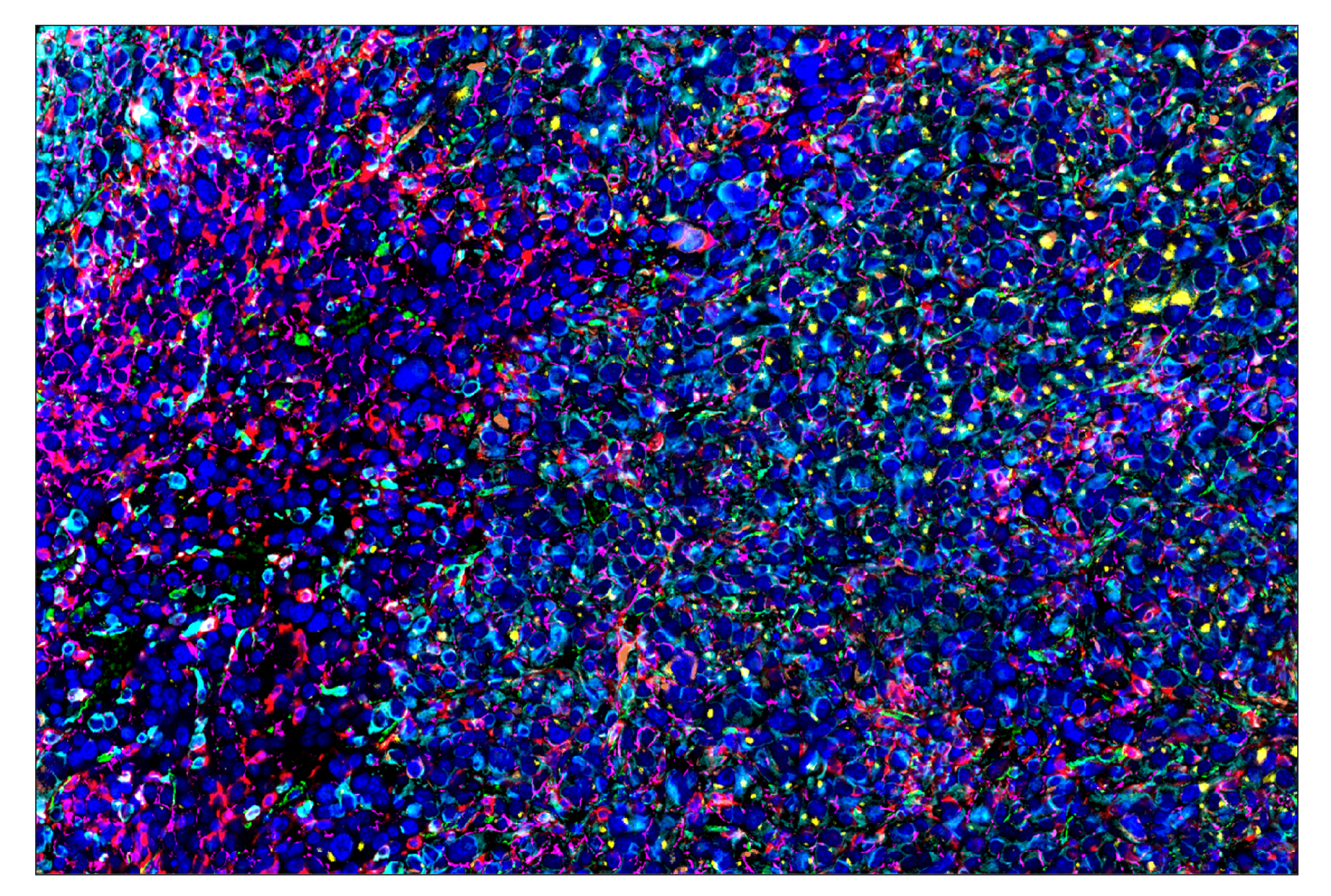
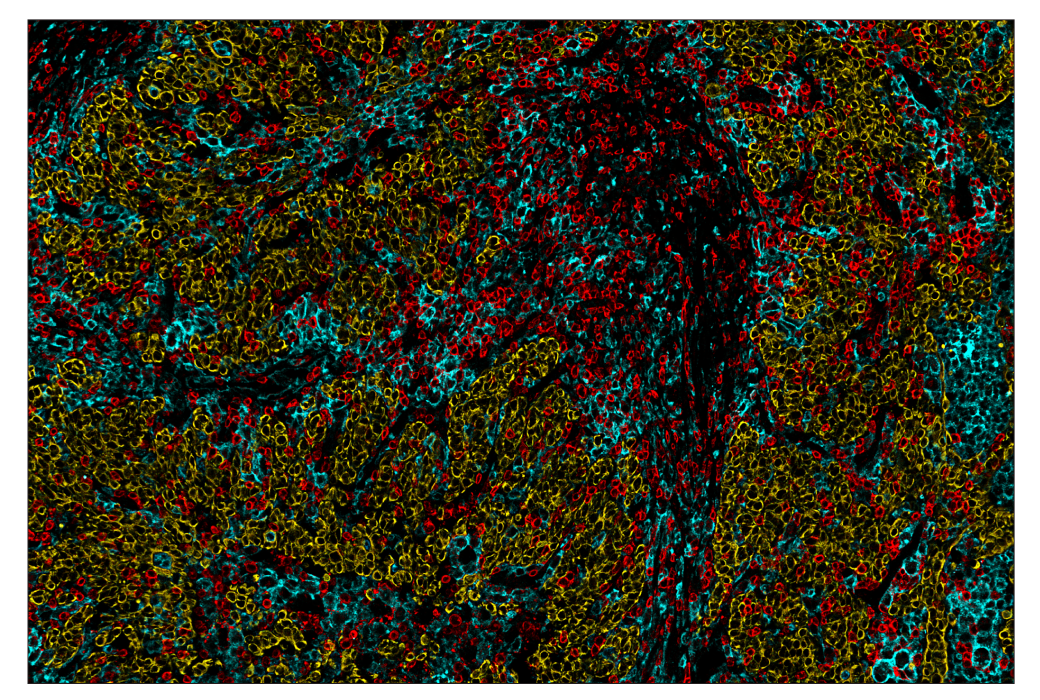
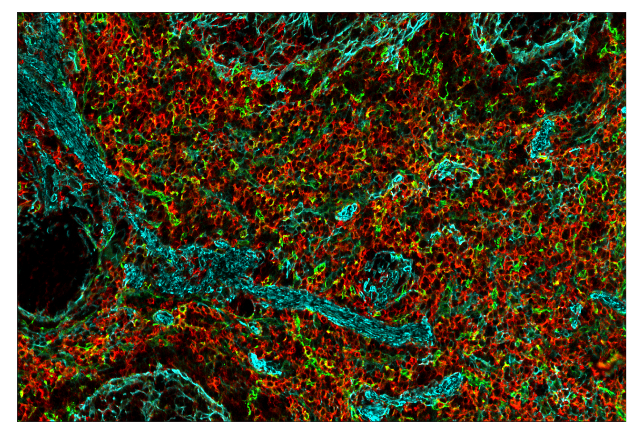
SignalStar multiplex immunohistochemistry (IHC) is an advanced technology for labeling multiple proteins simultaneously in tissue samples using specific primary antibodies and fluorescent detection reagents. This technology offers accuracy and reliability in visualizing and analyzing protein expression while maintaining spatial context and tissue architecture.
SignalStar Oligo-Antibody Pairs are compatible with the SignalStar Multiplex IHC Buffer Kits for use in fluorescent multiplex imaging experiments. This product includes the oligo-conjugated antibodies and complementary oligos required for labeling your target protein on up to 10 slides. SignalStar Multiplex IHC Buffer Kits are required to amplify and image the target signal. Multiple oligo-antibody pairs can be conveniently combined into a multiplex panel using the SignalStar Multiplex IHC Panel Builder. SignalStar Multiplex IHC Kits & Reagents are not compatible with all of Cell Signaling Technology® products and protocols that are recommended for use in immunohistochemical assays.
Specificity/Sensitivity
Source / Purification
Monoclonal antibody is produced by immunizing animals with a synthetic peptide corresponding to residues surrounding Pro413 of human SIRPα/SHPS1 protein.
Background
SHP-substrate 1 (SHPS1, SIRPα) is a single-pass membrane protein and member of both the immunoglobulin superfamily and the signal regulatory protein (SIRP) family. Following growth hormone stimulation or integrin binding, SHPS1 is phosphorylated at several tyrosine residues within its cytoplasmic tail. These phosphorylation events promote association between SHPS1 and multiple signaling proteins, including SHP-1, SHP-2, Grb2 and Shc via their SH2 domains (1-4). Recruitment of SHP-1 and SHP-2 results in SHPS1 dephosphorylation and suppression of tyrosine kinase signaling (1-3,5). The tyrosine kinase JAK2 associates with SHPS1 via its carboxy terminus and phosphorylates SHPS1 in response to extracellular stimuli (5). Research studies show that Src associates with and may phosphorylate SHPS1 in response to insulin (4). In macrophages, SHPS1 can form a complex with the Src pathway adaptor protein SKAP2, Fyn-binding protein FYB, and the tyrosine kinase PYK2 (6). The SHPS1 extracellular domain contains at least three IgG-like domains that interact with CD47, a ubiquitously expressed, integrin-associated protein that acts as a repressive cue in both immune and neuronal cells (7,8). The interaction between CD47 and SHPS1 on opposing cells can inhibit cellular migration (9), promote "tethering" between macrophages and target cells during engulfment (10), facilitate self versus non-self recognition (11), and maintain immune homeostasis (12). SHPS1 plays a critical role in modulating the immune response and inflammation, and may play a role in neuronal development (13,14). The interaction between SHPS1 and CD47 may be an exploitable target in cancer therapy (15-17).
- Kharitonenkov, A. et al. (1997) Nature 386, 181-6.
- Ochi, F. et al. (1997) Biochem Biophys Res Commun 239, 483-7.
- Takada, T. et al. (1998) J Biol Chem 273, 9234-42.
- Shen, X. et al. (2009) Mol Cell Proteomics 8, 1539-51.
- Stofega, M.R. et al. (2000) J Biol Chem 275, 28222-9.
- Timms, J.F. et al. (1999) Curr Biol 9, 927-30.
- Seiffert, M. et al. (1999) Blood 94, 3633-43.
- Vernon-Wilson, E.F. et al. (2000) Eur J Immunol 30, 2130-7.
- Motegi, S. et al. (2003) EMBO J 22, 2634-44.
- Tada, K. et al. (2003) J Immunol 171, 5718-26.
- van Beek, E.M. et al. (2005) J Immunol 175, 7781-7.
- Legrand, N. et al. (2011) Proc Natl Acad Sci U S A 108, 13224-9.
- Sarfati, M. et al. (2008) Curr Drug Targets 9, 842-50.
- Matozaki, T. et al. (2009) Trends Cell Biol 19, 72-80.
- Hara, K. et al. (2011) Cancer Res 71, 1229-34.
- Willingham, S.B. et al. (2012) Proc Natl Acad Sci U S A 109, 6662-7.
- Weiskopf, K. et al. (2013) Science 341, 88-91.
Species Reactivity
Species reactivity is determined by testing in at least one approved application (e.g., western blot).
Cross-Reactivity Key
H: human M: mouse R: rat Hm: hamster Mk: monkey Vir: virus Mi: mink C: chicken Dm: D. melanogaster X: Xenopus Z: zebrafish B: bovine Dg: dog Pg: pig Sc: S. cerevisiae Ce: C. elegans Hr: horse GP: Guinea Pig Rab: rabbit All: all species expected
Trademarks and Patents
Limited Uses
Except as otherwise expressly agreed in a writing signed by a legally authorized representative of CST, the following terms apply to Products provided by CST, its affiliates or its distributors. Any Customer's terms and conditions that are in addition to, or different from, those contained herein, unless separately accepted in writing by a legally authorized representative of CST, are rejected and are of no force or effect.
Products are labeled with For Research Use Only or a similar labeling statement and have not been approved, cleared, or licensed by the FDA or other regulatory foreign or domestic entity, for any purpose. Customer shall not use any Product for any diagnostic or therapeutic purpose, or otherwise in any manner that conflicts with its labeling statement. Products sold or licensed by CST are provided for Customer as the end-user and solely for research and development uses. Any use of Product for diagnostic, prophylactic or therapeutic purposes, or any purchase of Product for resale (alone or as a component) or other commercial purpose, requires a separate license from CST. Customer shall (a) not sell, license, loan, donate or otherwise transfer or make available any Product to any third party, whether alone or in combination with other materials, or use the Products to manufacture any commercial products, (b) not copy, modify, reverse engineer, decompile, disassemble or otherwise attempt to discover the underlying structure or technology of the Products, or use the Products for the purpose of developing any products or services that would compete with CST products or services, (c) not alter or remove from the Products any trademarks, trade names, logos, patent or copyright notices or markings, (d) use the Products solely in accordance with CST Product Terms of Sale and any applicable documentation, and (e) comply with any license, terms of service or similar agreement with respect to any third party products or services used by Customer in connection with the Products.