WB, IP
H M R
Endogenous
18
Rabbit IgG
#P61966
1174
Product Information
Product Usage Information
| Application | Dilution |
|---|---|
| Western Blotting | 1:1000 |
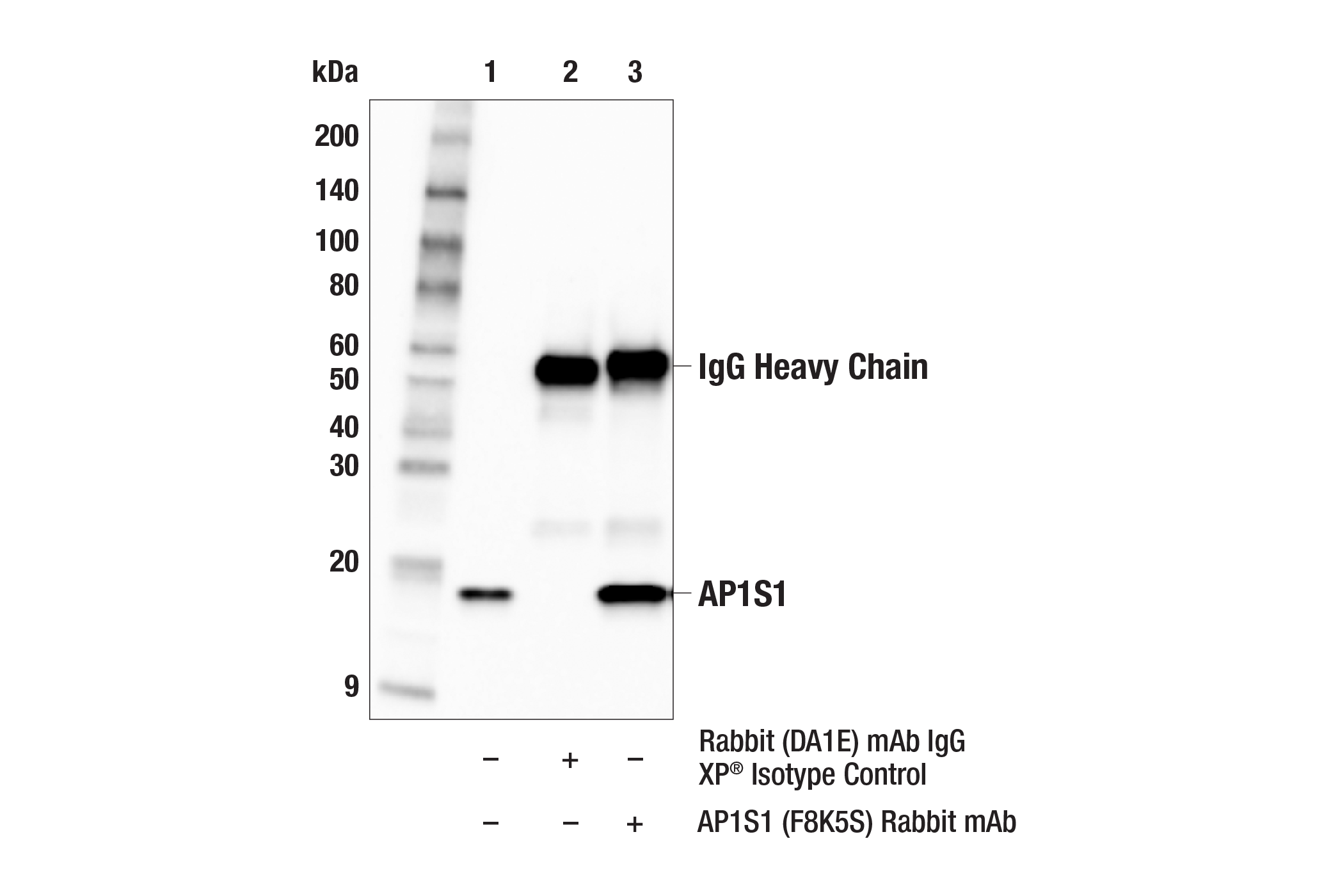
| Immunoprecipitation | 1:50 |
Storage
Specificity / Sensitivity
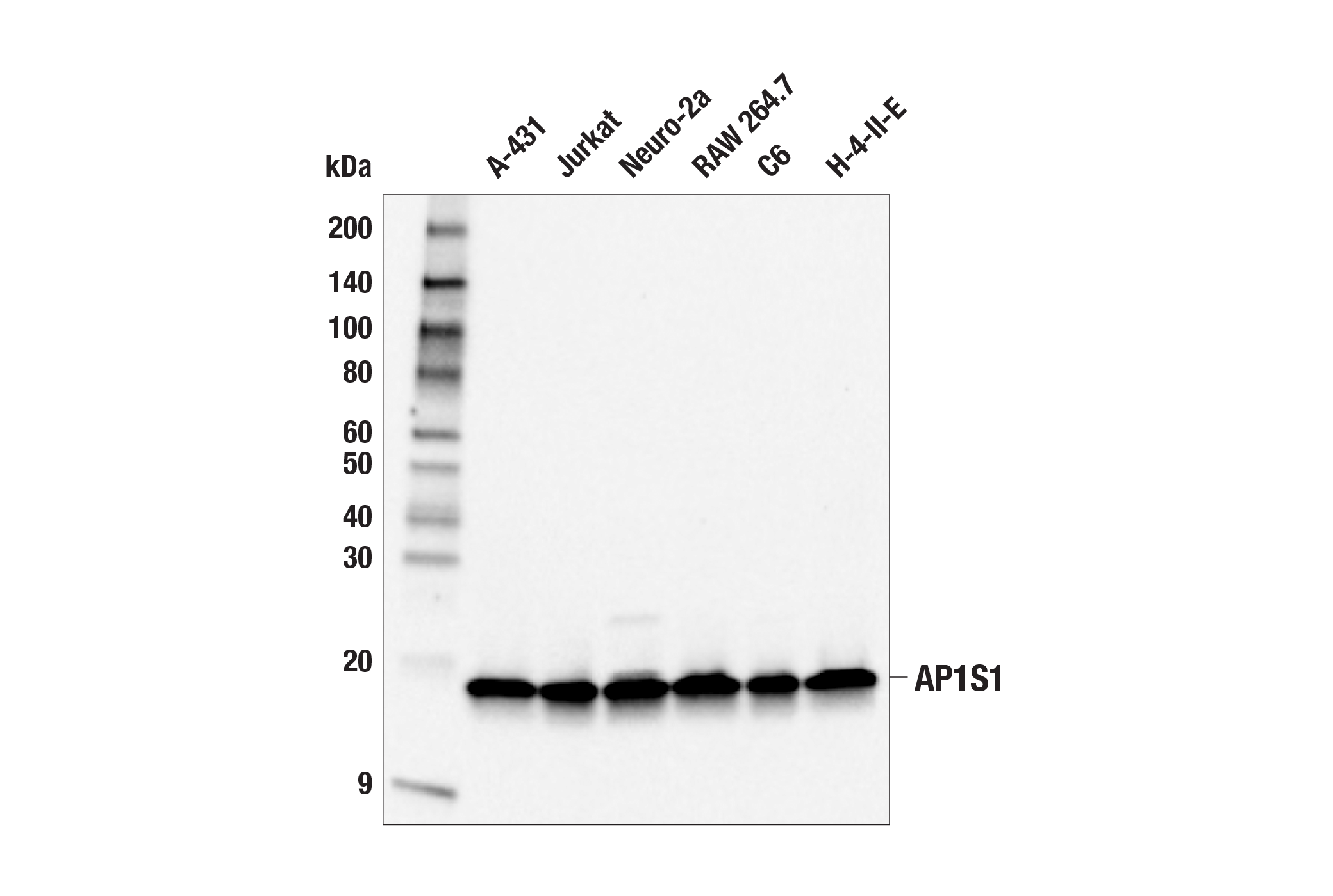
Species Reactivity:
Human, Mouse, Rat
Source / Purification
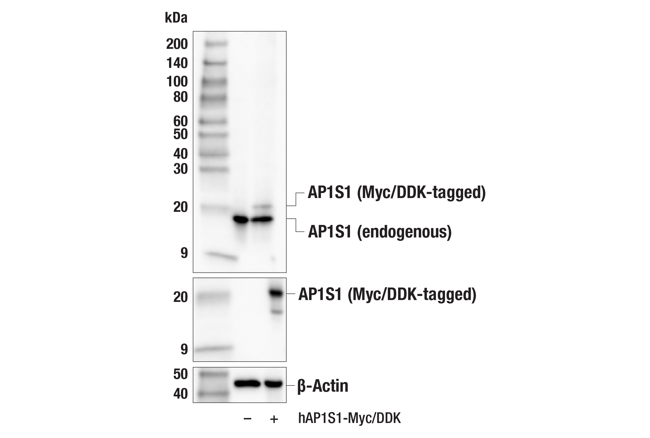
Monoclonal antibody is produced by immunizing animals with a synthetic peptide corresponding to residues near the carboxy terminus of human AP1S1 protein.
Background
The adaptor protein complex 1 (AP-1) is a heterotetrameric protein complex composed of two large adaptins (AP1G1 and AP1B1), a medium adaptin (AP1M1 or AP1M2), and one of several small adaptins (AP3S1, AP1S2, or AP1S3). AP-1 complexes containing AP1M1 are ubiquitously expressed, while complexes containing AP1M2 display a more restricted tissue expression pattern. Research studies have demonstrated that the AP-1 complex is localized to the trans-Golgi network and recycling endosomes where it regulates the bidirectional trafficking of cargo proteins between these subcellular compartments as well as the biogenesis of secretory vesicles (1-3).
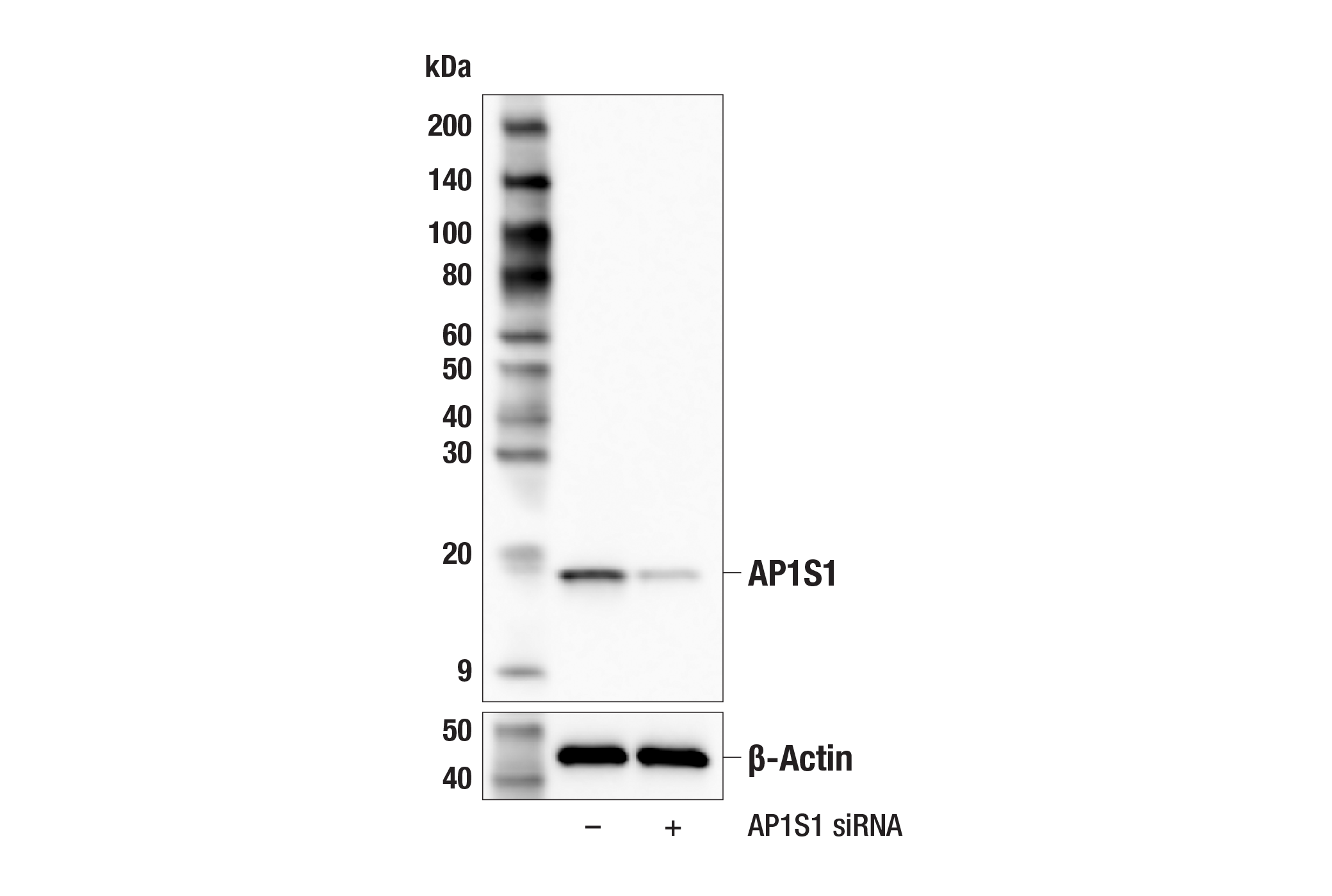
AP1S1 is one of three possible small adaptin subunits capable of being incorporated into the AP-1 heterotetrameric complex. Mutations in AP1S1 contribute to MEDNIK syndrome, which is characterized by a defect in copper metabolism due to aberrant sorting and trafficking of copper pumps ATP7A and ATP7B (4).
Species Reactivity
Species reactivity is determined by testing in at least one approved application (e.g., western blot).
Western Blot Buffer
IMPORTANT: For western blots, incubate membrane with diluted primary antibody in 5% w/v BSA, 1X TBS, 0.1% Tween® 20 at 4°C with gentle shaking, overnight.
Applications Key
WB: Western Blotting IP: Immunoprecipitation
Cross-Reactivity Key
H: human M: mouse R: rat Hm: hamster Mk: monkey Vir: virus Mi: mink C: chicken Dm: D. melanogaster X: Xenopus Z: zebrafish B: bovine Dg: dog Pg: pig Sc: S. cerevisiae Ce: C. elegans Hr: horse GP: Guinea Pig Rab: rabbit All: all species expected
Trademarks and Patents
Limited Uses
Except as otherwise expressly agreed in a writing signed by a legally authorized representative of CST, the following terms apply to Products provided by CST, its affiliates or its distributors. Any Customer's terms and conditions that are in addition to, or different from, those contained herein, unless separately accepted in writing by a legally authorized representative of CST, are rejected and are of no force or effect.
Products are labeled with For Research Use Only or a similar labeling statement and have not been approved, cleared, or licensed by the FDA or other regulatory foreign or domestic entity, for any purpose. Customer shall not use any Product for any diagnostic or therapeutic purpose, or otherwise in any manner that conflicts with its labeling statement. Products sold or licensed by CST are provided for Customer as the end-user and solely for research and development uses. Any use of Product for diagnostic, prophylactic or therapeutic purposes, or any purchase of Product for resale (alone or as a component) or other commercial purpose, requires a separate license from CST. Customer shall (a) not sell, license, loan, donate or otherwise transfer or make available any Product to any third party, whether alone or in combination with other materials, or use the Products to manufacture any commercial products, (b) not copy, modify, reverse engineer, decompile, disassemble or otherwise attempt to discover the underlying structure or technology of the Products, or use the Products for the purpose of developing any products or services that would compete with CST products or services, (c) not alter or remove from the Products any trademarks, trade names, logos, patent or copyright notices or markings, (d) use the Products solely in accordance with CST Product Terms of Sale and any applicable documentation, and (e) comply with any license, terms of service or similar agreement with respect to any third party products or services used by Customer in connection with the Products.