Revision 1
Cell Signaling Technology

Orders: 877-616-CELL (2355) [email protected]

Support: 877-678-TECH (8324)

Web: [email protected] cellsignal.com

3 Trask LaneDanversMassachusetts01923USA
For Research Use Only. Not for Use in Diagnostic Procedures.
:

:

:

:

UniProt ID:

#P16220

Entrez-Gene Id:

1385

Product Information

Storage

Supplied in 10 mM sodium HEPES (pH 7.5), 150 mM NaCl, 100 µg/ml BSA, 50% glycerol and less than 0.02% sodium azide. Store at –20°C. Do not aliquot the antibody.

Specificity / Sensitivity

Phospho-CREB (Ser133) (87G3) Rabbit mAb detects endogenous levels of CREB only when phosphorylated at Ser133. This antibody also detects the phosphorylated form of CREB-related protein ATF-1. CREB (48H2) Rabbit mAb detects endogenous levels of total CREB protein.

Source / Purification

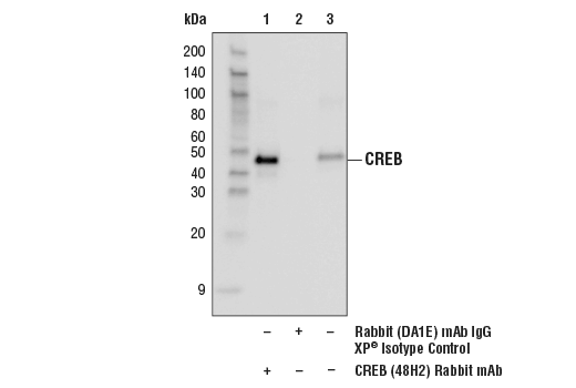
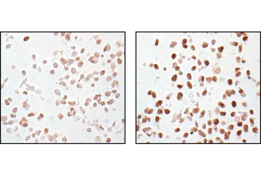
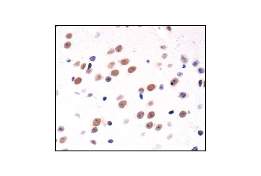
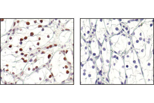
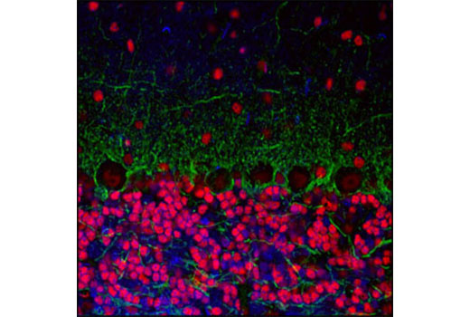
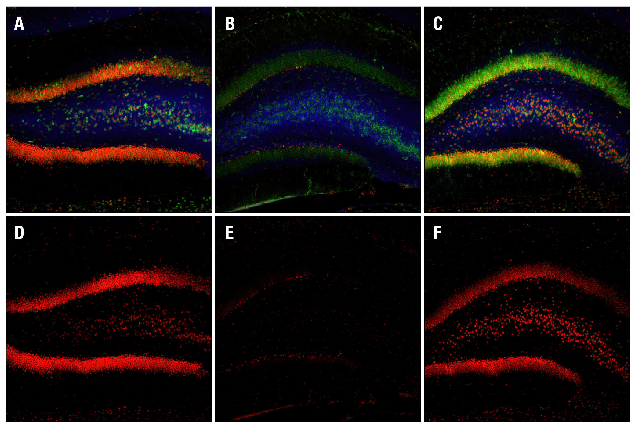
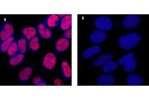
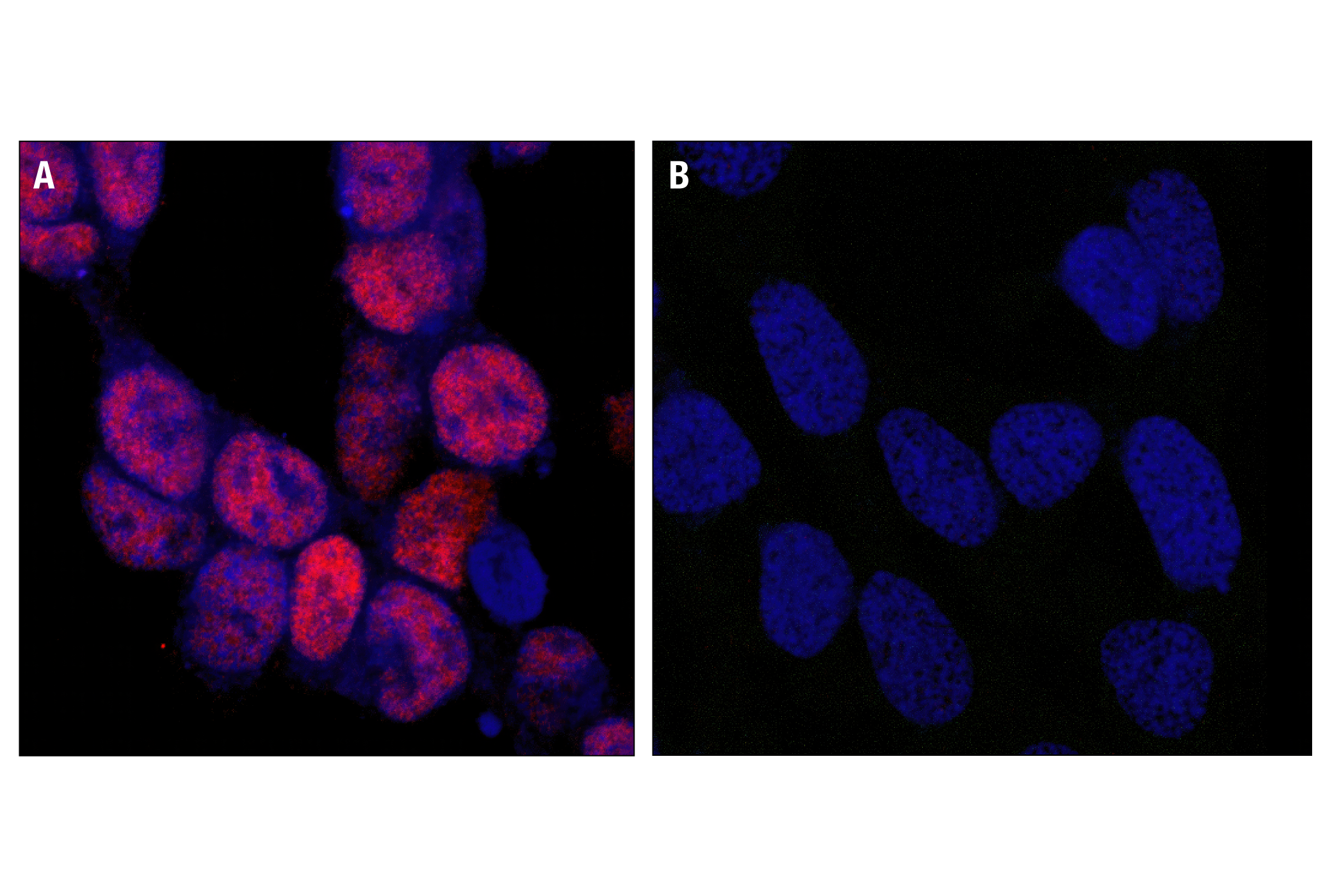
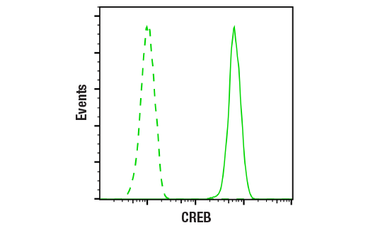
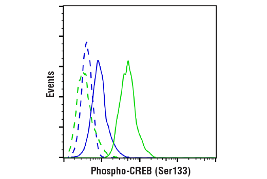
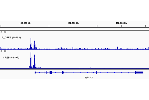
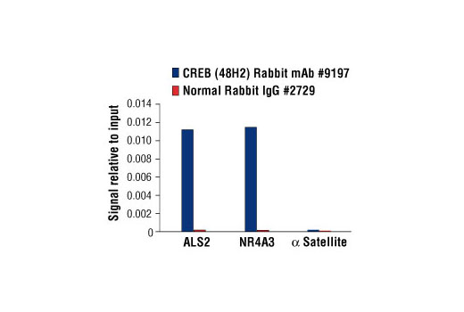
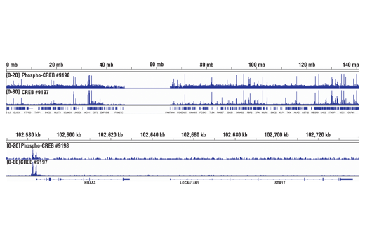
Monoclonal antibody is produced by immunizing animals with a synthetic phosphopeptide corresponding to residues surrounding Ser133 of human CREB (Phospho-CREB (Ser133) (87G3) Rabbit mAb), or with a synthetic peptide corresponding to residues near the carboxy-terminus of human CREB (CREB (48H2) Rabbit mAb).

Product Description

The PhosphoPlus CREB (Ser133) Antibody Kit provides reagents and protocols for the rapid analysis of the phosphorylation status of CREB at Ser133.

Background

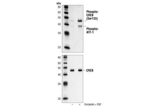
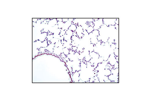
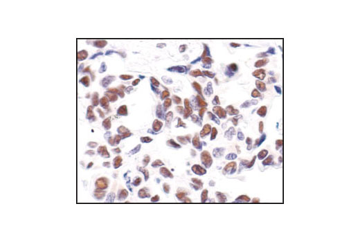
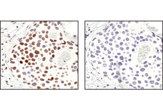
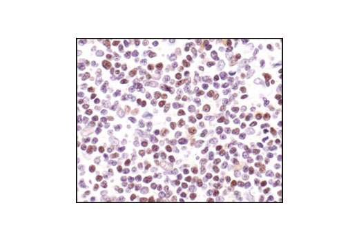
CREB is a bZIP transcription factor that activates target genes through cAMP response elements. CREB is able to mediate signals from numerous physiological stimuli, resulting in regulation of a broad array of cellular responses. While CREB is expressed in numerous tissues, it plays a large regulatory role in the nervous system. CREB is believed to play a key role in promoting neuronal survival, precursor proliferation, neurite outgrowth, and neuronal differentiation in certain neuronal populations (1-3). Additionally, CREB signaling is involved in learning and memory in several organisms (4-6). CREB is able to selectively activate numerous downstream genes through interactions with different dimerization partners. CREB is activated by phosphorylation at Ser133 by various signaling pathways, including Erk, Ca2+, and stress signaling. Some of the kinases involved in phosphorylating CREB at Ser133 are p90RSK, MSK, CaMKIV, and MAPKAPK-2 (7-9).

  1. Lonze, B.E. et al. (2002) Neuron 34, 371-85.
  2. Lee, M.M. et al. (1999) J Neurosci Res 55, 702-12.
  3. Redmond, L. et al. (2002) Neuron 34, 999-1010.
  4. Dash, P.K. et al. (1990) Nature 345, 718-21.
  5. Yin, J.C. et al. (1994) Cell 79, 49-58.
  6. Guzowski, J.F. and McGaugh, J.L. (1997) Proc Natl Acad Sci USA 94, 2693-8.
  7. Xing, J. et al. (1998) Mol Cell Biol 18, 1946-55.
  8. Ribar, T.J. et al. (2000) J Neurosci 20, RC107.
  9. Tan, Y. et al. (1996) EMBO J 15, 4629-42.

Species Reactivity

Species reactivity is determined by testing in at least one approved application (e.g., western blot).

Cross-Reactivity Key

H: human M: mouse R: rat Hm: hamster Mk: monkey Vir: virus Mi: mink C: chicken Dm: D. melanogaster X: Xenopus Z: zebrafish B: bovine Dg: dog Pg: pig Sc: S. cerevisiae Ce: C. elegans Hr: horse GP: Guinea Pig Rab: rabbit All: all species expected

Trademarks and Patents

Cell Signaling Technology is a trademark of Cell Signaling Technology, Inc.
PhosphoPlus is a registered trademark of Cell Signaling Technology, Inc.
All other trademarks are the property of their respective owners. Visit cellsignal.com/trademarks for more information.

Limited Uses

Except as otherwise expressly agreed in a writing signed by a legally authorized representative of CST, the following terms apply to Products provided by CST, its affiliates or its distributors. Any Customer's terms and conditions that are in addition to, or different from, those contained herein, unless separately accepted in writing by a legally authorized representative of CST, are rejected and are of no force or effect.

Products are labeled with For Research Use Only or a similar labeling statement and have not been approved, cleared, or licensed by the FDA or other regulatory foreign or domestic entity, for any purpose. Customer shall not use any Product for any diagnostic or therapeutic purpose, or otherwise in any manner that conflicts with its labeling statement. Products sold or licensed by CST are provided for Customer as the end-user and solely for research and development uses. Any use of Product for diagnostic, prophylactic or therapeutic purposes, or any purchase of Product for resale (alone or as a component) or other commercial purpose, requires a separate license from CST. Customer shall (a) not sell, license, loan, donate or otherwise transfer or make available any Product to any third party, whether alone or in combination with other materials, or use the Products to manufacture any commercial products, (b) not copy, modify, reverse engineer, decompile, disassemble or otherwise attempt to discover the underlying structure or technology of the Products, or use the Products for the purpose of developing any products or services that would compete with CST products or services, (c) not alter or remove from the Products any trademarks, trade names, logos, patent or copyright notices or markings, (d) use the Products solely in accordance with CST Product Terms of Sale and any applicable documentation, and (e) comply with any license, terms of service or similar agreement with respect to any third party products or services used by Customer in connection with the Products.

Revision 1
#9190

PhosphoPlus® CREB (Ser133) Antibody Kit

PhosphoPlus® CREB (Ser133) Antibody Kit: Image 1 Expand Image
CUT&RUN was performed with 293 cells treated with Forskolin #3828 (30 μM) for 1h and either CREB (48H2) Rabbit mAb or Phospho-CREB (Ser133) (87G3) Rabbit mAb #9198, using CUT&RUN Assay Kit #86652. DNA Libraries were prepared using SimpleChIP® ChIP-seq DNA Library Prep Kit for Illumina® #56795. The figure shows binding across NR4A3 gene.
PhosphoPlus® CREB (Ser133) Antibody Kit: Image 2 Expand Image
Simple Western™ analysis of lysates (0.1 mg/mL) from SK-N-MC cells treated with Forskolin (30uM, 30min) and IBMX (0.5mM, 30 min) using Phospho-CREB (Ser133) (87G3) Rabbit mAb #9198. The virtual lane view (left) shows the target band (as indicated) at 1:10 and 1:50 dilutions of primary antibody. This antibody also detects the phosphorylated form of the CREB-related protein, ATF-1 (as indicated). The corresponding electropherogram view (right) plots chemiluminescence by molecular weight along the capillary at 1:10 (blue line) and 1:50 (green line) dilutions of primary antibody. This experiment was performed under reducing conditions on the Jess™ ​​​​​​​Simple Western instrument from ProteinSimple, a BioTechne brand, using the 12-230 kDa separation module.
PhosphoPlus® CREB (Ser133) Antibody Kit: Image 3 Expand Image
After the primary antibody is bound to the target protein, a complex with HRP-linked secondary antibody is formed. The LumiGLO® is added and emits light during enzyme catalyzed decomposition.
PhosphoPlus® CREB (Ser133) Antibody Kit: Image 4 Expand Image
After the primary antibody is bound to the target protein, a complex with HRP-linked secondary antibody is formed. The LumiGLO® is added and emits light during enzyme catalyzed decomposition.
PhosphoPlus® CREB (Ser133) Antibody Kit: Image 5 Expand Image
Western blot analysis of extracts from SK-N-MC cells, untreated (-) or treated with Forskolin #3828 (30 μM) and IBMX (0.5 mM) for 30 min (+), using Phospho-CREB (Ser133) (87G3) Rabbit mAb #9198 (upper) and CREB (48H2) Rabbit mAb #9197 (lower).
PhosphoPlus® CREB (Ser133) Antibody Kit: Image 6 Expand Image
Western Blot analysis of extracts from SK-N-MC, COS, NIH/3T3, C6 and Drosophila S2 cells, using CREB (48H2) Rabbit mAb.
PhosphoPlus® CREB (Ser133) Antibody Kit: Image 7 Expand Image
Western blot analysis of extracts from SK-N-MC cells, untreated or forskolin- and FGF-treated, using Phospho-CREB (Ser133) (87G3) Rabbit mAb (upper) or CREB (48H2) Rabbit mAb #9197 (lower).
PhosphoPlus® CREB (Ser133) Antibody Kit: Image 8 Expand Image
Immunoprecipitation of CREB from SK-N-MC extracts. Lane 1 is CREB (48H2) Rabbit mAb, lane 2 is Rabbit (DA1E) mAb IgG XP® Isotype Control #3900, and lane 3 is 10% input. Western blot analysis was perfomed using CREB (86B10) Mouse mAb #9104. Anti-mouse IgG, HRP-linked Antibody #7076 was used as a secondary antibody.
PhosphoPlus® CREB (Ser133) Antibody Kit: Image 9 Expand Image
Immunohistochemical analysis of paraffin-embedded human lung carcinoma, showing nuclear staining, using Phospho-CREB (Ser133) (87G3) Rabbit mAb.
PhosphoPlus® CREB (Ser133) Antibody Kit: Image 10 Expand Image
Immunohistochemical analysis of paraffin-embedded human astrocytoma, using CREB (48H2) Rabbit mAb.
PhosphoPlus® CREB (Ser133) Antibody Kit: Image 11 Expand Image
Immunohistochemical analysis of paraffin-embedded mouse lung using Phospho-CREB (Ser133) (87G3) Rabbit mAb.
PhosphoPlus® CREB (Ser133) Antibody Kit: Image 12 Expand Image
Immunohistochemical analysis of paraffin-embedded human lung carcinoma, using CREB (48H2) Rabbit mAb.
PhosphoPlus® CREB (Ser133) Antibody Kit: Image 13 Expand Image
Immunohistochemical analysis of paraffin-embedded human breast carcinoma, using Phospho-CREB (Ser133) (87G3) Rabbit mAb in the presence of control peptide (left) or Phospho-CREB (Ser133) Blocking Peptide #1090 (right).
PhosphoPlus® CREB (Ser133) Antibody Kit: Image 14 Expand Image
Immunohistochemical analysis of paraffin-embedded human Non-Hodgkin's lymphoma, using CREB (48H2) Rabbit mAb.
PhosphoPlus® CREB (Ser133) Antibody Kit: Image 15 Expand Image
Immunohistochemical analysis of paraffin-embedded SK-N-MC cells, untreated (left) or IBMX- and forskolin-treated (right), showing induced nuclear staining, using Phospho-CREB (Ser133) (87G3) Rabbit mAb.
PhosphoPlus® CREB (Ser133) Antibody Kit: Image 16 Expand Image
Immunohistochemical analysis of paraffin-embedded mouse brain, using CREB (48H2) Rabbit mAb.
PhosphoPlus® CREB (Ser133) Antibody Kit: Image 17 Expand Image
Immunohistochemical analysis of paraffin-embedded human renal cell carcinoma, untreated (left) or lambda phosphatase-treated (right), using Phospho-CREB (Ser133) (87G3) Rabbit mAb.
PhosphoPlus® CREB (Ser133) Antibody Kit: Image 18 Expand Image
Confocal immunofluorescent analysis of mouse cerebellum labeled with CREB (48H2) Rabbit mAb (red) and Neurofilament-L (DA2) Mouse mAb #2835 (green). Blue pseudocolor =DRAQ5® #4084 (fluorescent DNA dye).
PhosphoPlus® CREB (Ser133) Antibody Kit: Image 19 Expand Image
Confocal immunofluorescent images of rat dentate gyrus, either sham-operated (left) or 15 min ischemia followed by 30 min (center) and 4 h (right) reperfusion, labeled with Phospho-CREB (Ser133) (87G3) Rabbit mAb (red), Neurofilament-L (DA2) Mouse mAb #2835 (blue) and Phospho-S6 Ribosomal Protein (Ser235/236) (2F9) Rabbit mAb (Alexa Fluor® 488 Conjugate) #4854.
PhosphoPlus® CREB (Ser133) Antibody Kit: Image 20 Expand Image
Confocal immunofluorescent analysis of SK-N-MC cells showing nuclear stain with CREB (48H2) Rabbit mAb (A, red) compared to an isotype control (B). Blue pseudocolor =DRAQ5® (fluorescent DNA dye).
PhosphoPlus® CREB (Ser133) Antibody Kit: Image 21 Expand Image
Confocal microscopic images of SK-N-MC cells showing nuclear stain after 25 minute treatment with Forskolin and IBMX using Phospho-CREB (Ser133) (87G3) Rabbit mAb (left, red) compared to untreated cells (right). Blue pseudocolor = DRAQ5® #4084 (fluorescent DNA dye).
PhosphoPlus® CREB (Ser133) Antibody Kit: Image 22 Expand Image
Flow cytometric analysis of Jurkat cells using CREB (48H2) Rabbit mAb (solid line) compared to concentration-matched Rabbit (DA1E) mAb IgG XP® Isotype Control #3900 (dashed line). Anti-rabbit IgG (H+L), F(ab')2 Fragment (Alexa Fluor® 488 Conjugate) #4412 was used as a secondary antibody.
PhosphoPlus® CREB (Ser133) Antibody Kit: Image 23 Expand Image
Flow cytometric analysis of Jurkat cells, untreated (blue) or treated with TPA #4174 (500nM) and Ionomycin #9995 (1uM,4hrs;green) using Phospho-CREB (Ser133) (87G3) Rabbit mAb (solid lines) or concentration-matched Rabbit (DA1E) mAb IgG XP® Isotype Control #3900 (dashed lines). Anti-rabbit IgG (H+L), F(ab')2 Fragment (Alexa Fluor® 488 Conjugate) #4412 was used as a secondary antibody.
PhosphoPlus® CREB (Ser133) Antibody Kit: Image 24 Expand Image
Chromatin immunoprecipitations were performed with cross-linked chromatin from 293 cells treated with Forskolin #3828 (30 μM) for 1h and either Phospho-CREB (Ser133) (87G3) Rabbit mAb or CREB (48H2) Rabbit mAb (#9197), using SimpleChIP® Enzymatic Chromatin IP Kit (Magnetic Beads) #9005. DNA Libraries were prepared using SimpleChIP® ChIP-seq DNA Library Prep Kit for Illumina® (ChIP-seq, CUT&RUN) #56795. The figure shows binding across NR4A3, a known target gene of both Phospho-CREB and CREB (see additional figure containing ChIP-qPCR data).
PhosphoPlus® CREB (Ser133) Antibody Kit: Image 25 Expand Image
Chromatin immunoprecipitations were performed with cross-linked chromatin from 293 cells, treated with Forskolin #3828 (30 μM) for 1h and either 10 μl of CREB (48H2) Rabbit mAb or 2 μl of Normal Rabbit IgG #2729, using SimpleChIP® Enzymatic Chromatin IP Kit (Magnetic Beads) #9003. The enriched DNA was quantified by real-time PCR using human ALS2 exon 1 primers, SimpleChIP® Human NR4A3 Promoter Primers #4829, and SimpleChIP® Human α Satellite Repeat Primers #4486. The amount of immunoprecipitated DNA in each sample is represented as signal relative to the total amount of input chromatin, which is equivalent to one.
PhosphoPlus® CREB (Ser133) Antibody Kit: Image 26 Expand Image
Chromatin immunoprecipitations were performed with cross-linked chromatin from 293 cells treated with Forskolin #3828 (30 μM) for 1h and either Phospho-CREB (Ser133) (87G3) Rabbit mAb or CREB (48H2) Rabbit mAb (#9197), using SimpleChIP® Enzymatic Chromatin IP Kit (Magnetic Beads) #9005. DNA Libraries were prepared using SimpleChIP® ChIP-seq DNA Library Prep Kit for Illumina® (ChIP-seq, CUT&RUN) #56795. The figure shows binding across chromosome 9 (upper), including NR4A3 (lower), a known target gene of both Phospho-CREB and CREB (see additional figure containing ChIP-qPCR data).
PhosphoPlus® CREB (Ser133) Antibody Kit: Image 27 Expand Image
Chromatin immunoprecipitations were performed with cross-linked chromatin from 293 cells treated with Forskolin #3828 (30 μM) for 1h and either Phospho-CREB (Ser133) (87G3) Rabbit mAb or Normal Rabbit IgG #2729 using SimpleChIP® Enzymatic Chromatin IP Kit (Magnetic Beads) #9003. The enriched DNA was quantified by real-time PCR using human ALS2 exon 1 primers, SimpleChIP® Human NR4A3 Promoter Primers #4829, and SimpleChIP® Human α Satellite Repeat Primers #4486. The amount of immunoprecipitated DNA in each sample is represented as signal relative to the total amount of input chromatin, which is equivalent to one.
PhosphoPlus® CREB (Ser133) Antibody Kit: Image 28 Expand Image
CUT&RUN was performed with 293 cells treated with Forskolin #3828 (30 μM) for 1h and Phospho-CREB (Ser133) (87G3) Rabbit mAb, using CUT&RUN Assay Kit #86652. DNA Libraries were prepared using SimpleChIP® ChIP-seq DNA Library Prep Kit for Illumina® (ChIP-seq, CUT&RUN) #56795. The figure shows binding across NR4A3 gene.
PhosphoPlus® CREB (Ser133) Antibody Kit: Image 29 Expand Image
CUT&RUN was performed with 293 cells treated with Forskolin #3828 (30 μM) for 1h and Phospho-CREB (Ser133) (87G3) Rabbit mAb, using CUT&RUN Assay Kit #86652. DNA Libraries were prepared using SimpleChIP® ChIP-seq DNA Library Prep Kit for Illumina® (ChIP-Seq, CUT&RUN) #56795. The figures show binding across chromosome 9 (upper), including NR4A3 (lower) gene.
PhosphoPlus® CREB (Ser133) Antibody Kit: Image 30 Expand Image
CUT&RUN was performed with 293 cells treated with Forskolin #3828 (30 μM) for 1h and either Phospho-CREB (Ser133) (87G3) Rabbit mAb or Rabbit (DA1E) mAb IgG XP® Isotype Control (CUT&RUN) #66362, using CUT&RUN Assay Kit #86652. The enriched DNA was quantified by real-time PCR using human ALS2 exon 1 primers and SimpleChIP® Human α Satellite Repeat Primers #4486. The amount of immunoprecipitated DNA in each sample is represented as signal relative to the total amount of input chromatin, which is equivalent to one.