| Product Includes | Product # | Quantity | Mol. Wt | Isotype/Source |
|---|---|---|---|---|
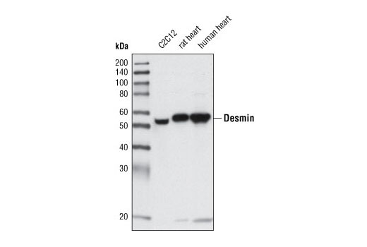
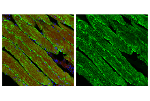
| Desmin (D93F5) XP® Rabbit mAb | 5332 | 20 µl | 53 kDa | Rabbit IgG |
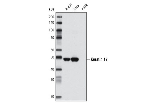
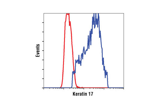
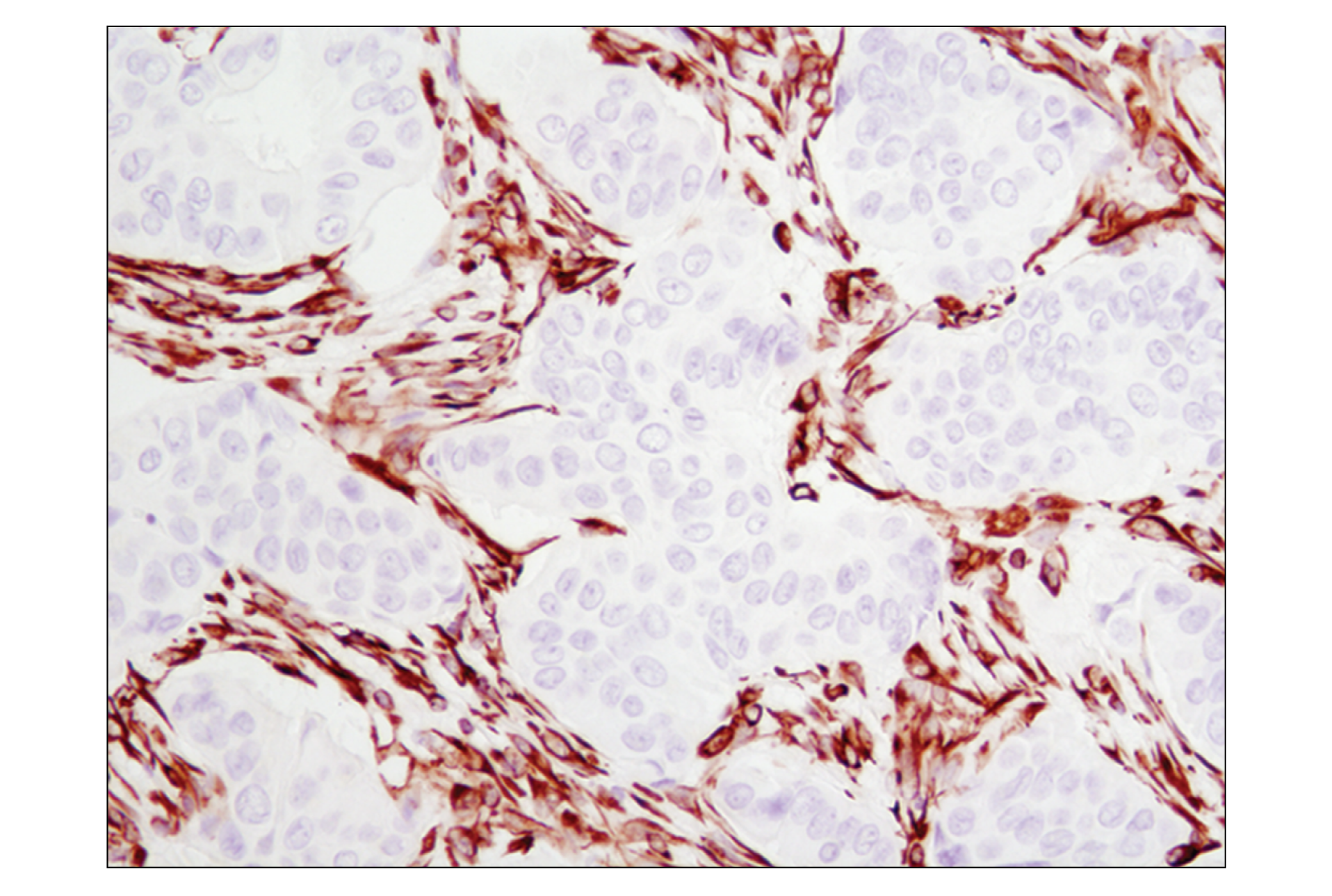
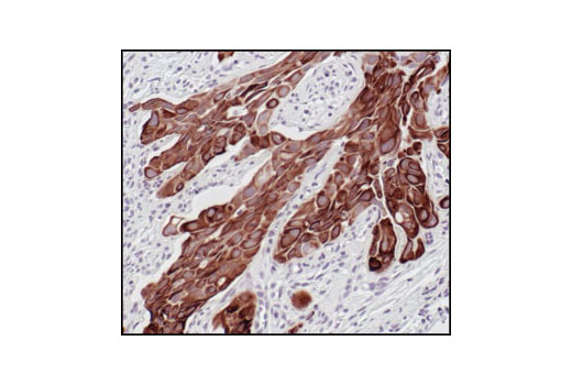
| Keratin 17 (D73C7) Rabbit mAb | 4543 | 20 µl | 48 kDa | Rabbit IgG |
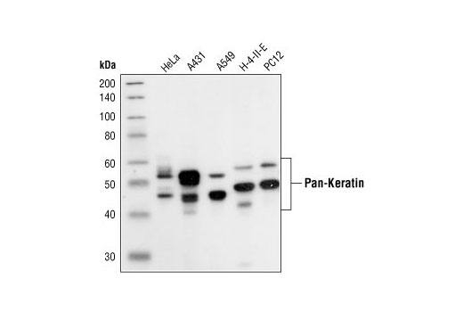
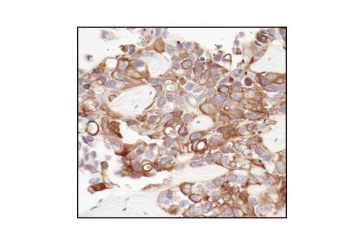
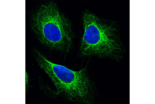
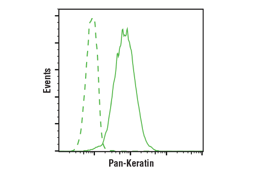
| Pan-Keratin (C11) Mouse mAb | 4545 | 20 µl | 46-58 kDa | Mouse IgG1 |
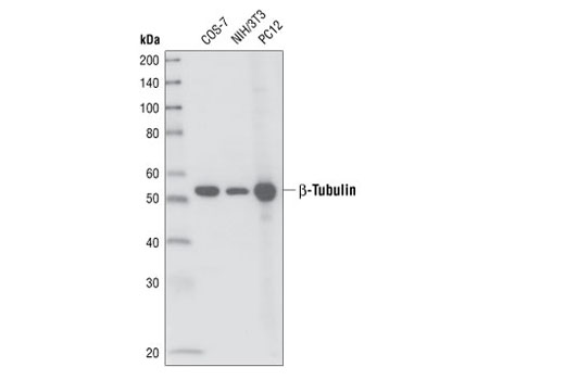
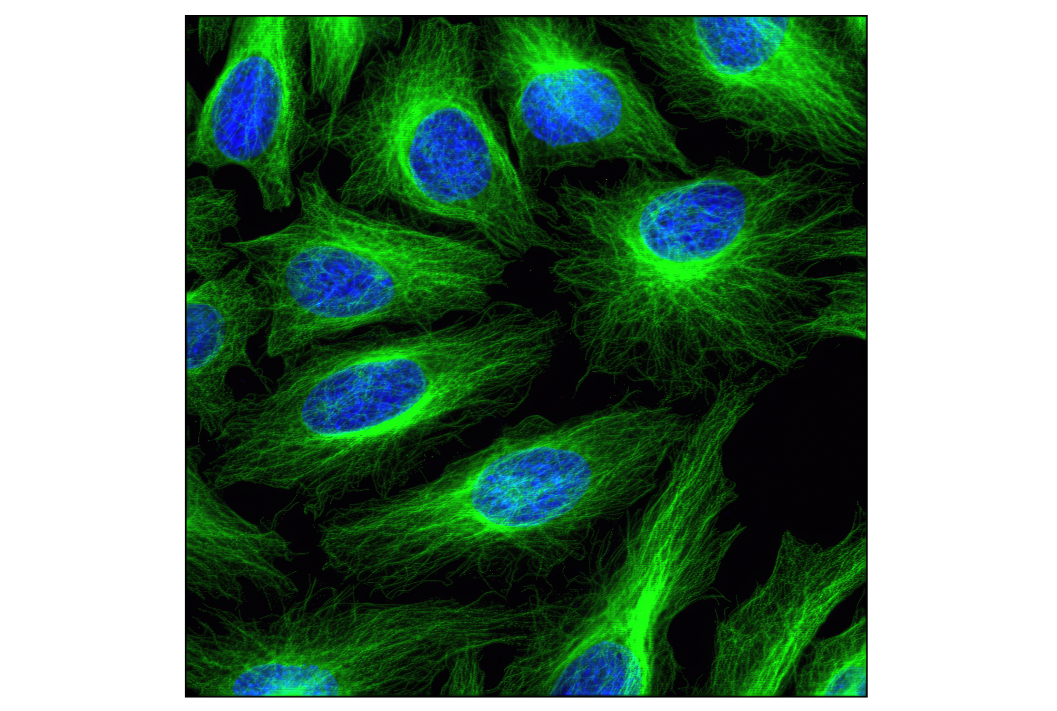
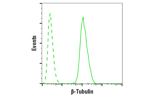
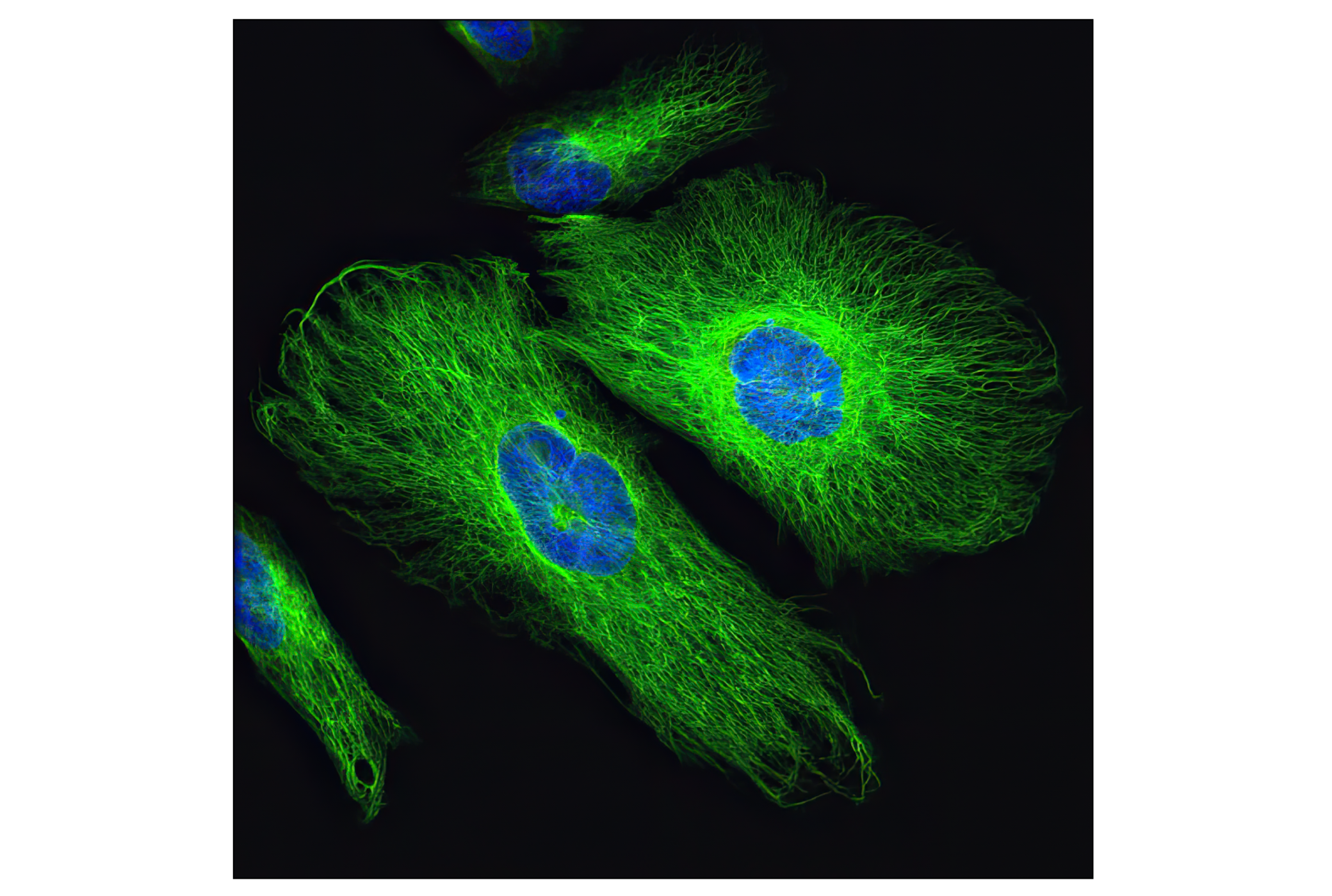
| β-Tubulin (9F3) Rabbit mAb | 2128 | 20 µl | 55 kDa | Rabbit IgG |
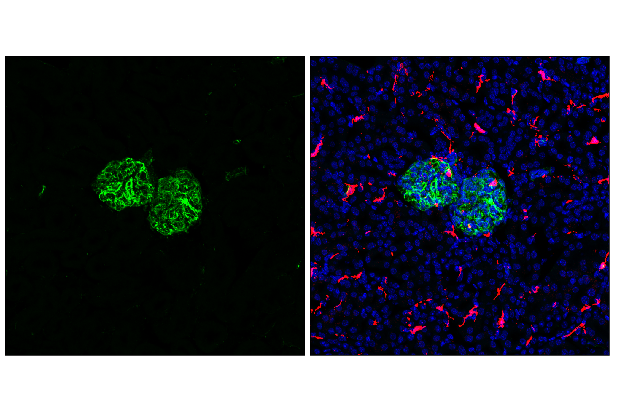
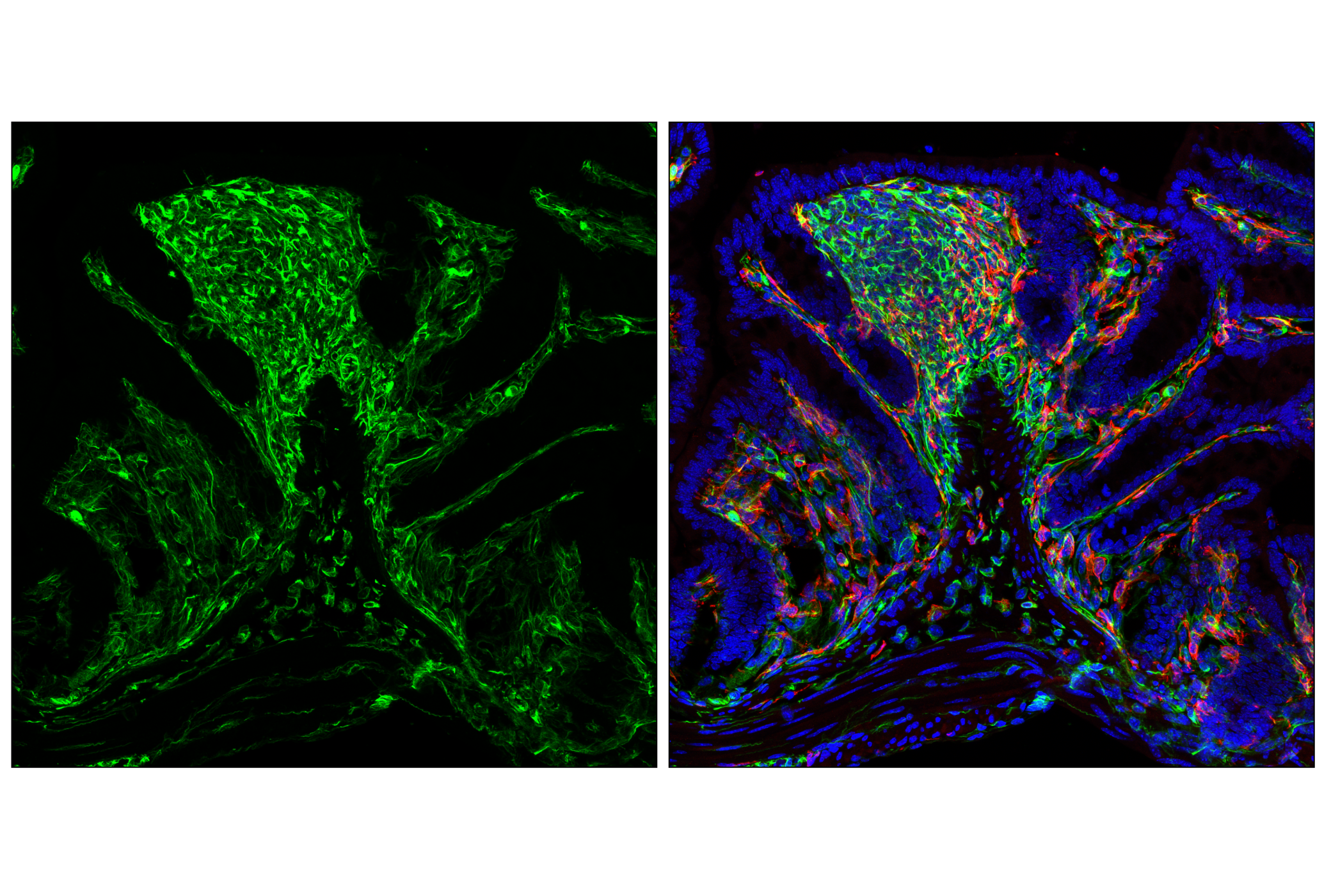
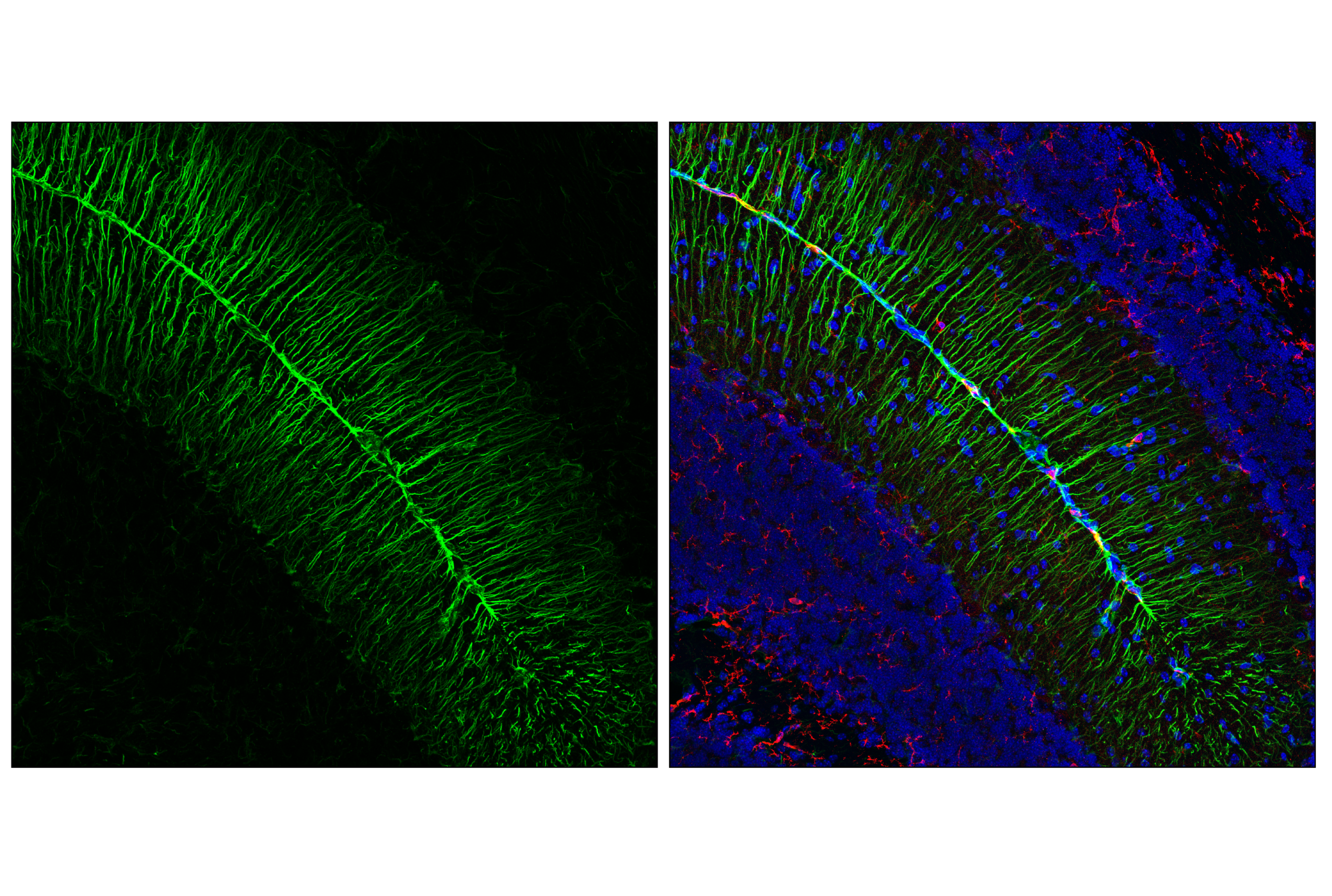
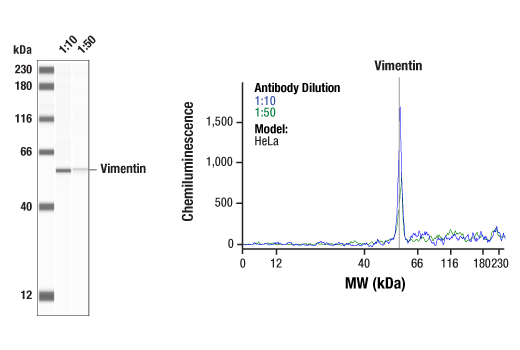
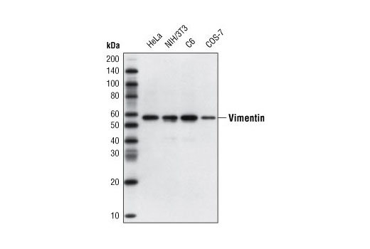
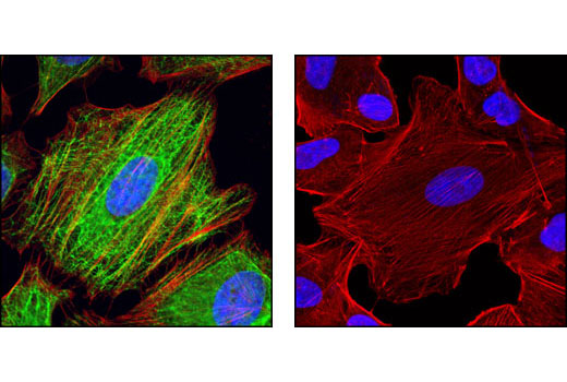
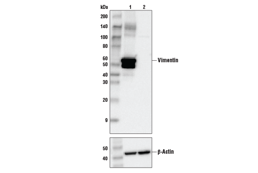
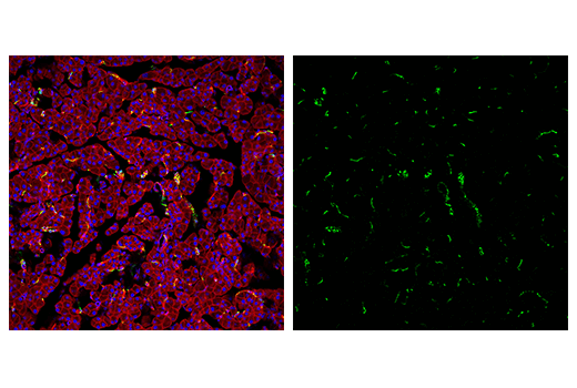
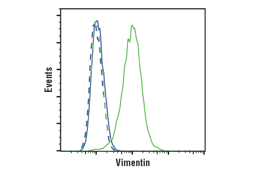
| Vimentin (D21H3) XP® Rabbit mAb | 5741 | 20 µl | 57 kDa | Rabbit IgG |
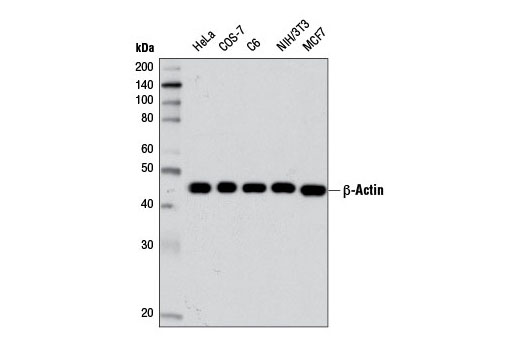
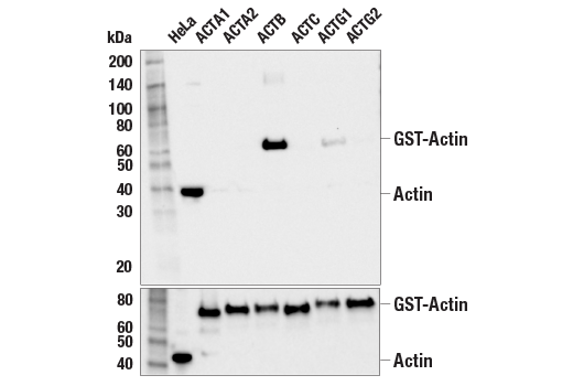
| β-Actin (D6A8) Rabbit mAb | 8457 | 20 µl | 45 kDa | Rabbit IgG |
| Anti-rabbit IgG, HRP-linked Antibody | 7074 | 100 µl | Goat | |
| Anti-mouse IgG, HRP-linked Antibody | 7076 | 100 µl | Horse |
Please visit cellsignal.com for individual component applications, species cross-reactivity, dilutions, protocols, and additional product information.
Description
The Cytoskeletal Marker Antibody Sampler Kit provides an economical means to evaluate the presence and status of select cytoskeleton associated proteins. The kit provides enough primary antibodies to perform two western blots per primary antibody.
Storage
Background
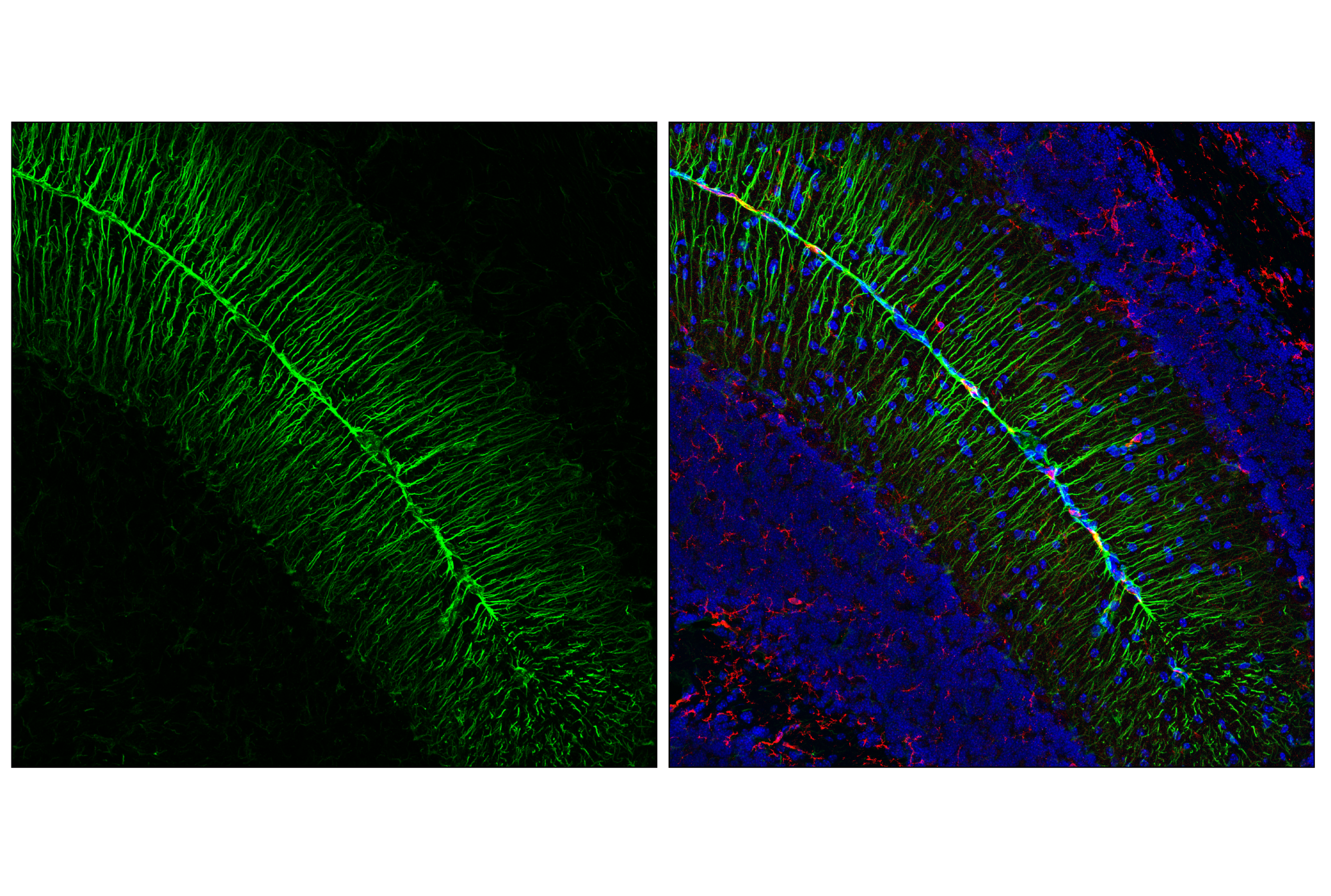
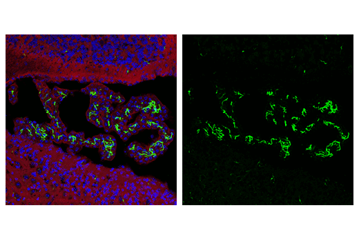
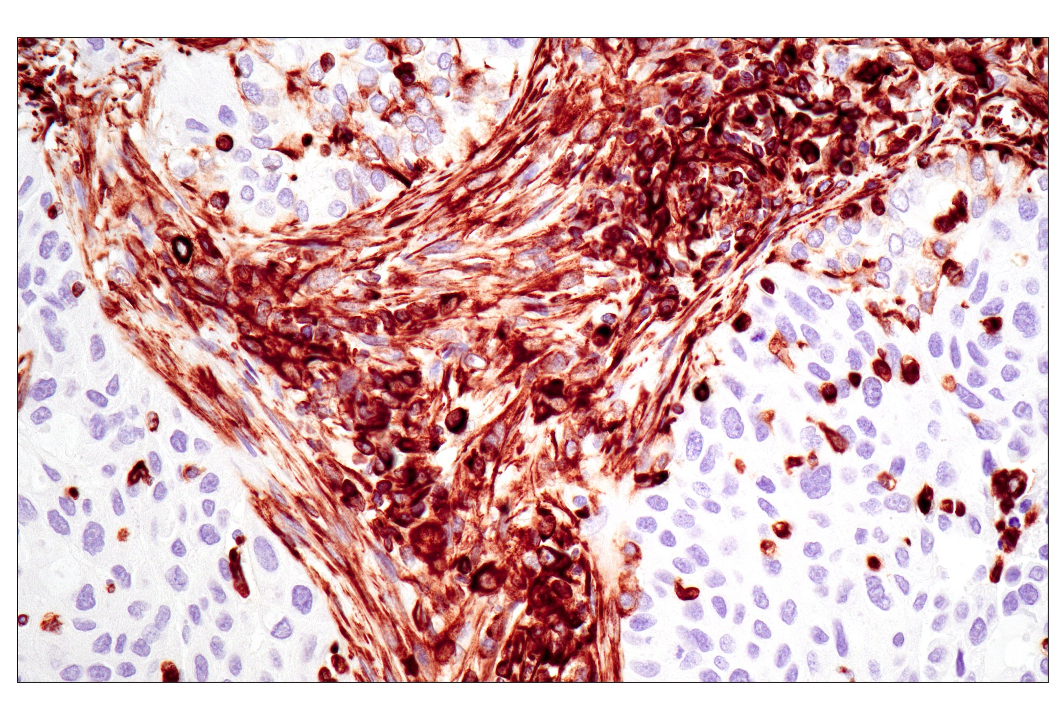
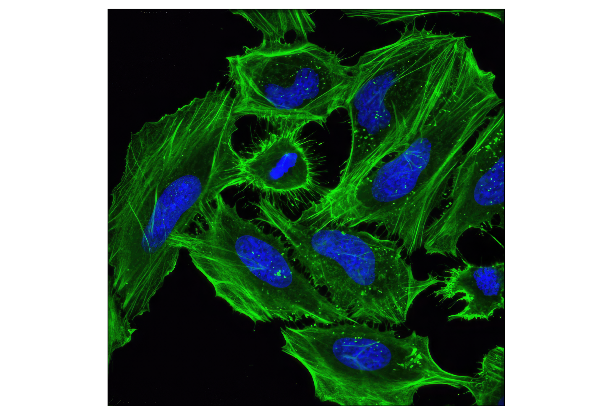
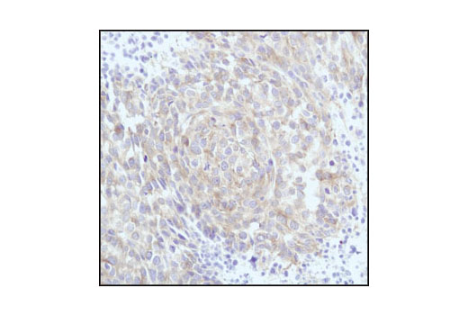
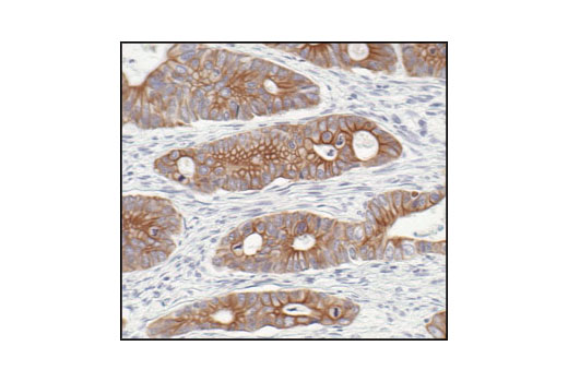
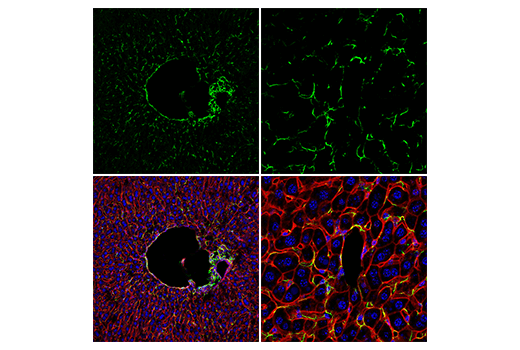
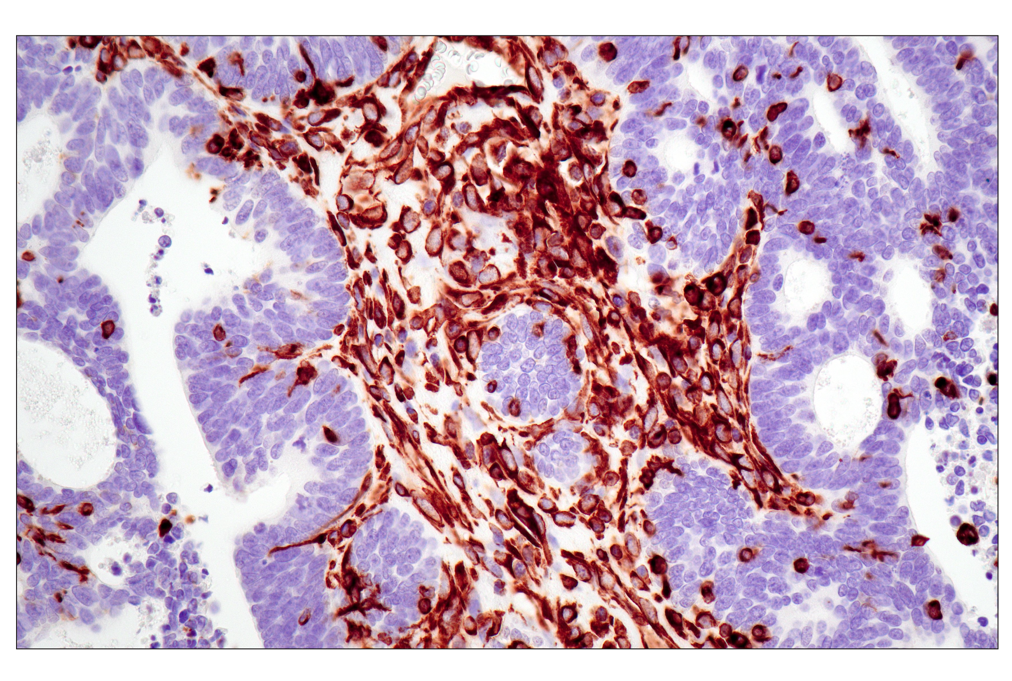
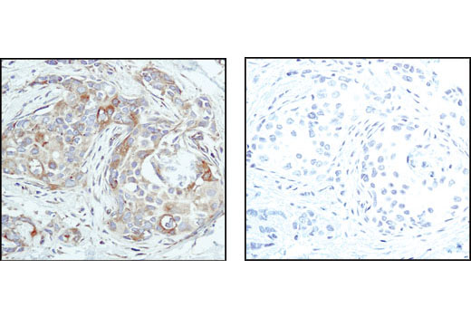
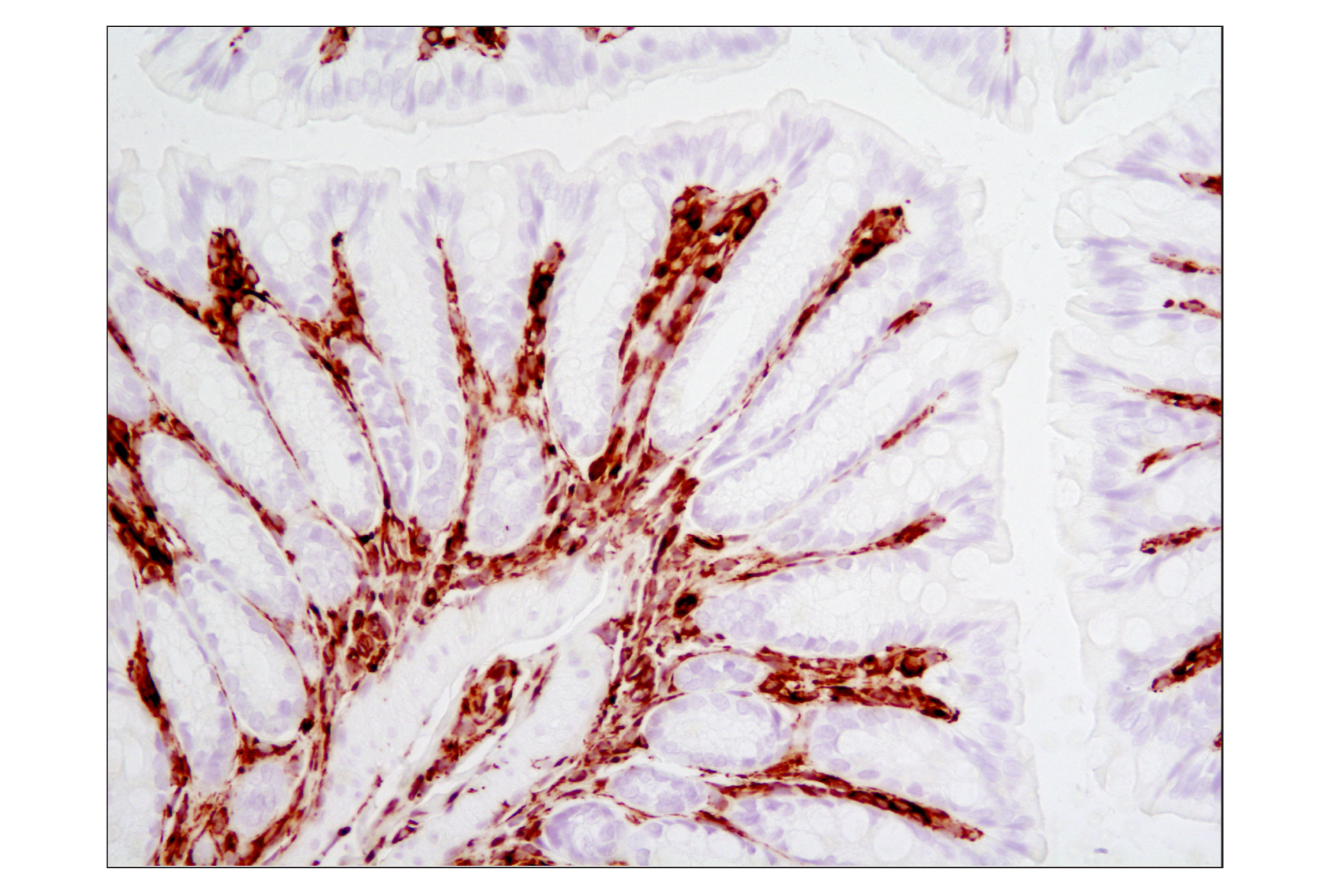
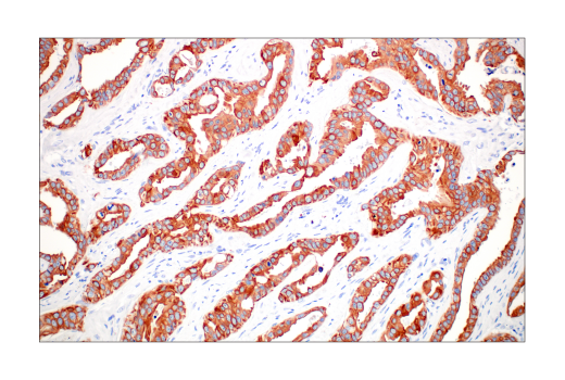
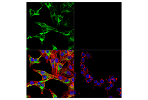
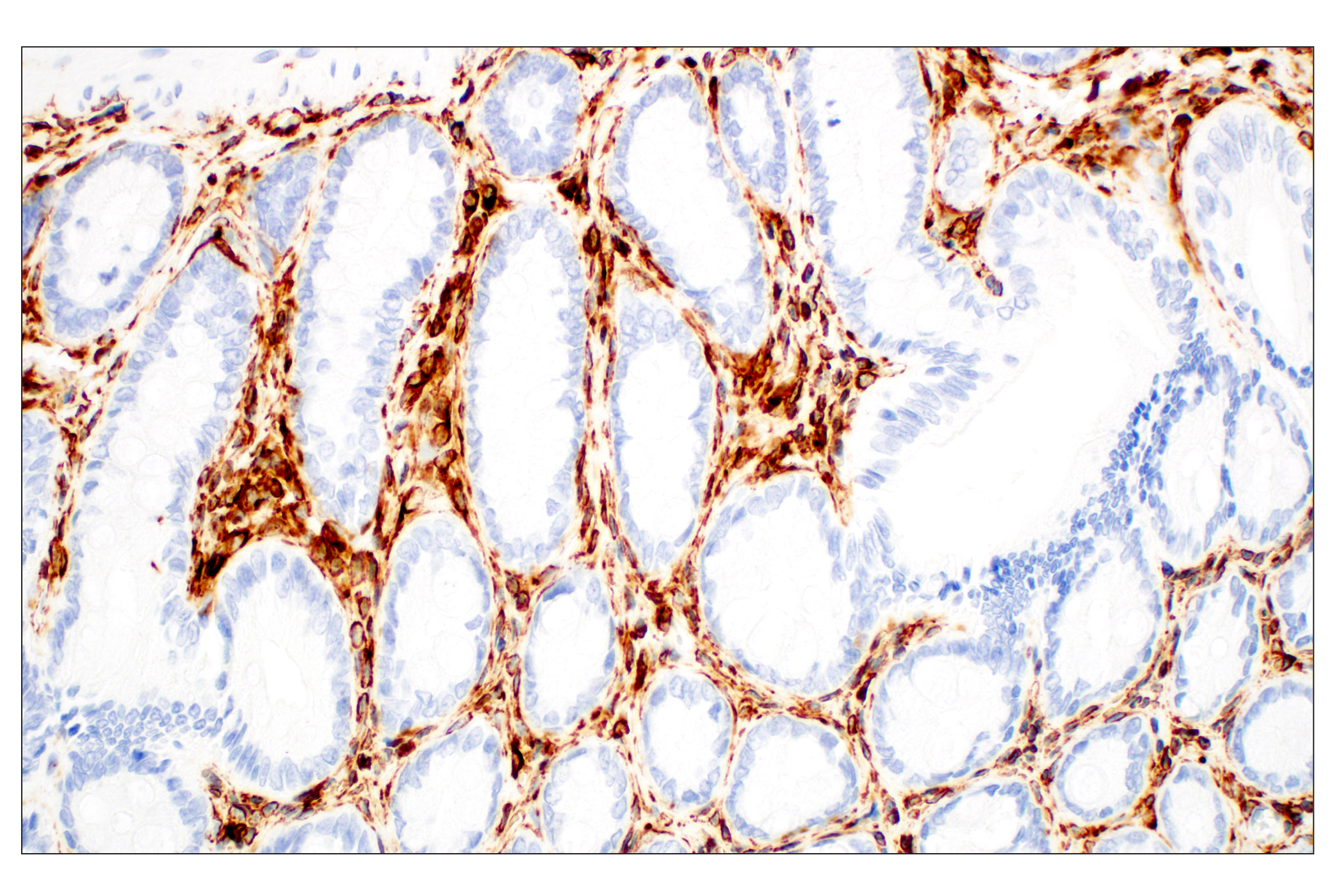
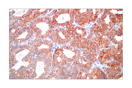
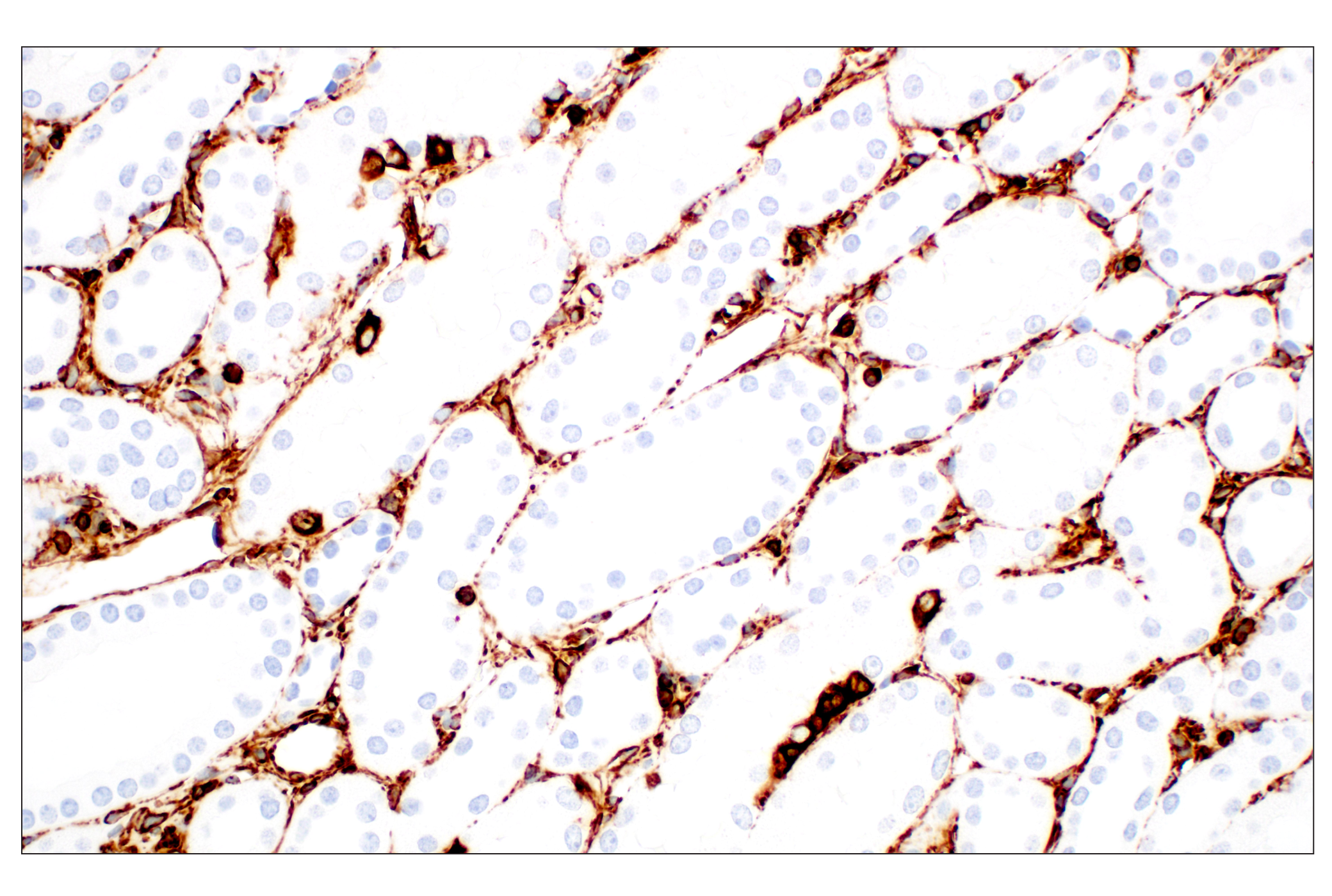
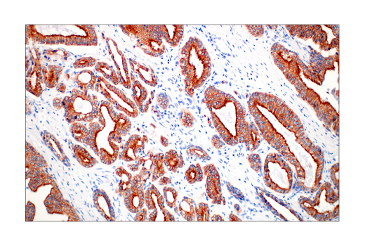
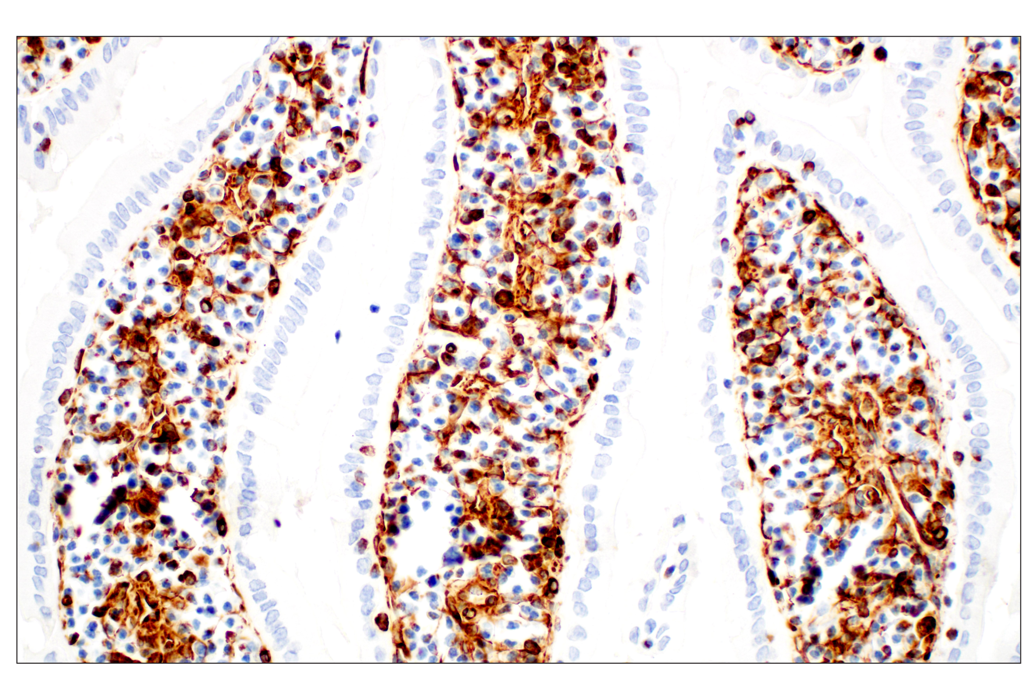
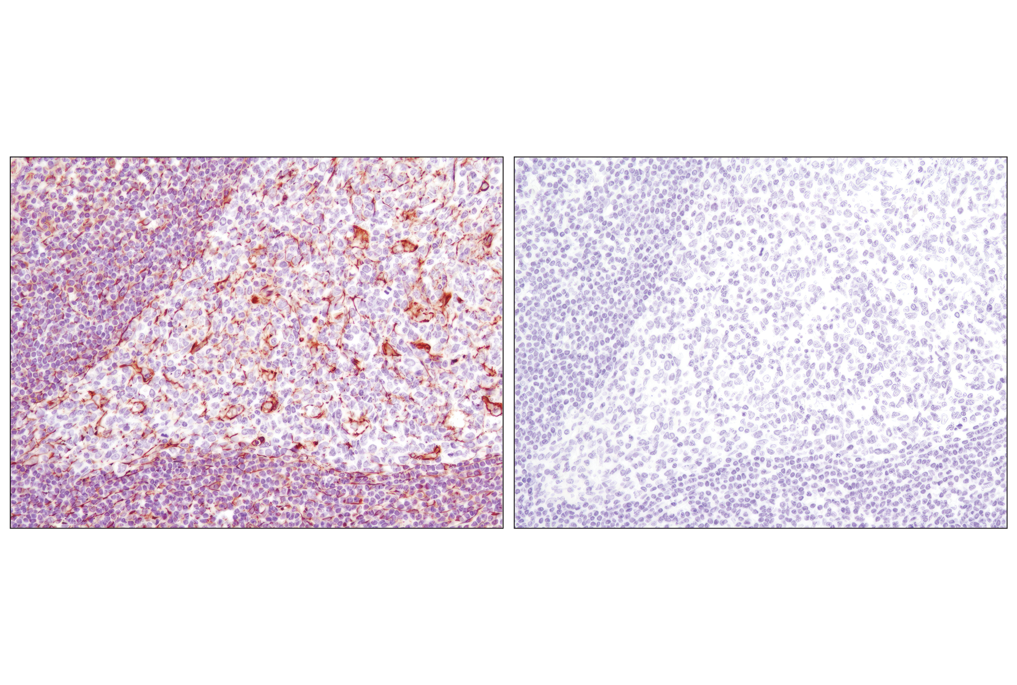
The cytoskeleton consists of three different types of cystosolic fibers: microtubules, microfilaments (actin) and intermediate filaments. Actin, a ubiquitous eukaryotic protein, is the major component of the cytoskeleton. At least six isoforms are known in mammals. Nonmuscle β- and γ-actin, also known as cytoplasmic actin, are predominantly expressed in nonmuscle cells, controlling cell structure and motility (1). Major types of intermediate filaments are distinguished in part by the tissue in which they are expressed, for example; cytokeratins (epithelial cells), vimentin (mesenchyme origin), and desmin (skeletal, visceral and certain vascular smooth muscle cells) (2). Keratin heterodimers composed of an acidic keratin (or type I keratin, keratins 9 to 23) and a basic keratin (or type II keratin, keratins 1 to 8) assemble to form intermediate filaments (3). Research studies have demonstrated that vimentin is present in sarcomas, but not carcinomas, and its expression is examined relative to other markers in order to distinguish between the two forms of neoplasm (4). Desmin is a myogenic marker expressed in early development that forms a network of filaments that extends across the myofibril and surrounds Z discs (5). α/β-tubulin heterodimers form the tubulin subunit that comprises the microtubule building block (6).
.
- Herman, I.M. (1993) Curr Opin Cell Biol 5, 48-55.
- Eng, L.F. et al. (2000) Neurochem Res 25, 1439-51.
- Moll, R. et al. (1982) Cell 31, 11-24.
- Leader, M. et al. (1987) Histopathology 11, 63-72.
- Li, Z. et al. (1996) Dev Biol 175, 362-6.
- Westermann, S. and Weber, K. (2003) Nat Rev Mol Cell Biol 4, 938-47.
Background References
Trademarks and Patents
Limited Uses
Except as otherwise expressly agreed in a writing signed by a legally authorized representative of CST, the following terms apply to Products provided by CST, its affiliates or its distributors. Any Customer's terms and conditions that are in addition to, or different from, those contained herein, unless separately accepted in writing by a legally authorized representative of CST, are rejected and are of no force or effect.
Products are labeled with For Research Use Only or a similar labeling statement and have not been approved, cleared, or licensed by the FDA or other regulatory foreign or domestic entity, for any purpose. Customer shall not use any Product for any diagnostic or therapeutic purpose, or otherwise in any manner that conflicts with its labeling statement. Products sold or licensed by CST are provided for Customer as the end-user and solely for research and development uses. Any use of Product for diagnostic, prophylactic or therapeutic purposes, or any purchase of Product for resale (alone or as a component) or other commercial purpose, requires a separate license from CST. Customer shall (a) not sell, license, loan, donate or otherwise transfer or make available any Product to any third party, whether alone or in combination with other materials, or use the Products to manufacture any commercial products, (b) not copy, modify, reverse engineer, decompile, disassemble or otherwise attempt to discover the underlying structure or technology of the Products, or use the Products for the purpose of developing any products or services that would compete with CST products or services, (c) not alter or remove from the Products any trademarks, trade names, logos, patent or copyright notices or markings, (d) use the Products solely in accordance with CST Product Terms of Sale and any applicable documentation, and (e) comply with any license, terms of service or similar agreement with respect to any third party products or services used by Customer in connection with the Products.