| Product Includes | Product # | Quantity | Mol. Wt | Isotype/Source |
|---|---|---|---|---|
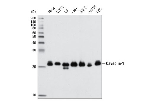
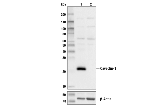
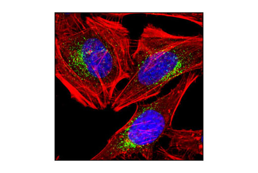
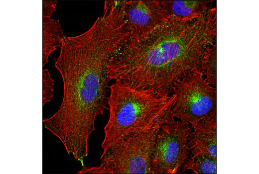
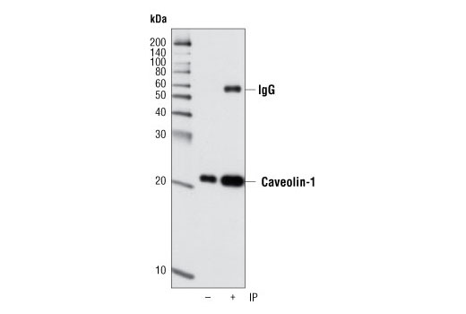
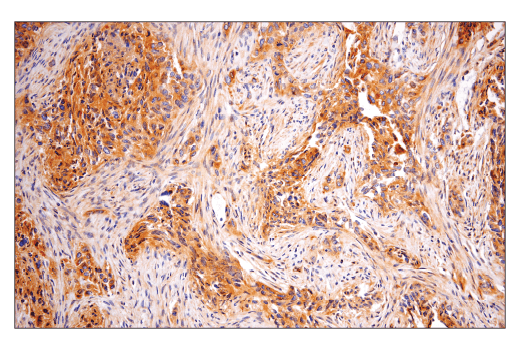
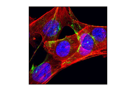
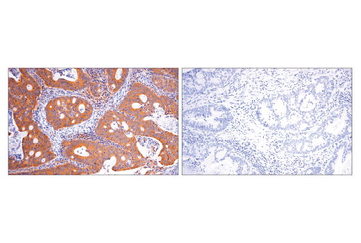
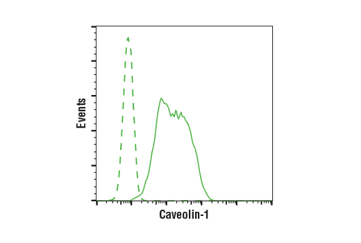
| Caveolin-1 (D46G3) XP® Rabbit mAb | 3267 | 20 µl | 21, 24 kDa | Rabbit IgG |
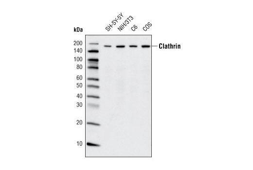
| Clathrin Heavy Chain (D3C6) XP® Rabbit mAb | 4796 | 20 µl | 190 kDa | Rabbit IgG |
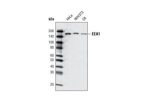
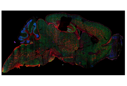
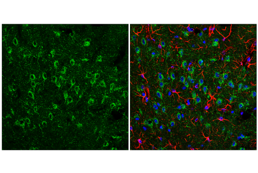
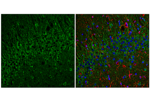
| EEA1 (C45B10) Rabbit mAb | 3288 | 20 µl | 170 kDa | Rabbit IgG |
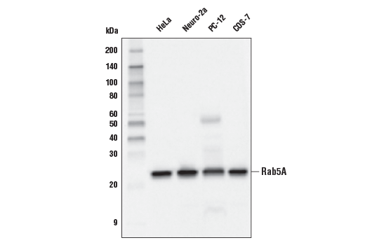
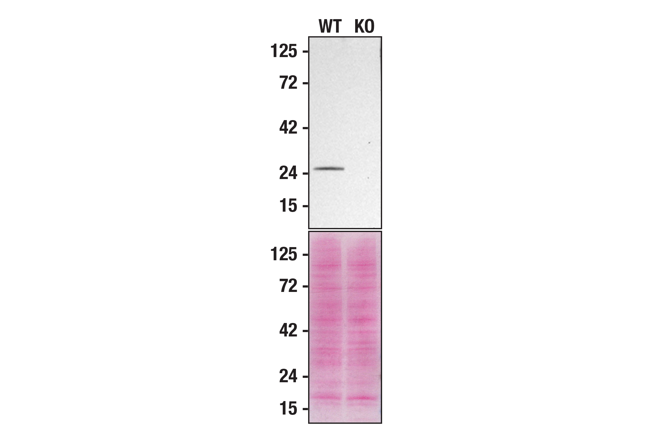
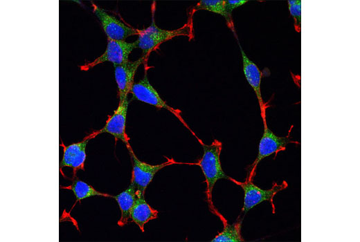
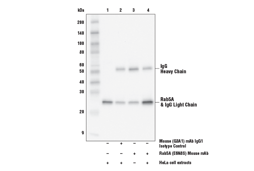
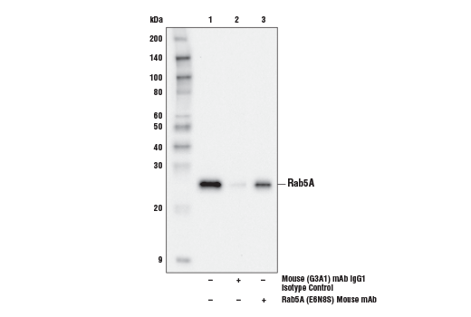
| Rab5A (E6N8S) Mouse mAb | 46449 | 20 µl | 25 kDa | Mouse IgG1 |
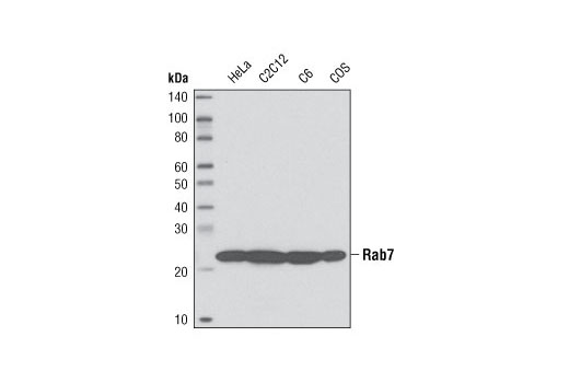
| Rab7 (D95F2) XP® Rabbit mAb | 9367 | 20 µl | 23 kDa | Rabbit IgG |
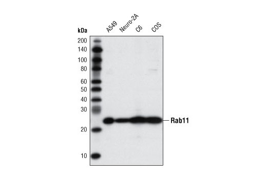
| Rab11 (D4F5) XP® Rabbit mAb | 5589 | 20 µl | 25 kDa | Rabbit IgG |
| Anti-rabbit IgG, HRP-linked Antibody | 7074 | 100 µl | Goat | |
| Anti-mouse IgG, HRP-linked Antibody | 7076 | 100 µl | Horse |
Please visit cellsignal.com for individual component applications, species cross-reactivity, dilutions, protocols, and additional product information.
Description
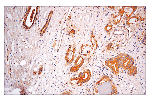
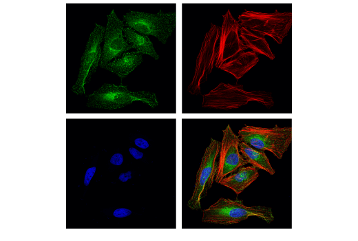
The Endosomal Marker Antibody Sampler Kit provides an economical means of distinguishing endosomes in the early, late, and recycling phases. The kit includes enough antibody to perform two western blot experiments with each primary antibody.
Storage
Background
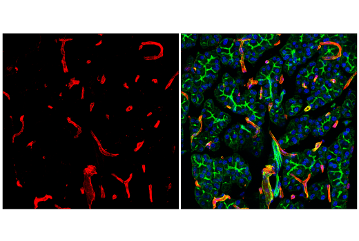
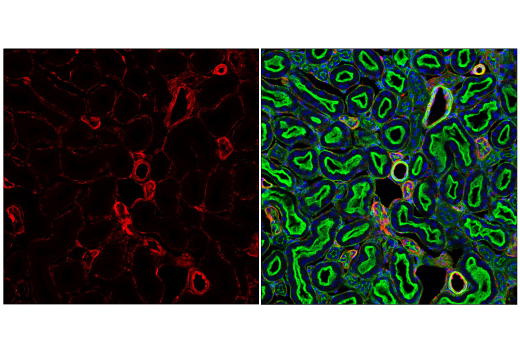
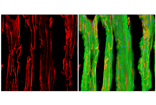
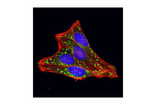
Endosomes are formed by the invagination of the plasma membrane to form vesicles in an effort to recycle components of the cell (1). Endosomes can be coated in clathrin when vesicles form at clathrin-coated pits (2). Caveolins are 21-24 kDa integral proteins that interact with cholesterol and are the main structural components of the cholesterol/sphingolipid-enriched plasma membrane caveolae (3). Each stage of endosome maturation is marked by a unique set of proteins. EEA1 is an early endosome marker that is essential for membrane fusion and trafficking (4). Members of the ras superfamily of small Rab GTPases, specifically Rab5, Rab7, and Rab11 are markers of the early, late and recycling endosomes (5).
Background References
Trademarks and Patents
Limited Uses
Except as otherwise expressly agreed in a writing signed by a legally authorized representative of CST, the following terms apply to Products provided by CST, its affiliates or its distributors. Any Customer's terms and conditions that are in addition to, or different from, those contained herein, unless separately accepted in writing by a legally authorized representative of CST, are rejected and are of no force or effect.
Products are labeled with For Research Use Only or a similar labeling statement and have not been approved, cleared, or licensed by the FDA or other regulatory foreign or domestic entity, for any purpose. Customer shall not use any Product for any diagnostic or therapeutic purpose, or otherwise in any manner that conflicts with its labeling statement. Products sold or licensed by CST are provided for Customer as the end-user and solely for research and development uses. Any use of Product for diagnostic, prophylactic or therapeutic purposes, or any purchase of Product for resale (alone or as a component) or other commercial purpose, requires a separate license from CST. Customer shall (a) not sell, license, loan, donate or otherwise transfer or make available any Product to any third party, whether alone or in combination with other materials, or use the Products to manufacture any commercial products, (b) not copy, modify, reverse engineer, decompile, disassemble or otherwise attempt to discover the underlying structure or technology of the Products, or use the Products for the purpose of developing any products or services that would compete with CST products or services, (c) not alter or remove from the Products any trademarks, trade names, logos, patent or copyright notices or markings, (d) use the Products solely in accordance with CST Product Terms of Sale and any applicable documentation, and (e) comply with any license, terms of service or similar agreement with respect to any third party products or services used by Customer in connection with the Products.