| Product Includes | Product # | Quantity | Mol. Wt | Isotype/Source |
|---|---|---|---|---|
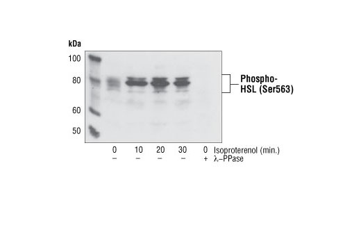
| Phospho-HSL (Ser563) Antibody | 4139 | 20 µl | 81, 83 kDa | Rabbit |
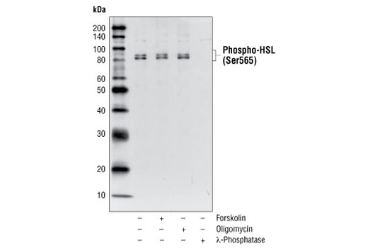
| Phospho-HSL (Ser565) Antibody | 4137 | 20 µl | 81, 83 kDa | Rabbit |
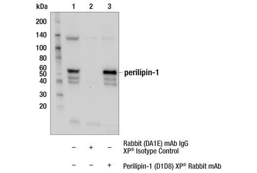
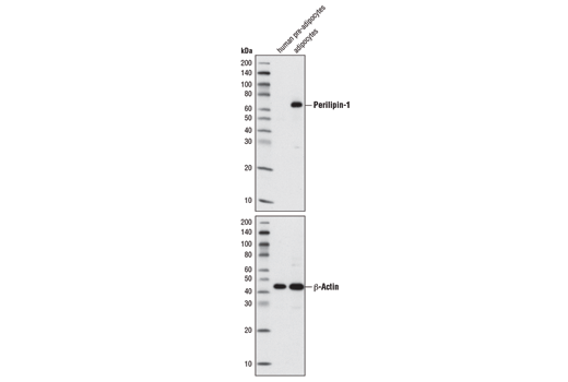
| Perilipin-1 (D1D8) XP® Rabbit mAb | 9349 | 20 µl | 62 kDa | Rabbit IgG |
| Anti-rabbit IgG, HRP-linked Antibody | 7074 | 100 µl | Goat | |
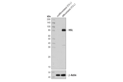
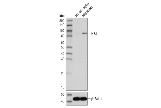
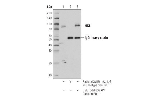
| HSL (D6W5S) XP® Rabbit mAb | 18381 | 20 µl | 81, 83 kDa | Rabbit IgG |
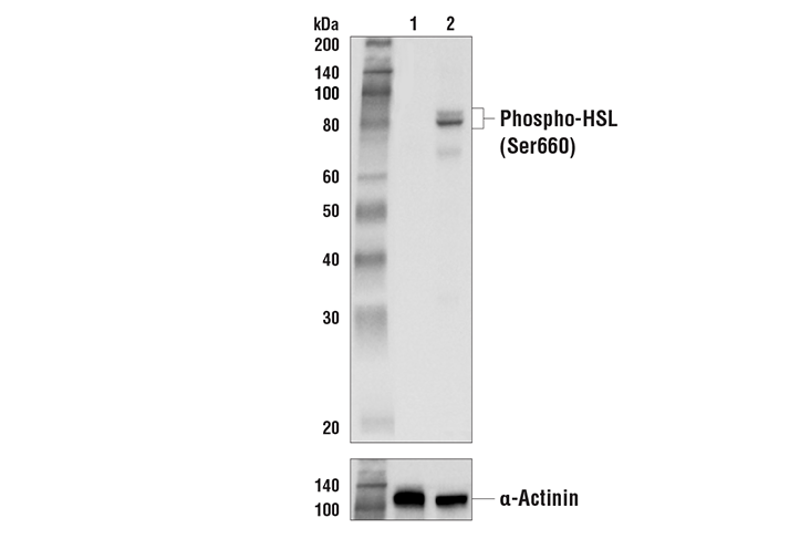
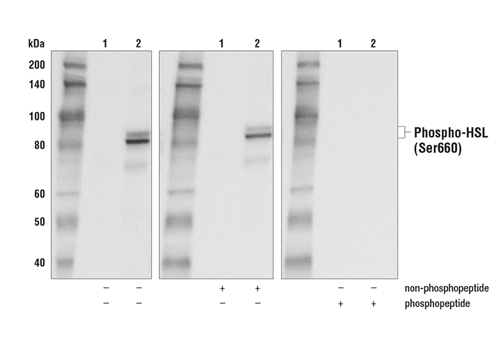
| Phospho-HSL (Ser660) Antibody | 45804 | 20 µl | 81, 83 kDa | Rabbit |
Please visit cellsignal.com for individual component applications, species cross-reactivity, dilutions, protocols, and additional product information.
Description
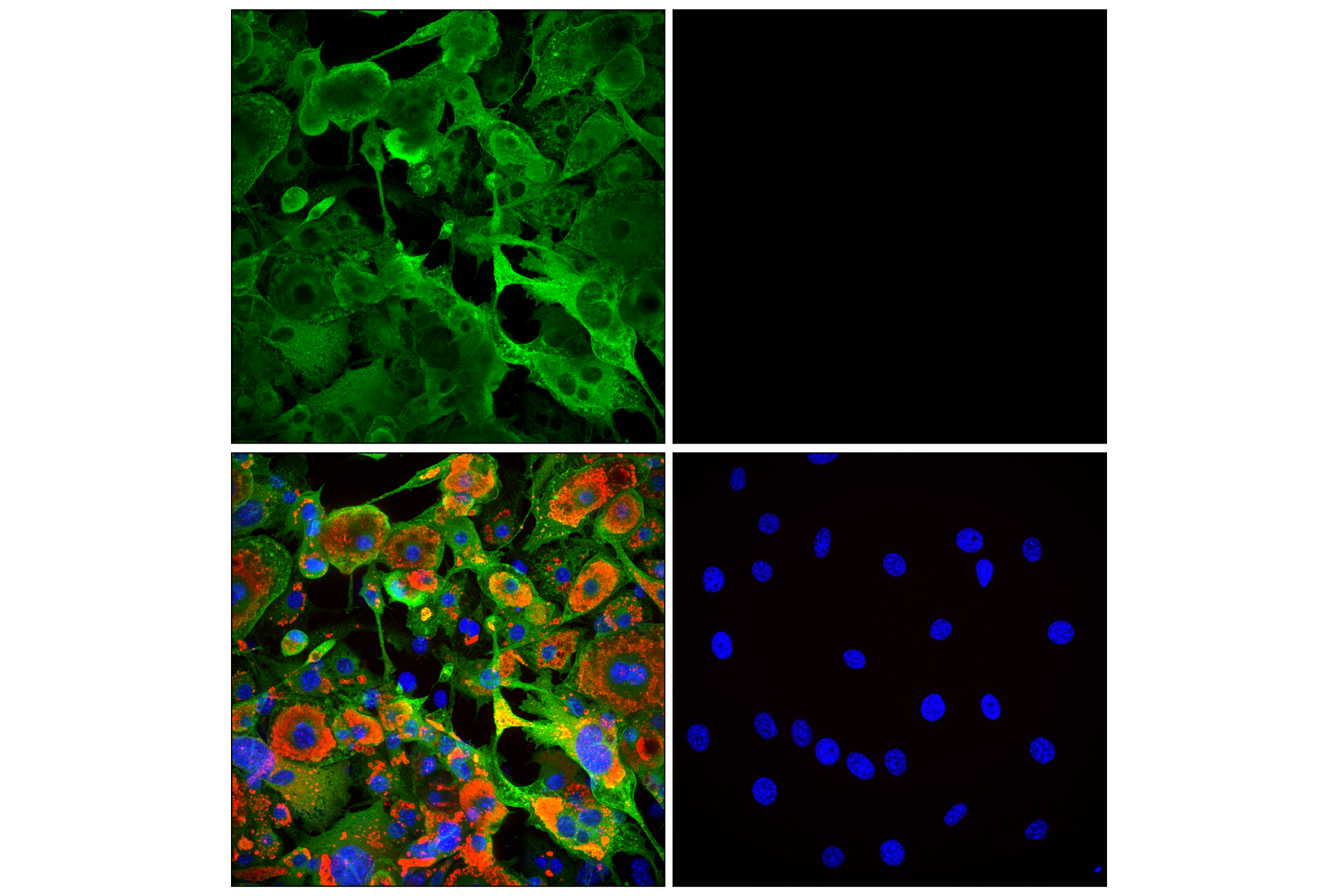
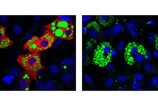
The Lipolysis Activation Antibody Sampler Kit provides an economical means to evaluate the activation status of multiple members of the lipolysis pathway, including phosphorylated HSL and perilipin. The kit includes enough antibody to perform two western mini-blot experiments with each primary antibody.
Storage
Background
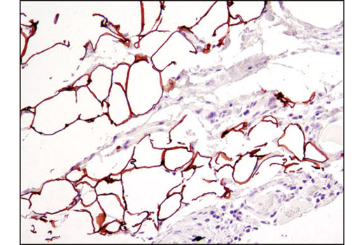
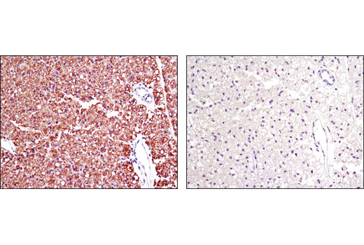
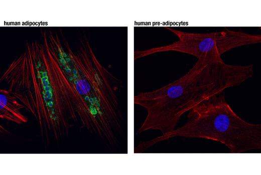
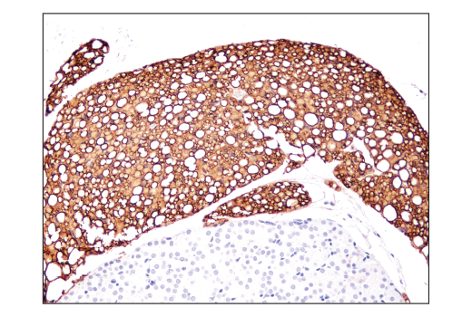
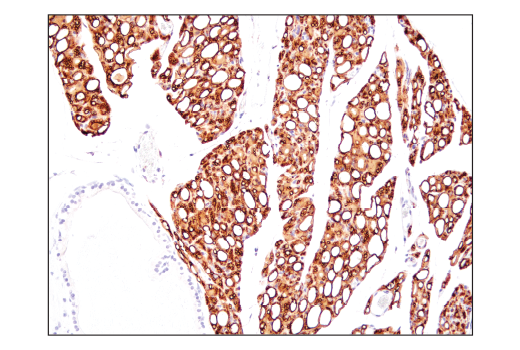
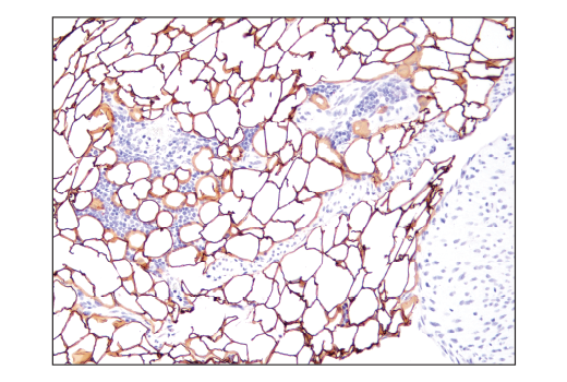
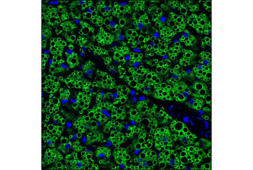
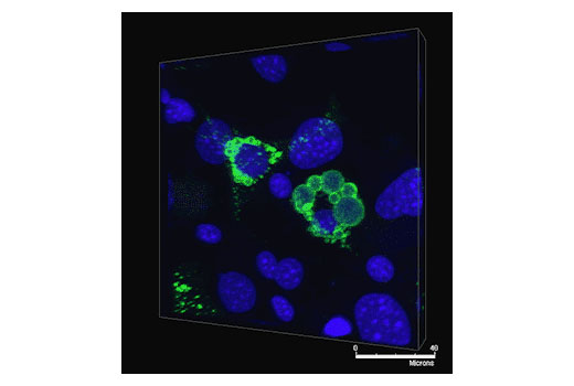
Triacylglycerol is stored in lipid droplets as a primary energy reserve. During lipolysis, triacylglycerols in adipocytes are hydrolyzed into free fatty acids and glycerol. Perilipin, localized at the periphery of lipid droplets, serves as a protective coating against lipases (1-3). Evidence suggests that PKA regulates lipolysis by phosphorylating perilipin and hormone-sensitive lipase (HSL) (1,2,4,5). Phosphorylation of perilipin results in the conformational change that exposes lipid droplets to endogenous lipases, such as HSL (2). Phosphorylation of HSL at Ser563, Ser659, and Ser660 by PKA stimulates HSL activity, which in turn catalyzes the hydrolysis of triacylglycerol (6,7).
- Greenberg, A.S. et al. (1991) J Biol Chem 266, 11341-6.
- Brasaemle, D.L. (2007) J Lipid Res 48, 2547-59.
- Ducharme, N.A. and Bickel, P.E. (2008) Endocrinology 149, 942-9.
- Egan, J.J. et al. (1990) J Biol Chem 265, 18769-75.
- Brasaemle, D.L. et al. (2009) Mol Cell Biochem 326, 15-21.
- Degerman, E. et al. (1990) Proc Natl Acad Sci U S A 87, 533-7.
- Anthonsen, M.W. et al. (1998) J Biol Chem 273, 215-21.
Background References
Trademarks and Patents
Limited Uses
Except as otherwise expressly agreed in a writing signed by a legally authorized representative of CST, the following terms apply to Products provided by CST, its affiliates or its distributors. Any Customer's terms and conditions that are in addition to, or different from, those contained herein, unless separately accepted in writing by a legally authorized representative of CST, are rejected and are of no force or effect.
Products are labeled with For Research Use Only or a similar labeling statement and have not been approved, cleared, or licensed by the FDA or other regulatory foreign or domestic entity, for any purpose. Customer shall not use any Product for any diagnostic or therapeutic purpose, or otherwise in any manner that conflicts with its labeling statement. Products sold or licensed by CST are provided for Customer as the end-user and solely for research and development uses. Any use of Product for diagnostic, prophylactic or therapeutic purposes, or any purchase of Product for resale (alone or as a component) or other commercial purpose, requires a separate license from CST. Customer shall (a) not sell, license, loan, donate or otherwise transfer or make available any Product to any third party, whether alone or in combination with other materials, or use the Products to manufacture any commercial products, (b) not copy, modify, reverse engineer, decompile, disassemble or otherwise attempt to discover the underlying structure or technology of the Products, or use the Products for the purpose of developing any products or services that would compete with CST products or services, (c) not alter or remove from the Products any trademarks, trade names, logos, patent or copyright notices or markings, (d) use the Products solely in accordance with CST Product Terms of Sale and any applicable documentation, and (e) comply with any license, terms of service or similar agreement with respect to any third party products or services used by Customer in connection with the Products.