| Product Includes | Product # | Quantity | Mol. Wt | Isotype/Source |
|---|---|---|---|---|
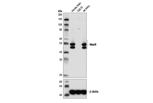
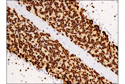
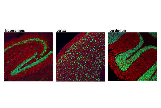
| NeuN (D4G4O) XP® Rabbit mAb | 24307 | 20 µl | 46-55 kDa | Rabbit IgG |
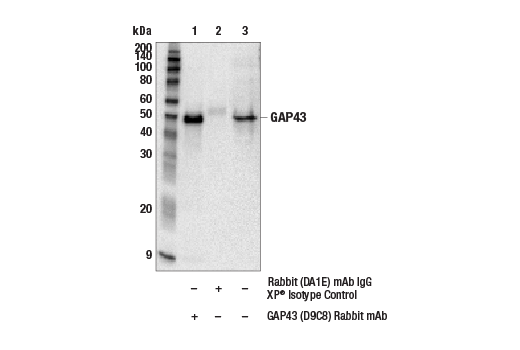
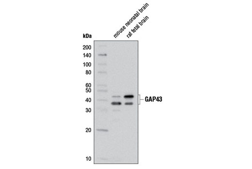
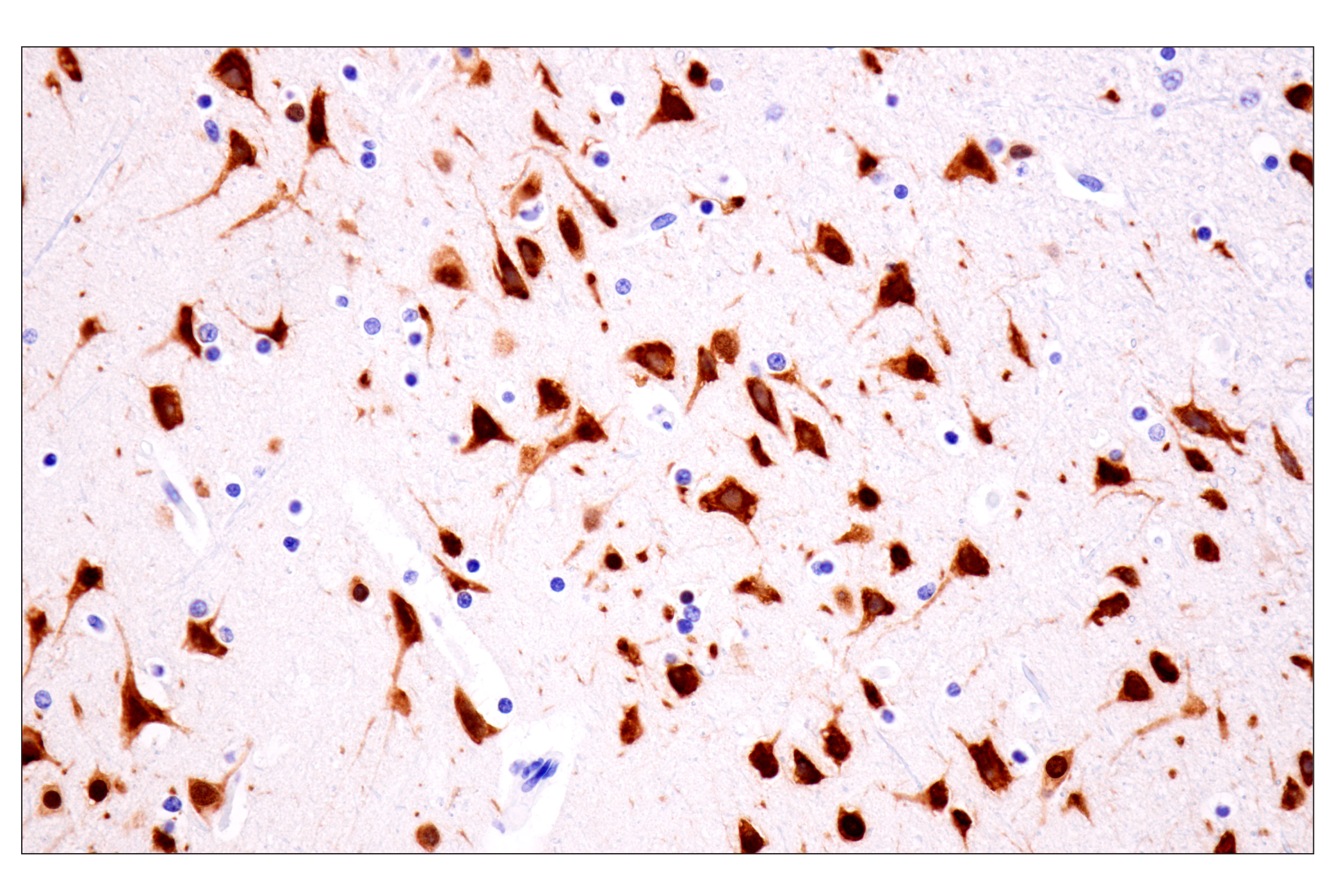
| GAP43 (D9C8) Rabbit mAb | 8945 | 20 µl | 38, 43 kDa | Rabbit IgG |
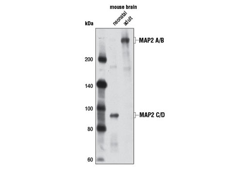
| MAP2 (D5G1) XP® Rabbit mAb | 8707 | 20 µl | 75, 82, 280 kDa | Rabbit IgG |
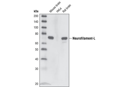
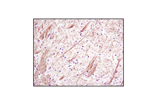
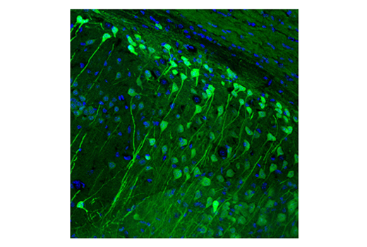
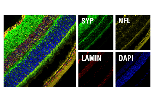
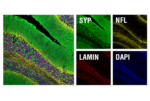
| Neurofilament-L (C28E10) Rabbit mAb | 2837 | 20 µl | 70 kDa | Rabbit IgG |
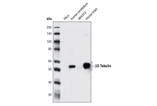
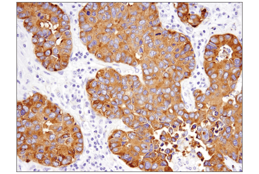
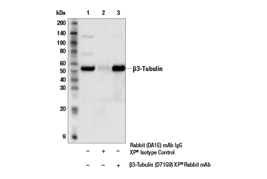
| β3-Tubulin (D71G9) XP® Rabbit mAb | 5568 | 20 µl | 55 kDa | Rabbit IgG |
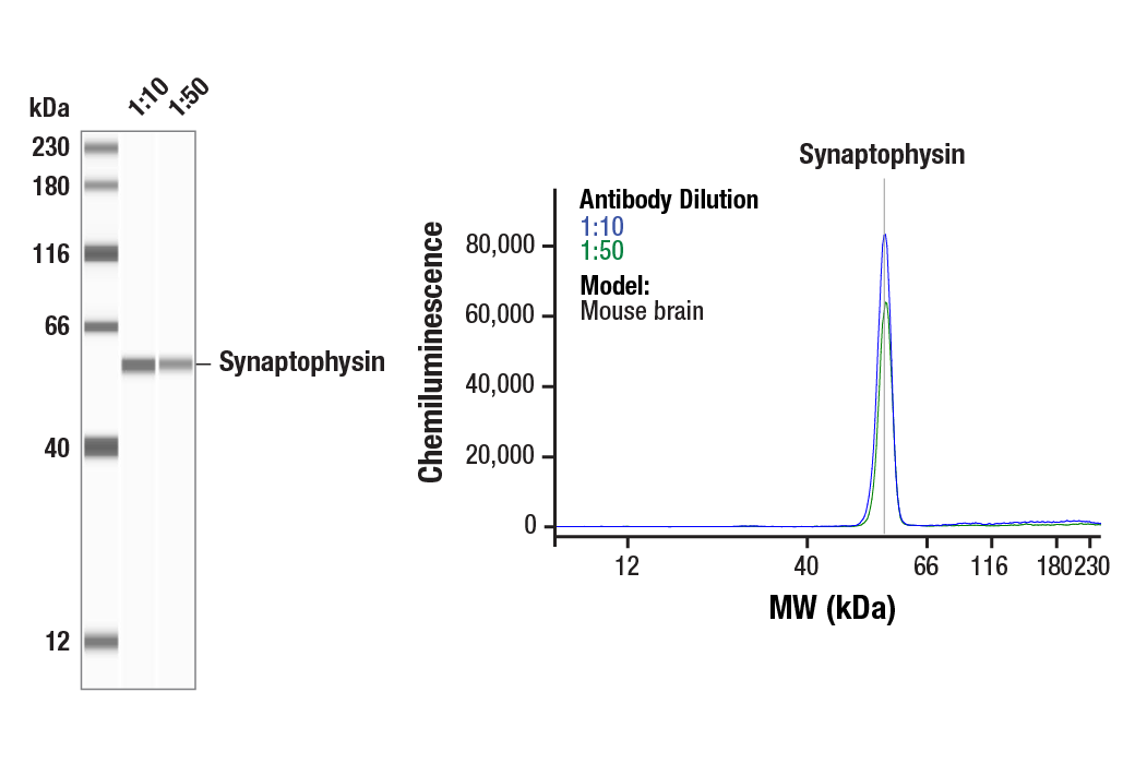
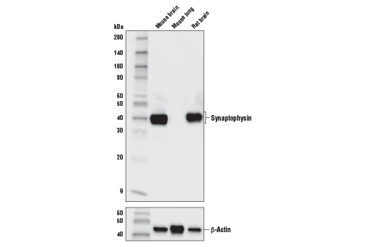
| Synaptophysin (D8F6H) XP® Rabbit mAb | 36406 | 20 µl | 38 kDa | Rabbit IgG |
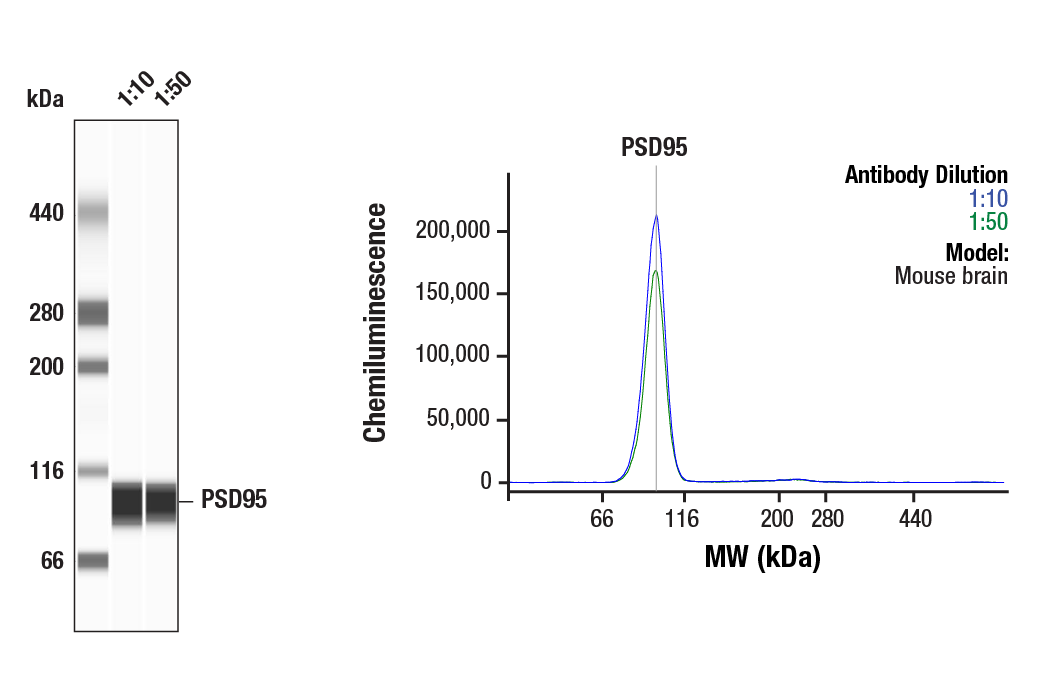
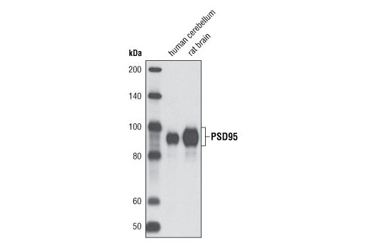
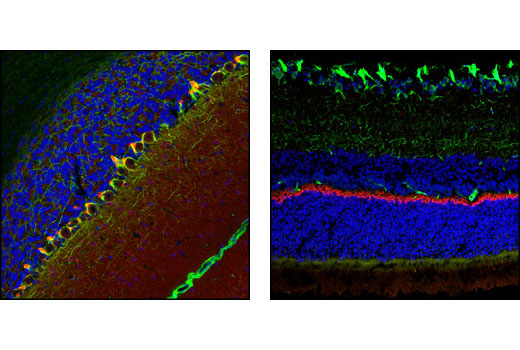
| PSD95 (D27E11) XP® Rabbit mAb | 3450 | 20 µl | 95 kDa | Rabbit IgG |
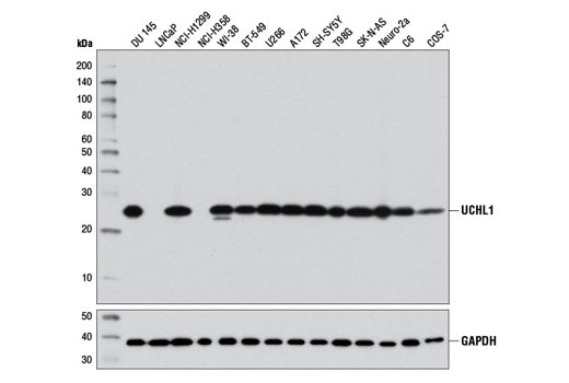
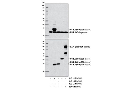
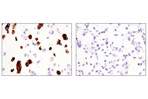
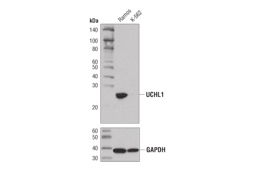
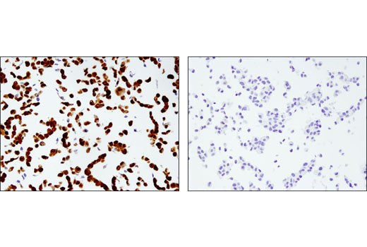
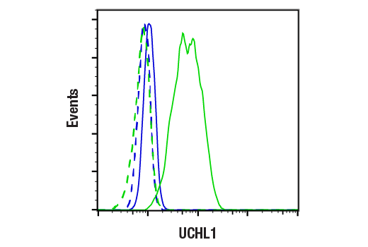
| UCHL1 (D3T2E) XP® Rabbit mAb | 13179 | 20 µl | 27 kDa | Rabbit IgG |
| Anti-rabbit IgG, HRP-linked Antibody | 7074 | 100 µl | Goat |
Please visit cellsignal.com for individual component applications, species cross-reactivity, dilutions, protocols, and additional product information.
Description
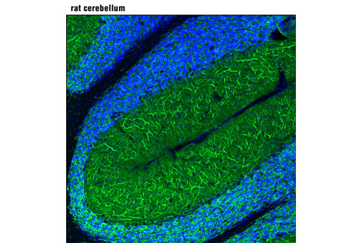
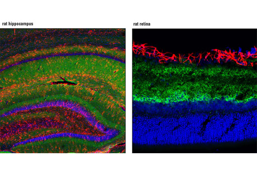
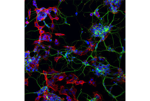
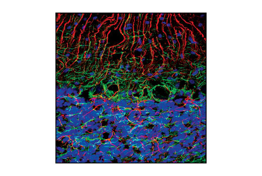
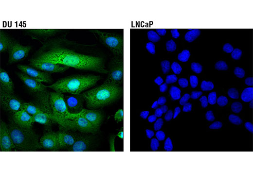
The Mature Neuron Marker Antibody Sampler Kit provides an economical means for detecting mature neuron proteins by western and labeling mature neuronal structures by immunofluorescence (IF). This kit includes enough primary antibodies to perform two western blot experiments or at least forty IF tests per primary antibody.
Storage
Background
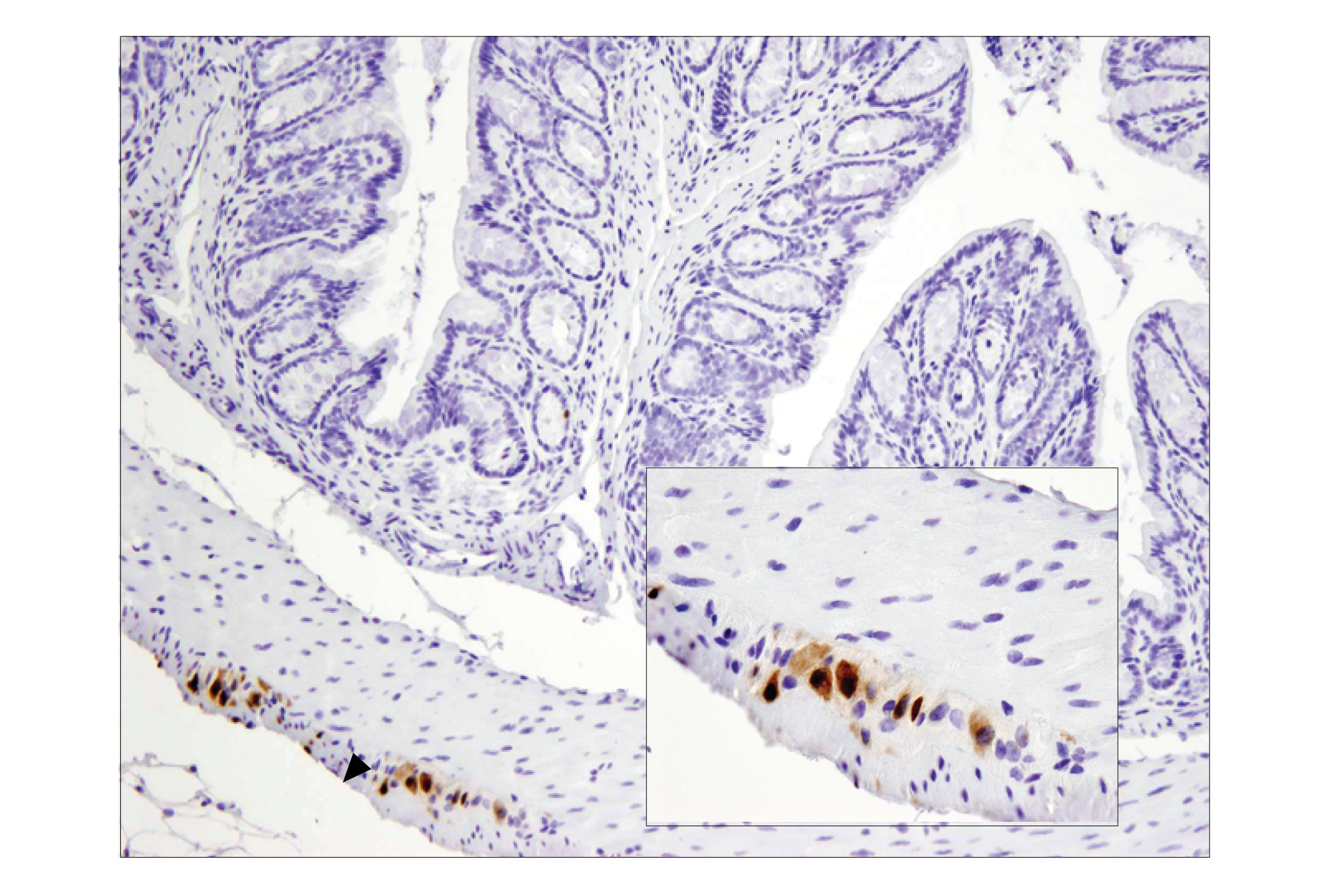
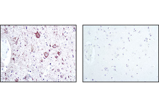
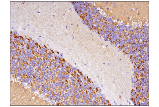
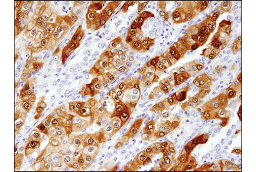
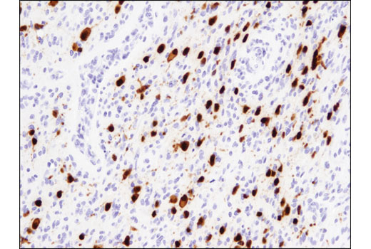
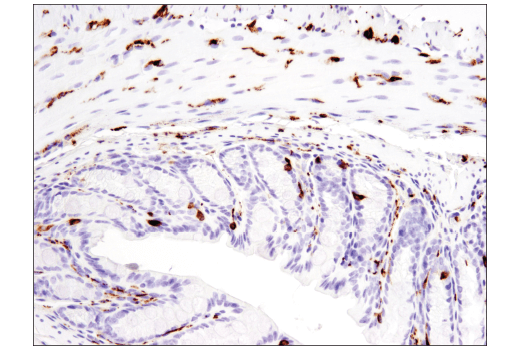
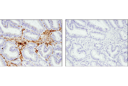
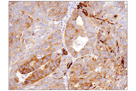
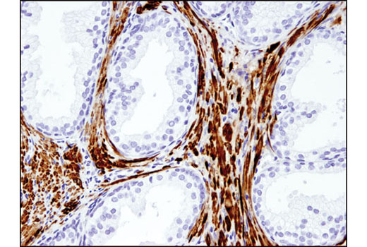
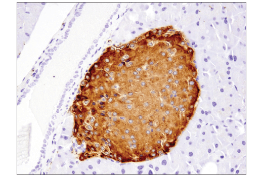
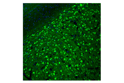
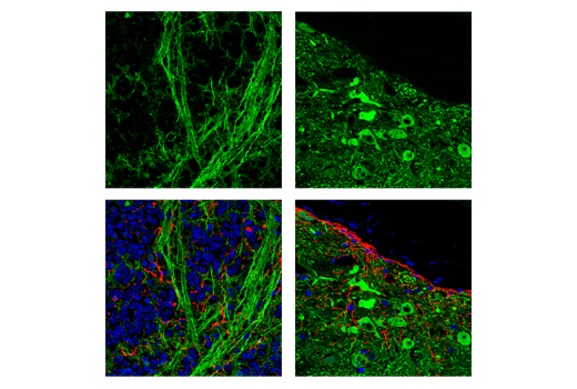
The antibodies in this kit serve to characterize and identify mature neurons. Neural stem cells differentiate into mature post-mitotic neurons that are incapable of cellular division. Several neuron-enriched markers can be used to identify mature neurons. Neuronal nuclei (NeuN, Fox-3, RBFOX3) is a nuclear protein expressed in most post-mitotic neurons of the central and peripheral nervous systems. NeuN is not detected in Purkinje cells, sympathetic ganglion cells, Cajal-Retzius cells, INL retinal cells, inferior olivary, or dentate nucleus neurons (1). This neuronal protein was originally identified by immunoreactivity with a monoclonal antibody also called NeuN. Using MS-analysis, NeuN was later identified as the Fox-3 gene product, which contains an RNA recognition motif and functions as a splicing regulator (2). As neurons mature, they develop elaborate processes like axons and dendrites that are necessary to drive core neuronal functions, including synaptic transmission.
GAP43 is a nervous system specific, growth-associated protein enriched in growth cones and areas of high plasticity (3). GAP43 is integral to growth cone formation, neurite outgrowth, and the development of a functional cerebral cortex (4). The cytoskeleton, which is important in generating neuronal processes, consists of three types of cytosolic fibers: actin microfilaments, intermediate filaments, and microtubules. β3-tubulin is one of six β-tubulin isoforms that make up the building blocks of microtubules (5). Microtubule-associated protein 2 (MAP2) is a neuronal phosphoprotein that regulates the structure and stability of microtubules, neuronal morphogenesis, cytoskeleton dynamics, and organelle trafficking in axons and dendrites (6). MAP2 is preferentially localized to dendrites in cultured neurons (7). Neurofilaments are the major intermediate filaments found in neurons and consist of light (NFL), medium (NFM), and heavy (NFH) subunits (8). Similar in structure to other intermediate filament proteins, neurofilaments have a globular amino-terminal head, a central α-helical rod domain, and a carboxy-terminal tail. A heterotetrameric unit (NFL-NFM and NFL-NFH) forms a protofilament, with eight protofilaments comprising the typical 10 nm intermediate filament (9). Neurofilaments are critical for radial axon growth and determine axon caliber, serving as markers for neuronal axons.
Mature neurons function as cellular mediators of synaptic transmission. Synaptophysin is a neuronal synaptic vesicle glycoprotein (10). Synaptophysin is responsible for targeting synaptobrevin 2/VAMP2 to synaptic vesicles, and is a critical component and marker for the presynaptic fusion complex (11). Postsynaptic Density protein 95 (PSD95) is a member of the membrane-associated guanylate kinase (MAGUK) family of proteins. These family members consist of an amino-terminal variable segment followed by three PDZ domains, an SH3 domain, and an inactive guanylate kinase (GK) domain. PSD95 is a scaffolding protein involved in the assembly and function of mature postsynaptic density complexes (12,13).
Several cellular processes are required to support dynamic functions existing in mature neurons, including protein regulation by protein ubiquitination. Ubiquitin C-terminal hydrolase L1 (UCH-L1) is a deubiquitinating enzyme that is selectively and abundantly expressed in the brain, and its activity is required for normal synaptic function (14).
- Mullen, R.J. et al. (1992) Development 116, 201-11.
- Kim, K.K. et al. (2009) J Biol Chem 284, 31052-61.
- Biewenga, J.E. et al. (1996) Acta Biochim Pol 43, 327-38.
- Aigner, L. and Caroni, P. (1993) J Cell Biol 123, 417-29.
- Jiang, Y.Q. and Oblinger, M.M. (1992) J Cell Sci 103 (Pt 3), 643-51.
- Sánchez, C. et al. (2000) Prog Neurobiol 61, 133-68.
- Caceres, A. et al. (1984) Brain Res 315, 314-8.
- Al-Chalabi, A. and Miller, C.C. (2003) Bioessays 25, 346-55.
- Cohlberg, J.A. et al. (1995) J Biol Chem 270, 9334-9.
- Wiedenmann, B. and Franke, W.W. (1985) Cell 41, 1017-28.
- Bonanomi, D. et al. (2007) Biochem J 404, 525-34.
- Cao, J. et al. (2005) J Cell Biol 168, 117-26.
- Chetkovich, D.M. et al. (2002) J Neurosci 22, 6415-25.
- Gong, B. et al. (2006) Cell 126, 775-88.
Background References
Trademarks and Patents
Limited Uses
Except as otherwise expressly agreed in a writing signed by a legally authorized representative of CST, the following terms apply to Products provided by CST, its affiliates or its distributors. Any Customer's terms and conditions that are in addition to, or different from, those contained herein, unless separately accepted in writing by a legally authorized representative of CST, are rejected and are of no force or effect.
Products are labeled with For Research Use Only or a similar labeling statement and have not been approved, cleared, or licensed by the FDA or other regulatory foreign or domestic entity, for any purpose. Customer shall not use any Product for any diagnostic or therapeutic purpose, or otherwise in any manner that conflicts with its labeling statement. Products sold or licensed by CST are provided for Customer as the end-user and solely for research and development uses. Any use of Product for diagnostic, prophylactic or therapeutic purposes, or any purchase of Product for resale (alone or as a component) or other commercial purpose, requires a separate license from CST. Customer shall (a) not sell, license, loan, donate or otherwise transfer or make available any Product to any third party, whether alone or in combination with other materials, or use the Products to manufacture any commercial products, (b) not copy, modify, reverse engineer, decompile, disassemble or otherwise attempt to discover the underlying structure or technology of the Products, or use the Products for the purpose of developing any products or services that would compete with CST products or services, (c) not alter or remove from the Products any trademarks, trade names, logos, patent or copyright notices or markings, (d) use the Products solely in accordance with CST Product Terms of Sale and any applicable documentation, and (e) comply with any license, terms of service or similar agreement with respect to any third party products or services used by Customer in connection with the Products.