| Product Includes | Product # | Quantity | Mol. Wt | Isotype/Source |
|---|---|---|---|---|
| Anti-rabbit IgG, HRP-linked Antibody | 7074 | 100 µl | Goat | |
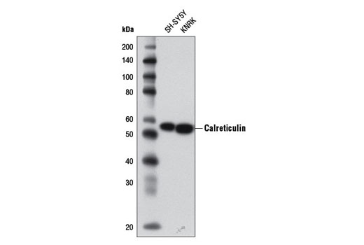
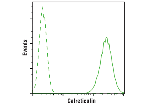
| Calreticulin (D3E6) XP® Rabbit mAb | 12238 | 20 µl | 55 kDa | Rabbit IgG |
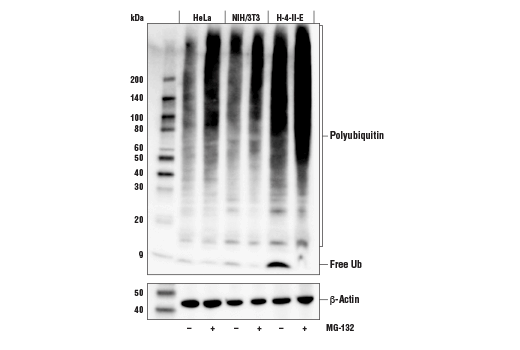
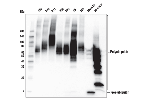
| Ubiquitin (E4I2J) Rabbit mAb | 43124 | 20 µl | Rabbit IgG | |
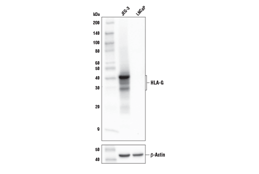
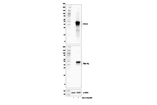
| HLA-G (E8N9C) XP® Rabbit mAb | 79769 | 20 µl | 30-40 kDa | Rabbit IgG |
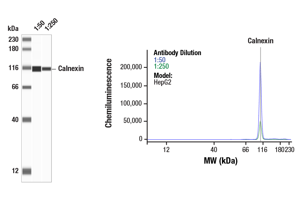
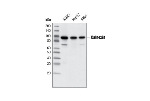
| Calnexin (C5C9) Rabbit mAb | 2679 | 20 µl | 90 kDa | Rabbit IgG |
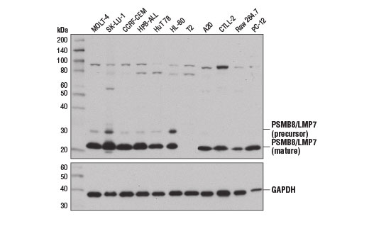
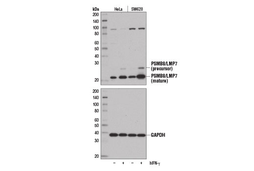
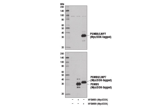
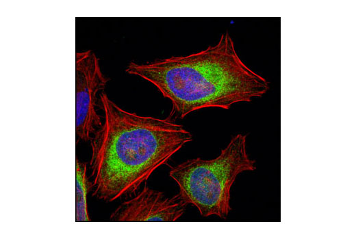
| PSMB8/LMP7 (D1K7X) Rabbit mAb | 13635 | 20 µl | 23, 28 kDa | Rabbit IgG |
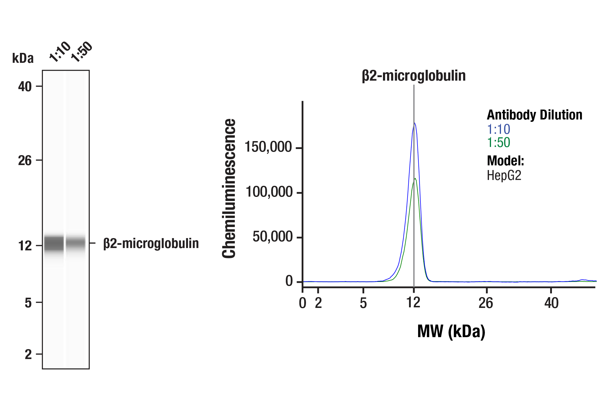
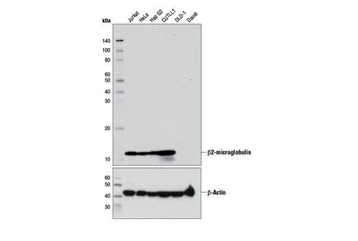
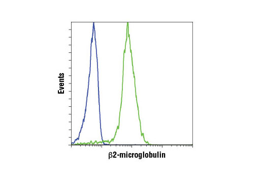
| β2-microglobulin (D8P1H) Rabbit mAb | 12851 | 20 µl | 12 kDa | Rabbit IgG |
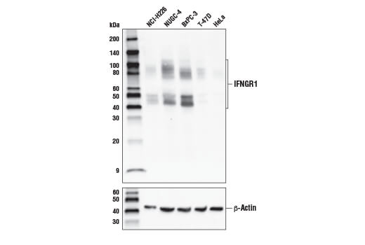
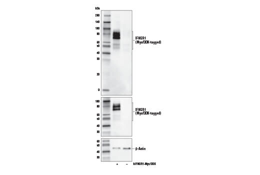
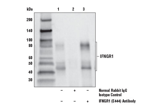
| IFNGR1 (E444) Antibody | 10405 | 20 µl | 45-90 kDa | Rabbit |
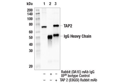
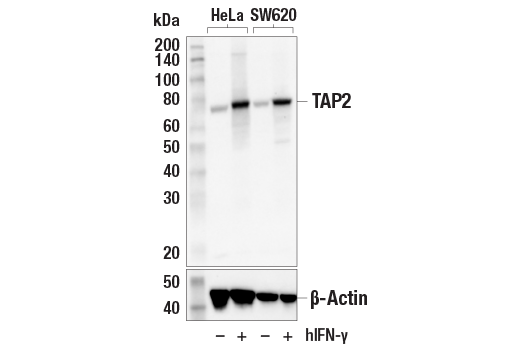
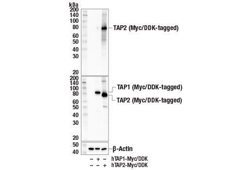
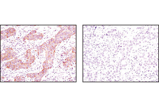
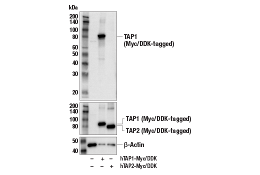
| TAP2 (E8G5I) Rabbit mAb | 25657 | 20 µl | 72 kDa | Rabbit IgG |
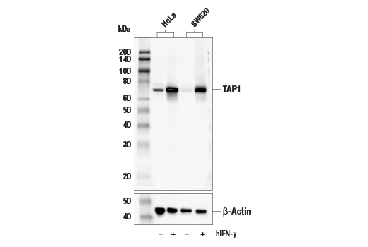
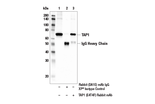
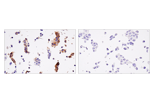
| TAP1 (E4T4F) Rabbit mAb | 49671 | 20 µl | 68 kDa | Rabbit IgG |
Please visit cellsignal.com for individual component applications, species cross-reactivity, dilutions, protocols, and additional product information.
Description
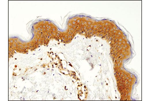
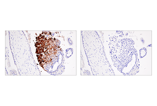
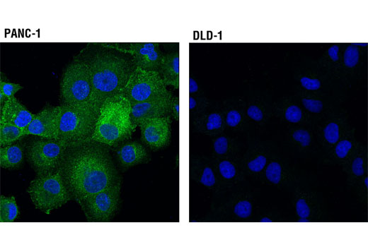
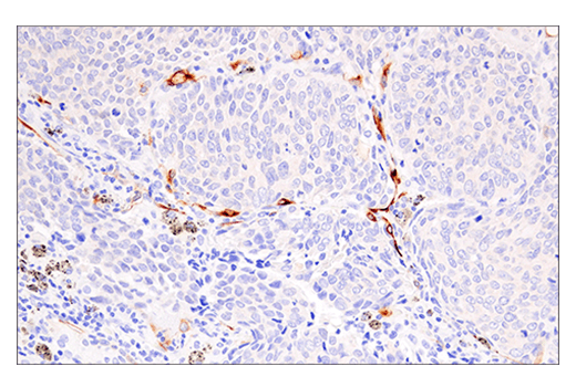
The MHC Class I Antigen Processing and Presentation Antibody Sampler Kit provides an economical means to examine key proteins associated with the processing and presentation of MHC class I-restricted antigens. The provided antibodies allow monitoring of total protein levels. The kit includes enough antibodies to perform two western blot experiments with each primary antibody.
Storage
Background
The predominant function of class I MHC/β2-microglobulin dimers, which are expressed on the surface of most nucleated cell types, is to modulate the adaptive immune response by presenting proteolytic peptide fragments from cytosolic proteins to cytotoxic CD8+ T cells. In order for self and nonself peptides to be presented by MHC class I molecules, the peptide fragments must first be derived from polyubiquitinated proteins that undergo degradation via the ubiquitin-proteasome system. In the context of inflammatory processes, the enzymatic core of the proteasome can be shaped by IFNγ signaling to contain subunits, such as PSMB8/LMP7, which enhance the presentation of antigenic peptides by antigen presenting cells (1). The resulting cytosolic peptide fragments generated through ubiquitin-dependent proteasomal degradation are then transported into the ER lumen via the peptide transporters, TAP1 and TAP2, where the activity of multiple chaperone proteins, such as calnexin and calreticulin, facilitate loading onto class I MHC/β2-microglobulin dimers for transport to the Golgi and eventually, the cell surface (2-6). Defects in the expression of multiple components of the class I antigen presenting machinery have been observed in both solid and liquid tumors, which serves as a mechanism of tumor-immune evasion (7).
- Ferrington, D.A. and Gregerson, D.S. (2012) Prog Mol Biol Transl Sci 109, 75-112.
- Antoniou, A.N. et al. (2003) Curr Opin Immunol 15, 75-81.
- Jensen, P.E. (2007) Nat Immunol 8, 1041-8.
- Kloetzel, P.M. (2001) Nat Rev Mol Cell Biol 2, 179-87.
- Sant, A. and Yewdell, J. (2003) Curr Opin Immunol 15, 66-8.
- Yewdell, J.W. (2005) Immunol Rev 207, 8-18.
- Seliger, B. (2008) Cancer Immunol Immunother 57, 1719-26.
Background References
Trademarks and Patents
Limited Uses
Except as otherwise expressly agreed in a writing signed by a legally authorized representative of CST, the following terms apply to Products provided by CST, its affiliates or its distributors. Any Customer's terms and conditions that are in addition to, or different from, those contained herein, unless separately accepted in writing by a legally authorized representative of CST, are rejected and are of no force or effect.
Products are labeled with For Research Use Only or a similar labeling statement and have not been approved, cleared, or licensed by the FDA or other regulatory foreign or domestic entity, for any purpose. Customer shall not use any Product for any diagnostic or therapeutic purpose, or otherwise in any manner that conflicts with its labeling statement. Products sold or licensed by CST are provided for Customer as the end-user and solely for research and development uses. Any use of Product for diagnostic, prophylactic or therapeutic purposes, or any purchase of Product for resale (alone or as a component) or other commercial purpose, requires a separate license from CST. Customer shall (a) not sell, license, loan, donate or otherwise transfer or make available any Product to any third party, whether alone or in combination with other materials, or use the Products to manufacture any commercial products, (b) not copy, modify, reverse engineer, decompile, disassemble or otherwise attempt to discover the underlying structure or technology of the Products, or use the Products for the purpose of developing any products or services that would compete with CST products or services, (c) not alter or remove from the Products any trademarks, trade names, logos, patent or copyright notices or markings, (d) use the Products solely in accordance with CST Product Terms of Sale and any applicable documentation, and (e) comply with any license, terms of service or similar agreement with respect to any third party products or services used by Customer in connection with the Products.