| Product Includes | Product # | Quantity | Mol. Wt | Isotype/Source |
|---|---|---|---|---|
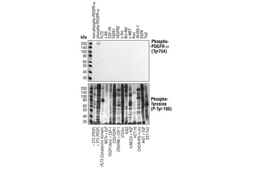
| Phospho-PDGF Receptor α (Tyr754) (23B2) Rabbit mAb | 2992 | 20 µl | 190 kDa | Rabbit |
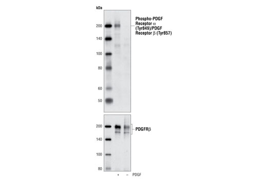
| Phospho-PDGF Receptor α (Tyr849)/PDGF Receptor β (Tyr857) (C43E9) Rabbit mAb | 3170 | 20 µl | 190 kDa | Rabbit IgG |
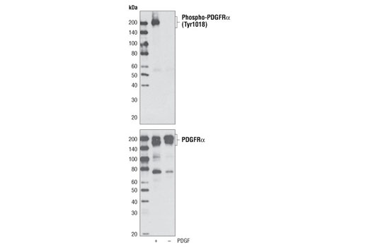
| Phospho-PDGF Receptor α (Tyr1018) Antibody | 4547 | 20 µl | 190 kDa | Rabbit |
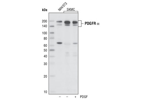
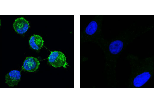
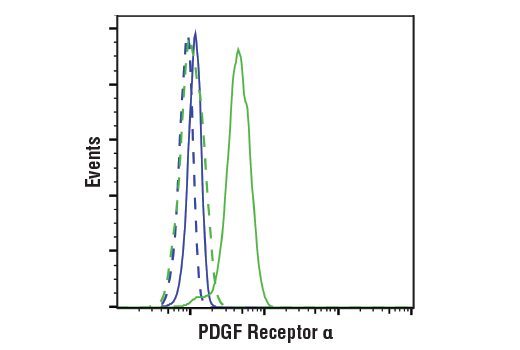
| PDGF Receptor α (D1E1E) XP® Rabbit mAb | 3174 | 20 µl | 190 kDa | Rabbit IgG |
| Anti-rabbit IgG, HRP-linked Antibody | 7074 | 100 µl | Goat |
Please visit cellsignal.com for individual component applications, species cross-reactivity, dilutions, protocols, and additional product information.
Description
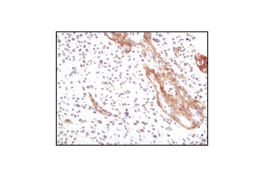
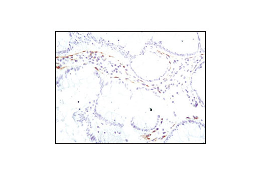
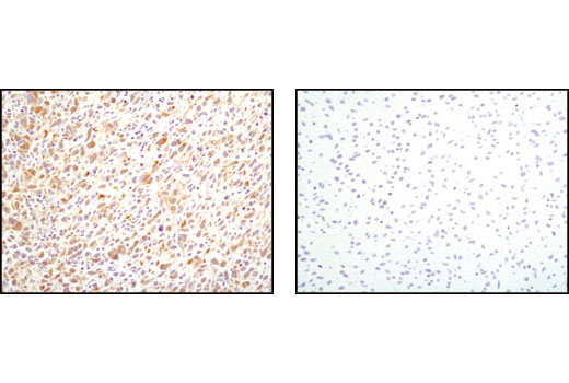
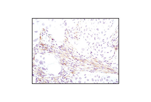
The PDGF Receptor α Antibody Sampler Kit provides an economical means of evaluating total PDGF receptor α protein (PDGFRα) levels as well as PDGFRα phosphorylated at specific sites. The kit contains enough primary and secondary antibody to perform two western blots with each antibody.
Storage
Background
Platelet derived growth factor (PDGF) family proteins exist as several disulphide-bonded, dimeric isoforms (PDGF AA, PDGF AB, PDGF BB, PDGF CC, and PDGF DD) that bind in a specific pattern to two closely related receptor tyrosine kinases, PDGF receptor α (PDGFRα) and PDGF receptor β (PDGFRβ). PDGFRα and PDGFRβ share 75% to 85% sequence homology between their two intracellular kinase domains, while the kinase insert and carboxy-terminal tail regions display a lower level (27% to 28%) of homology (1). PDGFRα homodimers bind all PDGF isoforms except those containing PDGF D. PDGFRβ homodimers bind PDGF BB and DD isoforms, as well as the PDGF AB heterodimer. The heteromeric PDGF receptor α/β binds PDGF B, C, and D homodimers, as well as the PDGF AB heterodimer (2). PDGFRα and PDGFRβ can each form heterodimers with EGFR, which is also activated by PDGF (3). Various cells differ in the total number of receptors present and in the receptor subunit composition, which may account for responsive differences among cell types to PDGF binding (4). Ligand binding induces receptor dimerization and autophosphorylation, followed by binding and activation of cytoplasmic SH2 domain-containing signal transduction molecules, such as GRB2, Src, GAP, PI3 kinase, PLCγ, and NCK. A number of different signaling pathways are initiated by activated PDGF receptors and lead to control of cell growth, actin reorganization, migration, and differentiation (5). Tyr751 in the kinase-insert region of PDGFRβ is the docking site for PI3 kinase (6). Phosphorylated pentapeptides derived from Tyr751 of PDGFRβ (pTyr751-Val-Pro-Met-Leu) inhibit the association of the carboxy-terminal SH2 domain of the p85 subunit of PI3 kinase with PDGFRβ (7). Tyr740 is also required for PDGFRβ-mediated PI3 kinase activation (8).
- Deuel, T.F. et al. (1988) Biofactors 1, 213-217.
- Bergsten, E. et al. (2001) Nat. Cell Biol. 3, 512-516.
- Betsholtz, C. et al. (2001) Bioessays 23, 494-507.
- Coughlin, S.R. et al. (1988) Prog. Clin. Biol. Res. 266, 39-45.
- Ostman, A. and Heldin, C.H. (2001) Adv. Cancer Res. 80, 1-38.
- Panayotou, G. et al. (1992) EMBO J. 11, 4261-4272.
- Ramalingam, K. et al. (1995) Bioorg. Med. Chem. 3, 1263-1272.
- Kashishian, A. et al. (1992) EMBO J. 11, 1373-1382.
Background References
Trademarks and Patents
Limited Uses
Except as otherwise expressly agreed in a writing signed by a legally authorized representative of CST, the following terms apply to Products provided by CST, its affiliates or its distributors. Any Customer's terms and conditions that are in addition to, or different from, those contained herein, unless separately accepted in writing by a legally authorized representative of CST, are rejected and are of no force or effect.
Products are labeled with For Research Use Only or a similar labeling statement and have not been approved, cleared, or licensed by the FDA or other regulatory foreign or domestic entity, for any purpose. Customer shall not use any Product for any diagnostic or therapeutic purpose, or otherwise in any manner that conflicts with its labeling statement. Products sold or licensed by CST are provided for Customer as the end-user and solely for research and development uses. Any use of Product for diagnostic, prophylactic or therapeutic purposes, or any purchase of Product for resale (alone or as a component) or other commercial purpose, requires a separate license from CST. Customer shall (a) not sell, license, loan, donate or otherwise transfer or make available any Product to any third party, whether alone or in combination with other materials, or use the Products to manufacture any commercial products, (b) not copy, modify, reverse engineer, decompile, disassemble or otherwise attempt to discover the underlying structure or technology of the Products, or use the Products for the purpose of developing any products or services that would compete with CST products or services, (c) not alter or remove from the Products any trademarks, trade names, logos, patent or copyright notices or markings, (d) use the Products solely in accordance with CST Product Terms of Sale and any applicable documentation, and (e) comply with any license, terms of service or similar agreement with respect to any third party products or services used by Customer in connection with the Products.