| Product Includes | Product # | Quantity | Mol. Wt | Isotype/Source |
|---|---|---|---|---|
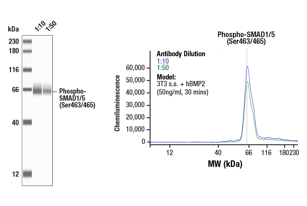
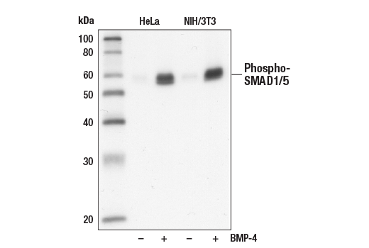
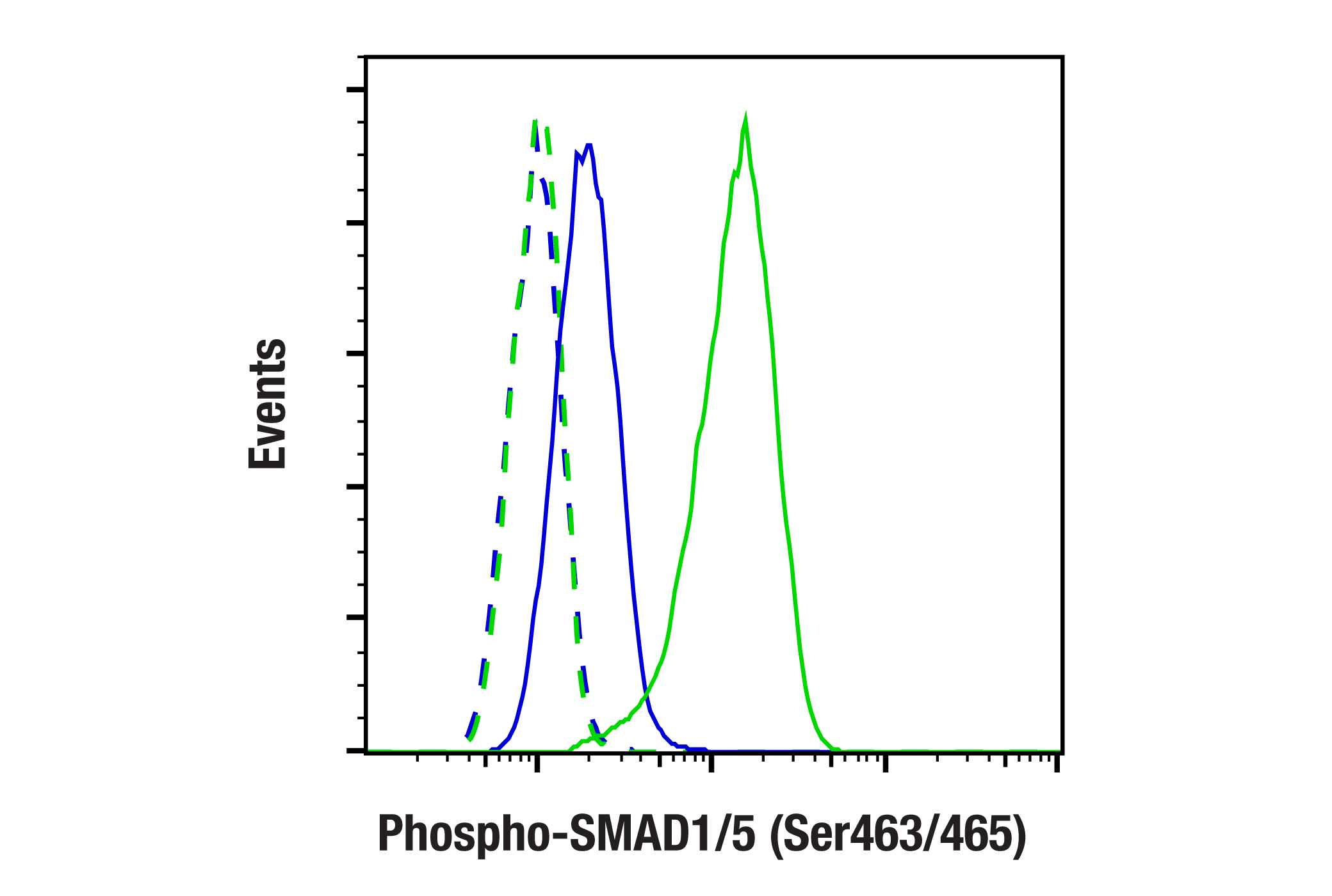
| Phospho-SMAD1/5 (Ser463/465) (41D10) Rabbit mAb | 9516 | 40 µl | 60 kDa | Rabbit |
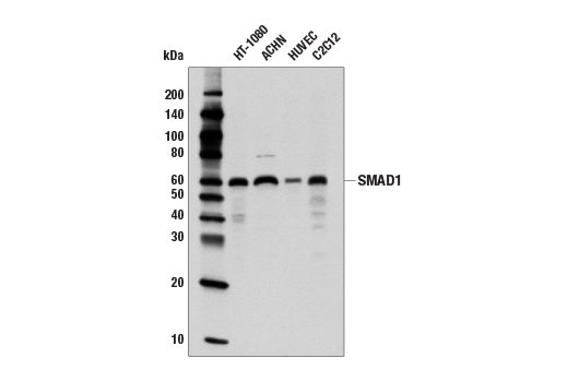
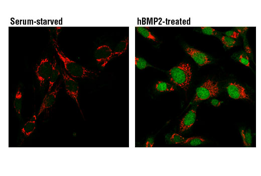
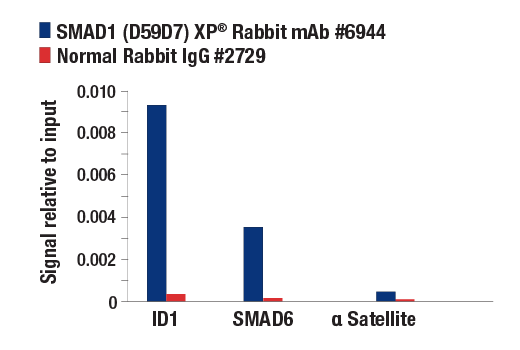
| SMAD1 (D59D7) XP® Rabbit mAb | 6944 | 40 µl | 60 kDa | Rabbit IgG |
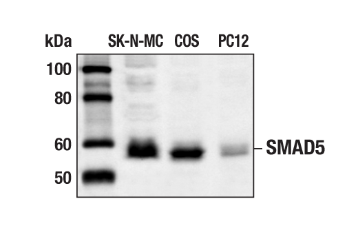
| SMAD5 Antibody | 9517 | 40 µl | 60 kDa | Rabbit |
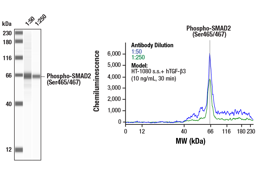
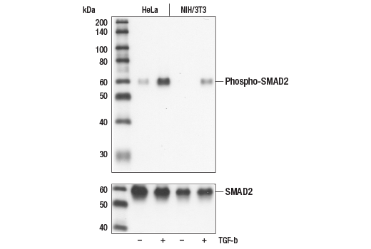
| Phospho-SMAD2 (Ser465/467) (138D4) Rabbit mAb | 3108 | 40 µl | 60 kDa | Rabbit IgG |
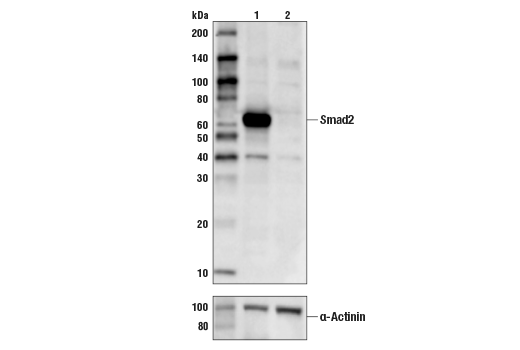
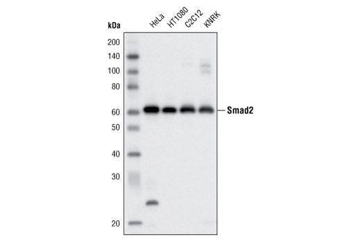
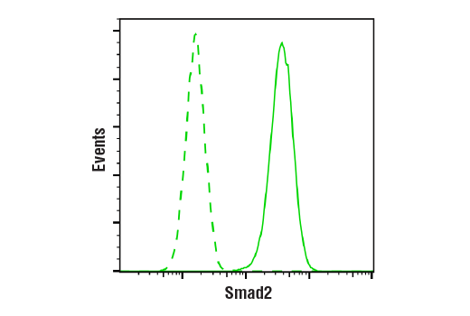
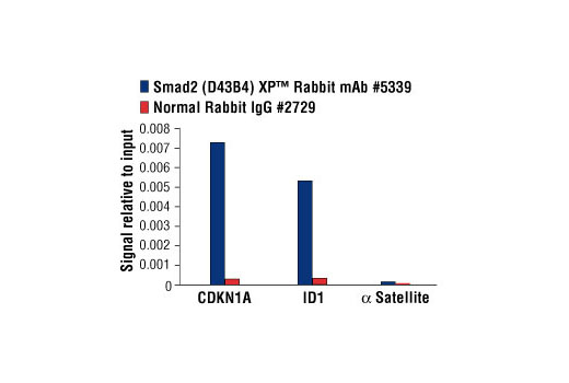
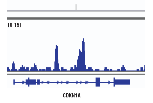
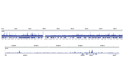
| Smad2 (D43B4) XP® Rabbit mAb | 5339 | 40 µl | 60 kDa | Rabbit IgG |
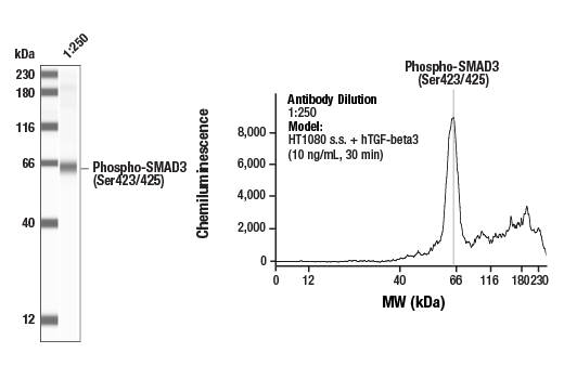
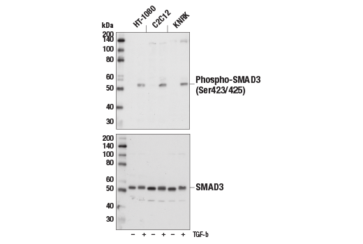
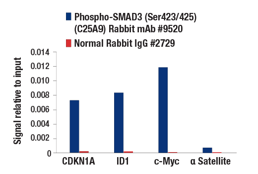
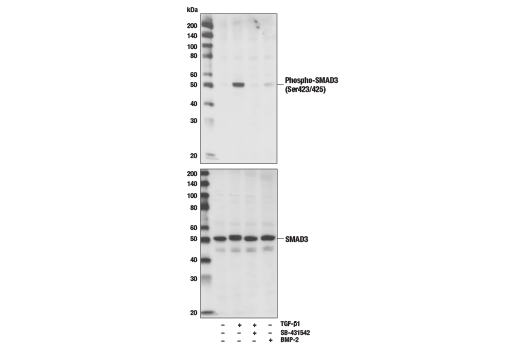
| Phospho-SMAD3 (Ser423/425) (C25A9) Rabbit mAb | 9520 | 40 µl | 52 kDa | Rabbit IgG |
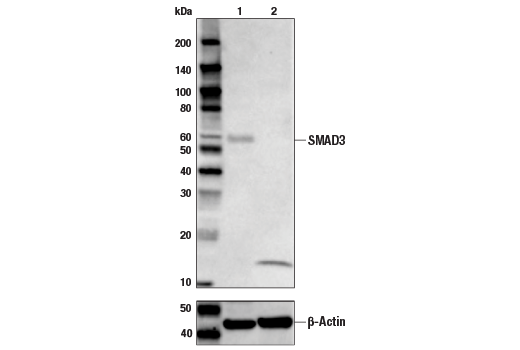
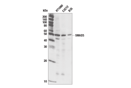
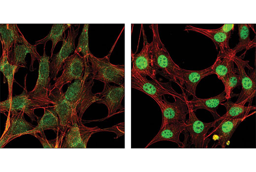
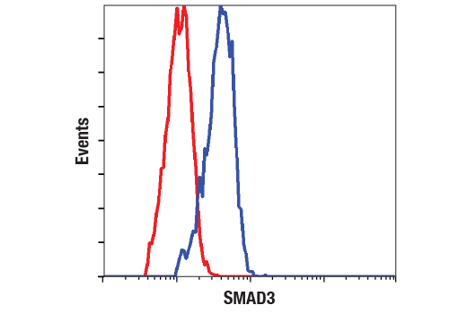
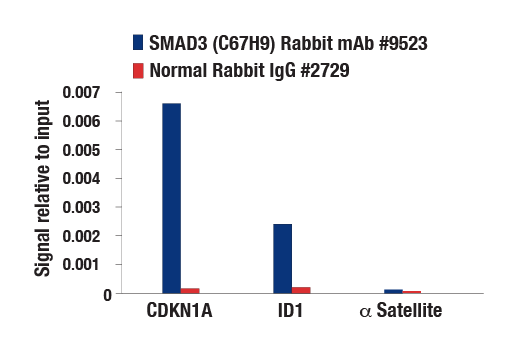
| SMAD3 (C67H9) Rabbit mAb | 9523 | 40 µl | 52 kDa | Rabbit IgG |
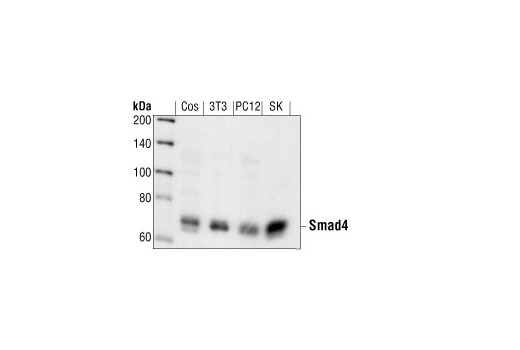
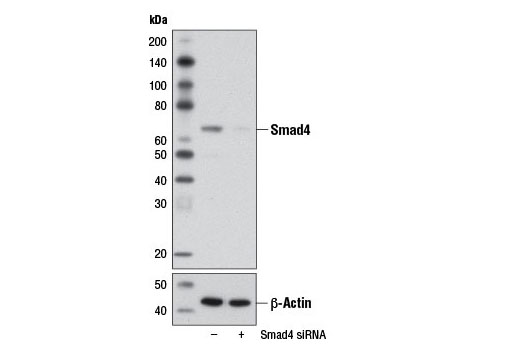
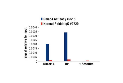
| Smad4 Antibody | 9515 | 40 µl | 70 kDa | Rabbit |
| Smad6 Antibody | 9519 | 40 µl | 62 kDa | Rabbit |
| Anti-rabbit IgG, HRP-linked Antibody | 7074 | 100 µl | Goat |
Please visit cellsignal.com for individual component applications, species cross-reactivity, dilutions, protocols, and additional product information.
Description
The Phospho-Smad Antibody Sampler Kit contains reagents to investigate the activation of the TGF-β and BMP signaling pathways. The kit contains enough primary and secondary antibodies to perform four Western blot experiments per primary antibody.
Storage
Background
Members of the SMAD family of signal transduction molecules are components of a critical intracellular pathway that transmit TGF-β signals from the cell surface into the nucleus. Three distinct classes of SMADs have been defined: the receptor-regulated SMADs (R-SMADs), which include SMAD1, 2, 3, 5, and 9; the common-mediator SMAD (co-SMAD), SMAD4; and the antagonistic or inhibitory SMADs (I-SMADs), SMAD6 and 7 (1-5). Activated type I receptors associate with specific R-SMADs and phosphorylate them on a conserved carboxy-terminal SSXS motif. The phosphorylated R-SMADs dissociate from the receptor and form a heteromeric complex with SMAD4, initiating translocation of the heteromeric SMAD complex to the nucleus. Once in the nucleus, SMADs recruit a variety of DNA binding proteins that function to regulate transcriptional activity (6-8).
- Heldin, C.H. et al. (1997) Nature 390, 465-71.
- Attisano, L. and Wrana, J.L. (1998) Curr Opin Cell Biol 10, 188-94.
- Derynck, R. et al. (1998) Cell 95, 737-40.
- Massagué, J. (1998) Annu Rev Biochem 67, 753-91.
- Whitman, M. (1998) Genes Dev 12, 2445-62.
- Wrana, J.L. (2000) Sci STKE 2000, re1.
- Attisano, L. and Wrana, J.L. (2002) Science 296, 1646-7.
- Moustakas, A. et al. (2001) J Cell Sci 114, 4359-69.
Background References
Trademarks and Patents
Limited Uses
Except as otherwise expressly agreed in a writing signed by a legally authorized representative of CST, the following terms apply to Products provided by CST, its affiliates or its distributors. Any Customer's terms and conditions that are in addition to, or different from, those contained herein, unless separately accepted in writing by a legally authorized representative of CST, are rejected and are of no force or effect.
Products are labeled with For Research Use Only or a similar labeling statement and have not been approved, cleared, or licensed by the FDA or other regulatory foreign or domestic entity, for any purpose. Customer shall not use any Product for any diagnostic or therapeutic purpose, or otherwise in any manner that conflicts with its labeling statement. Products sold or licensed by CST are provided for Customer as the end-user and solely for research and development uses. Any use of Product for diagnostic, prophylactic or therapeutic purposes, or any purchase of Product for resale (alone or as a component) or other commercial purpose, requires a separate license from CST. Customer shall (a) not sell, license, loan, donate or otherwise transfer or make available any Product to any third party, whether alone or in combination with other materials, or use the Products to manufacture any commercial products, (b) not copy, modify, reverse engineer, decompile, disassemble or otherwise attempt to discover the underlying structure or technology of the Products, or use the Products for the purpose of developing any products or services that would compete with CST products or services, (c) not alter or remove from the Products any trademarks, trade names, logos, patent or copyright notices or markings, (d) use the Products solely in accordance with CST Product Terms of Sale and any applicable documentation, and (e) comply with any license, terms of service or similar agreement with respect to any third party products or services used by Customer in connection with the Products.