| Product Includes | Product # | Quantity | Mol. Wt | Isotype/Source |
|---|---|---|---|---|
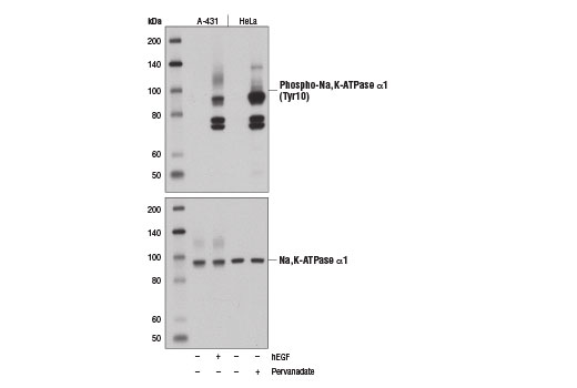
| Phospho-Na,K-ATPase α1 (Tyr10) (E1Y9C) Rabbit mAb | 13566 | 20 µl | 100 kDa | Rabbit IgG |
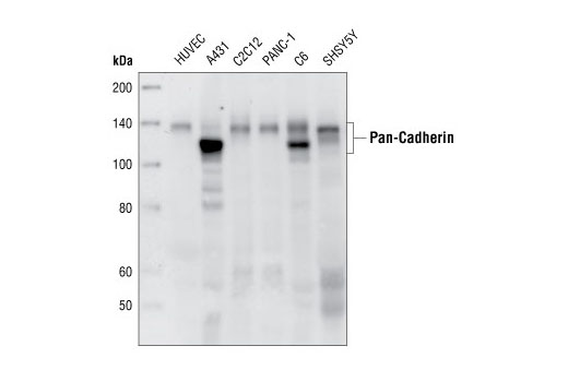
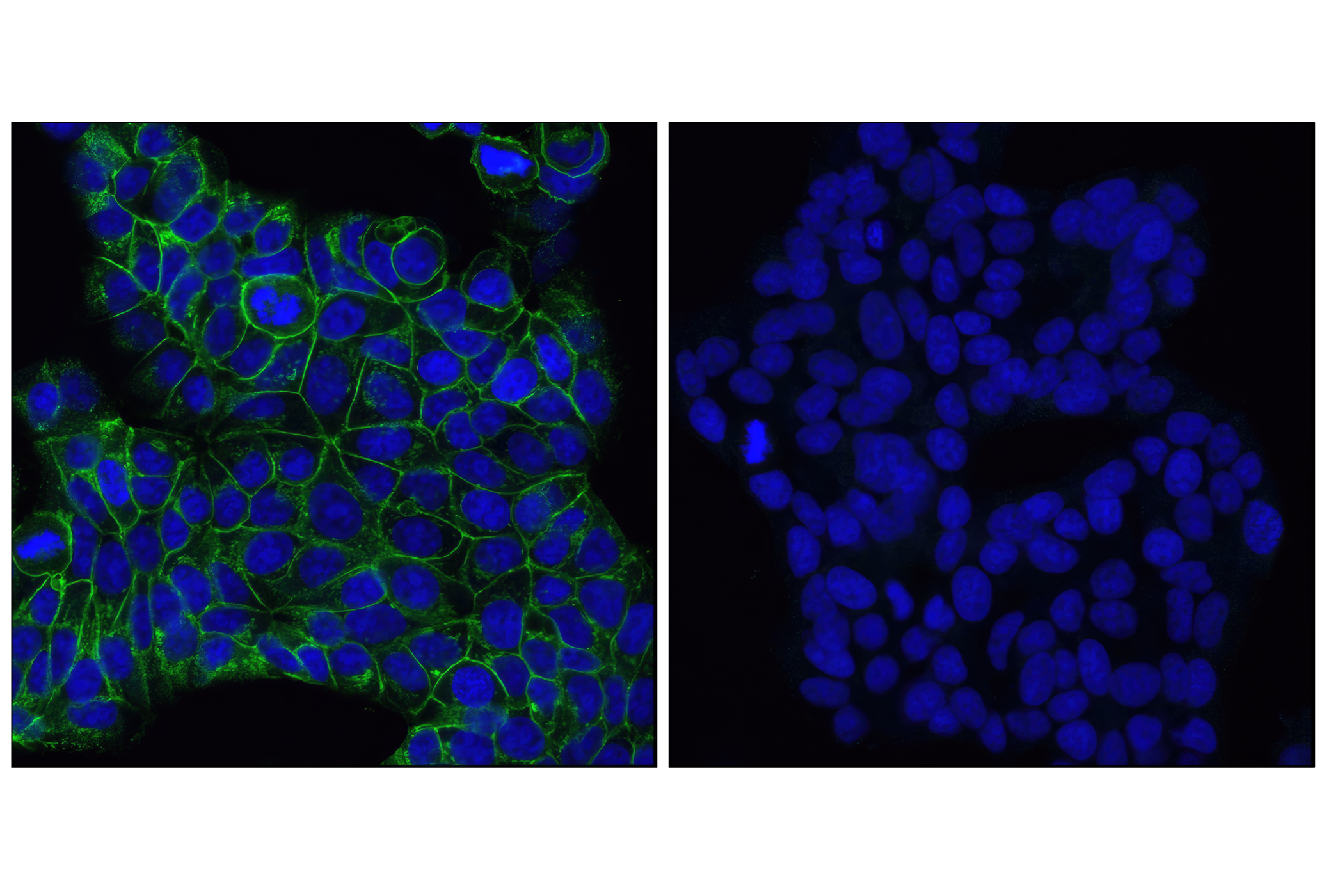
| Pan-Cadherin (28E12) Rabbit mAb | 4073 | 20 µl | 130-150 kDa | Rabbit IgG |
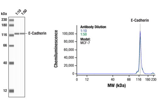
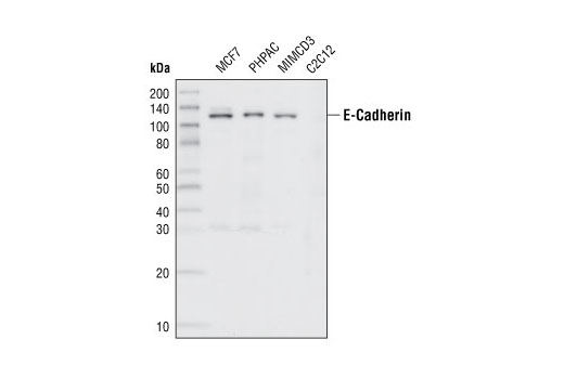
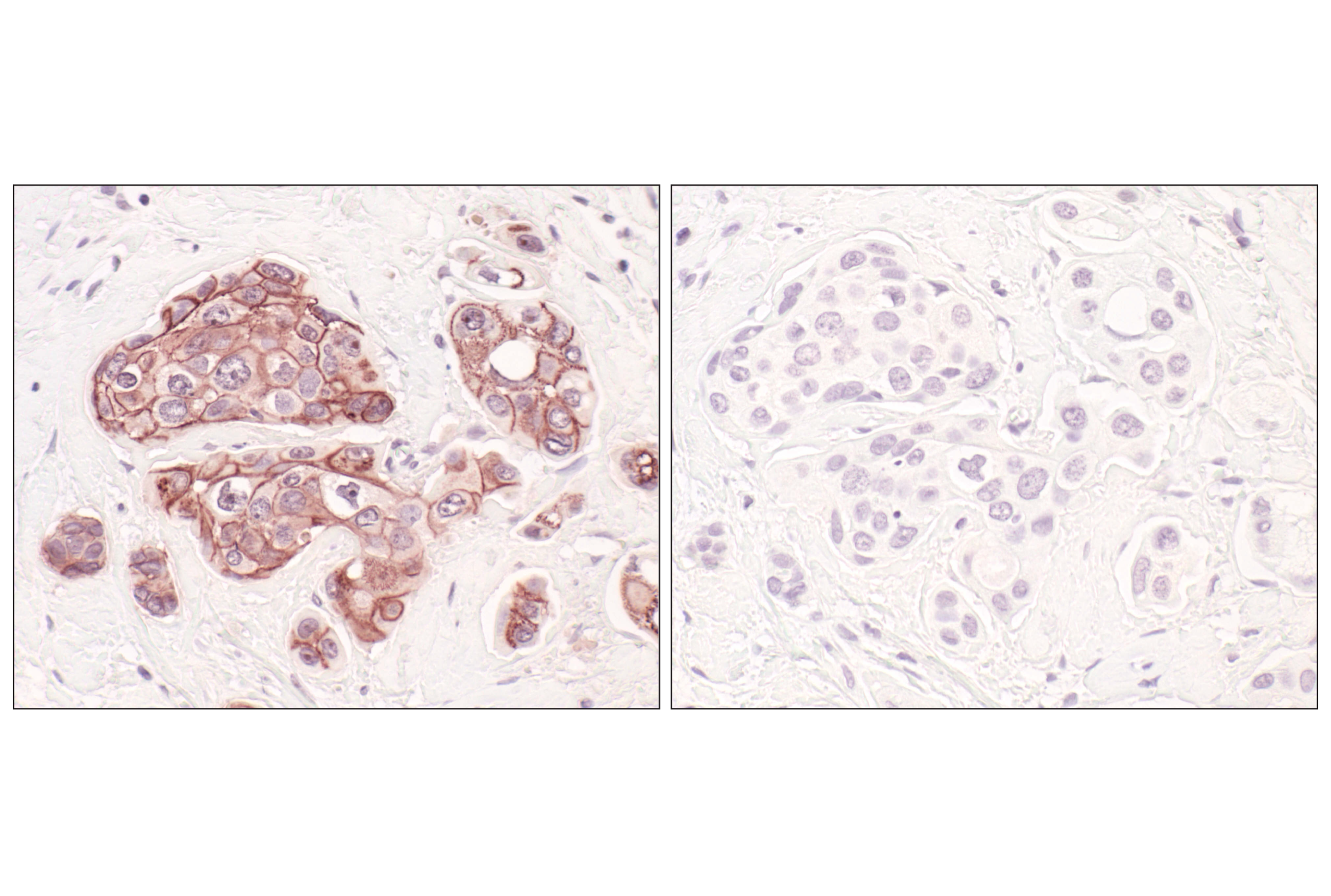
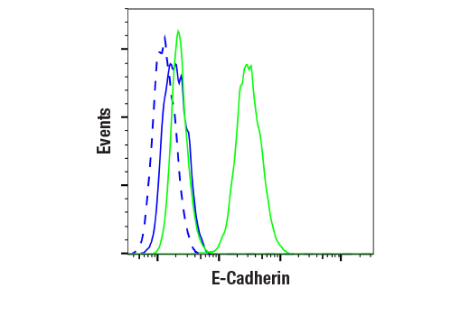
| E-Cadherin (24E10) Rabbit mAb | 3195 | 20 µl | 135 kDa | Rabbit IgG |
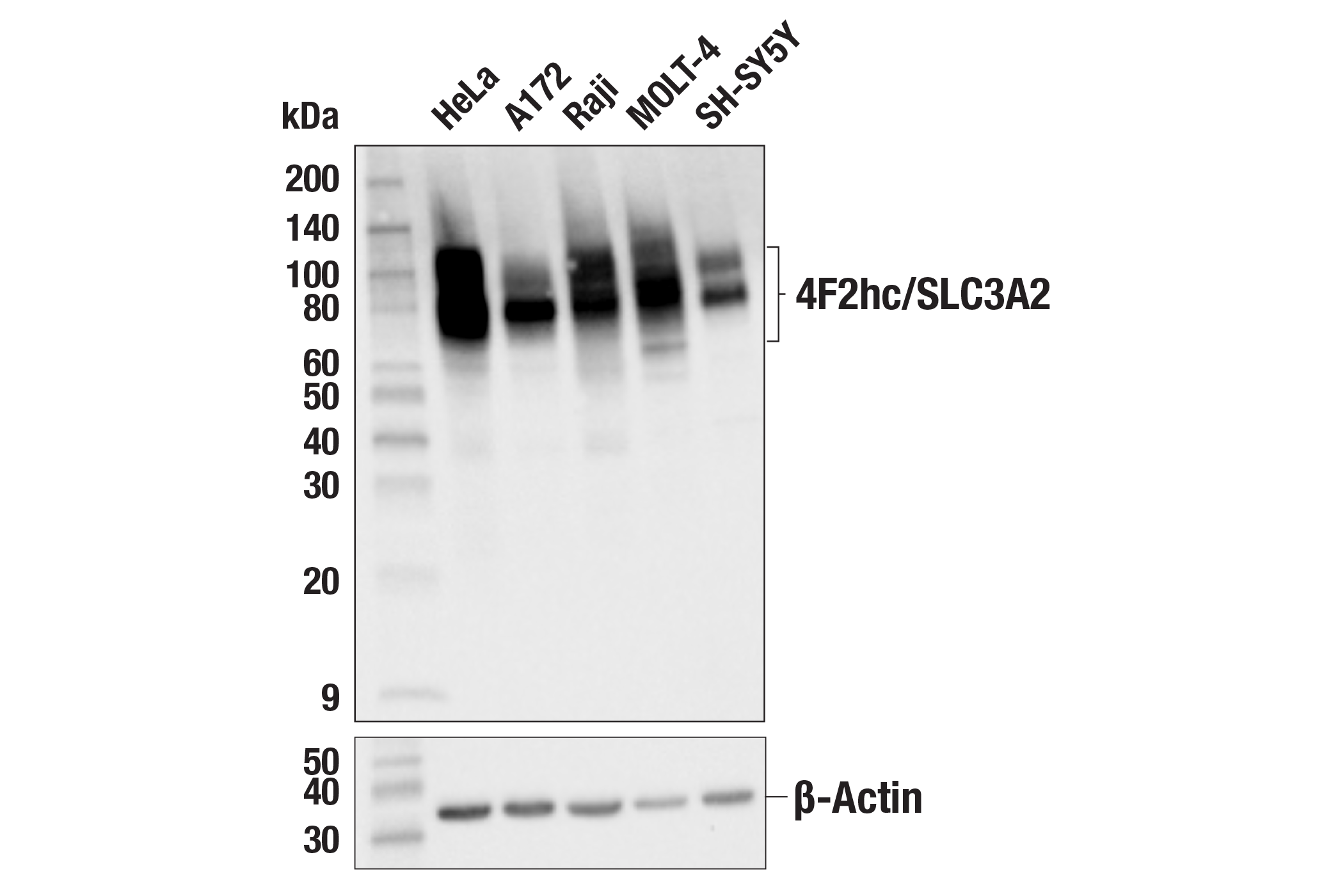
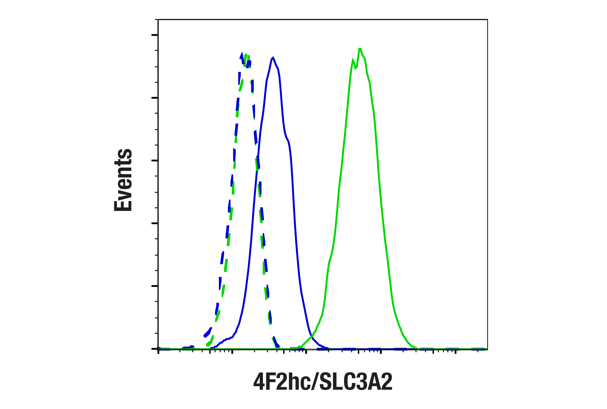
| 4F2hc/SLC3A2 (D3F9D) XP® Rabbit mAb | 47213 | 20 µl | 75-120 kDa | Rabbit IgG |
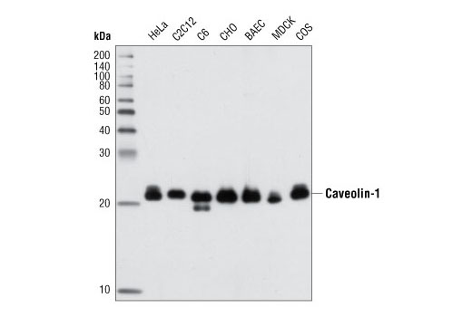
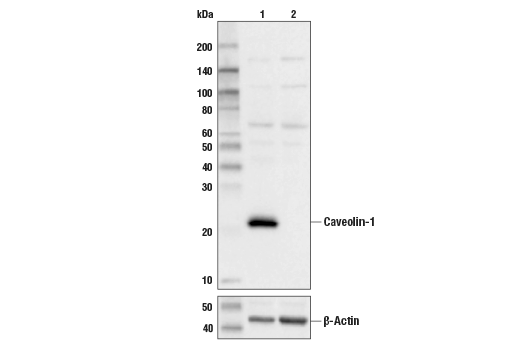
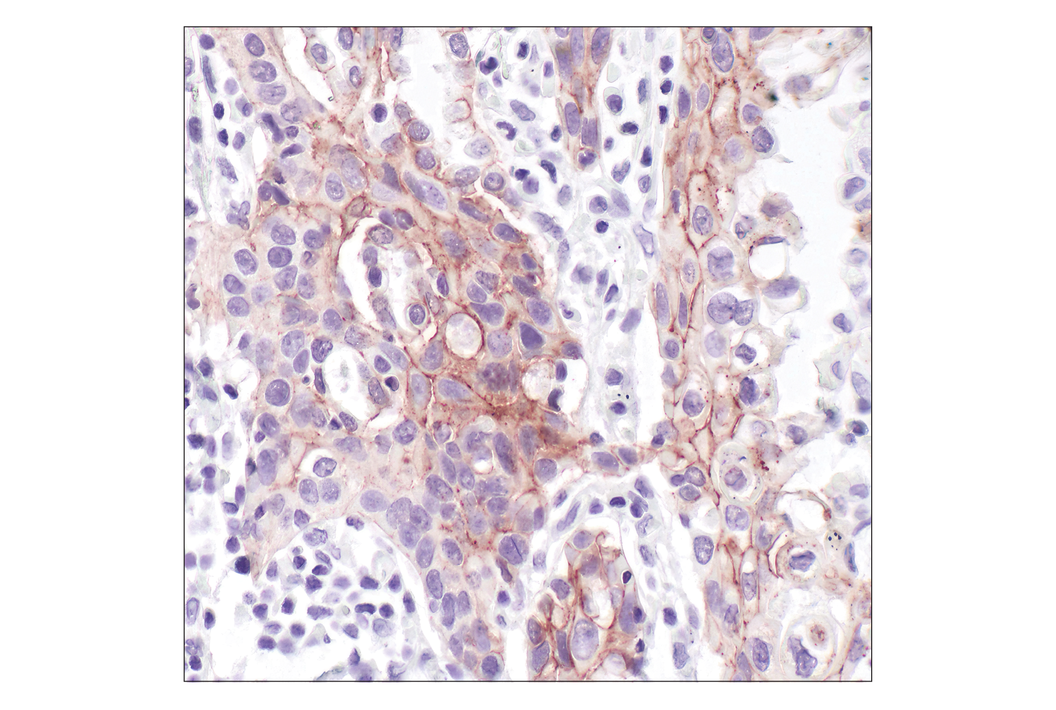
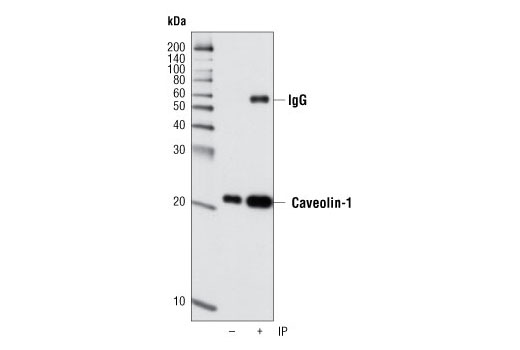
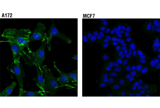
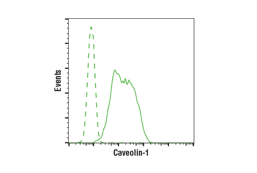
| Caveolin-1 (D46G3) XP® Rabbit mAb | 3267 | 20 µl | 21, 24 kDa | Rabbit IgG |
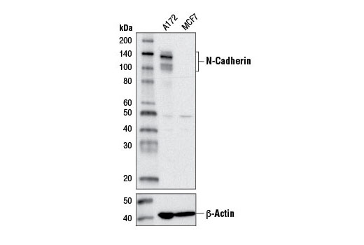
| N-Cadherin (D4R1H) XP® Rabbit mAb | 13116 | 20 µl | 140 kDa | Rabbit IgG |
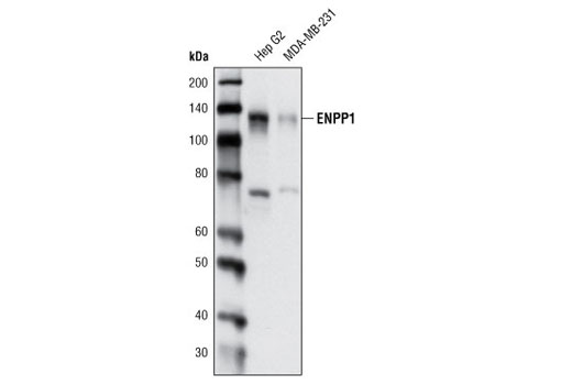
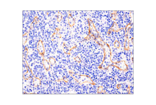
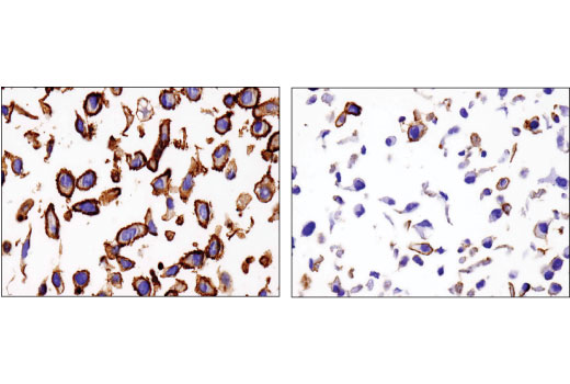
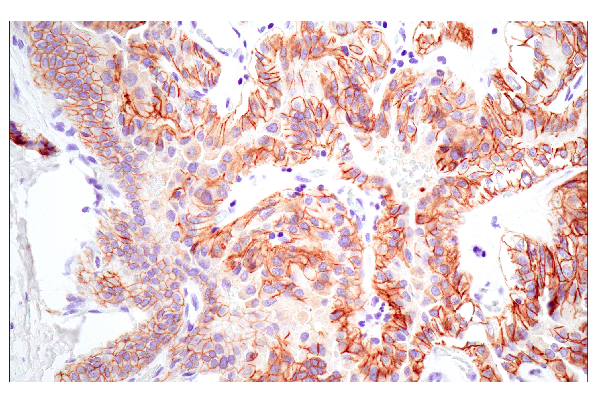
| ENPP1 (D37B7) Rabbit mAb | 5342 | 20 µl | 140 kDa | Rabbit IgG |
| Anti-rabbit IgG, HRP-linked Antibody | 7074 | 100 µl | Goat | |
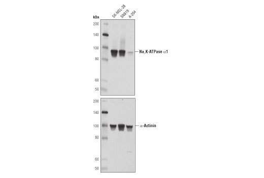
| Na,K-ATPase α1 (D4Y7E) Rabbit mAb | 23565 | 20 µl | 100 kDa | Rabbit IgG |
Please visit cellsignal.com for individual component applications, species cross-reactivity, dilutions, protocols, and additional product information.
Description
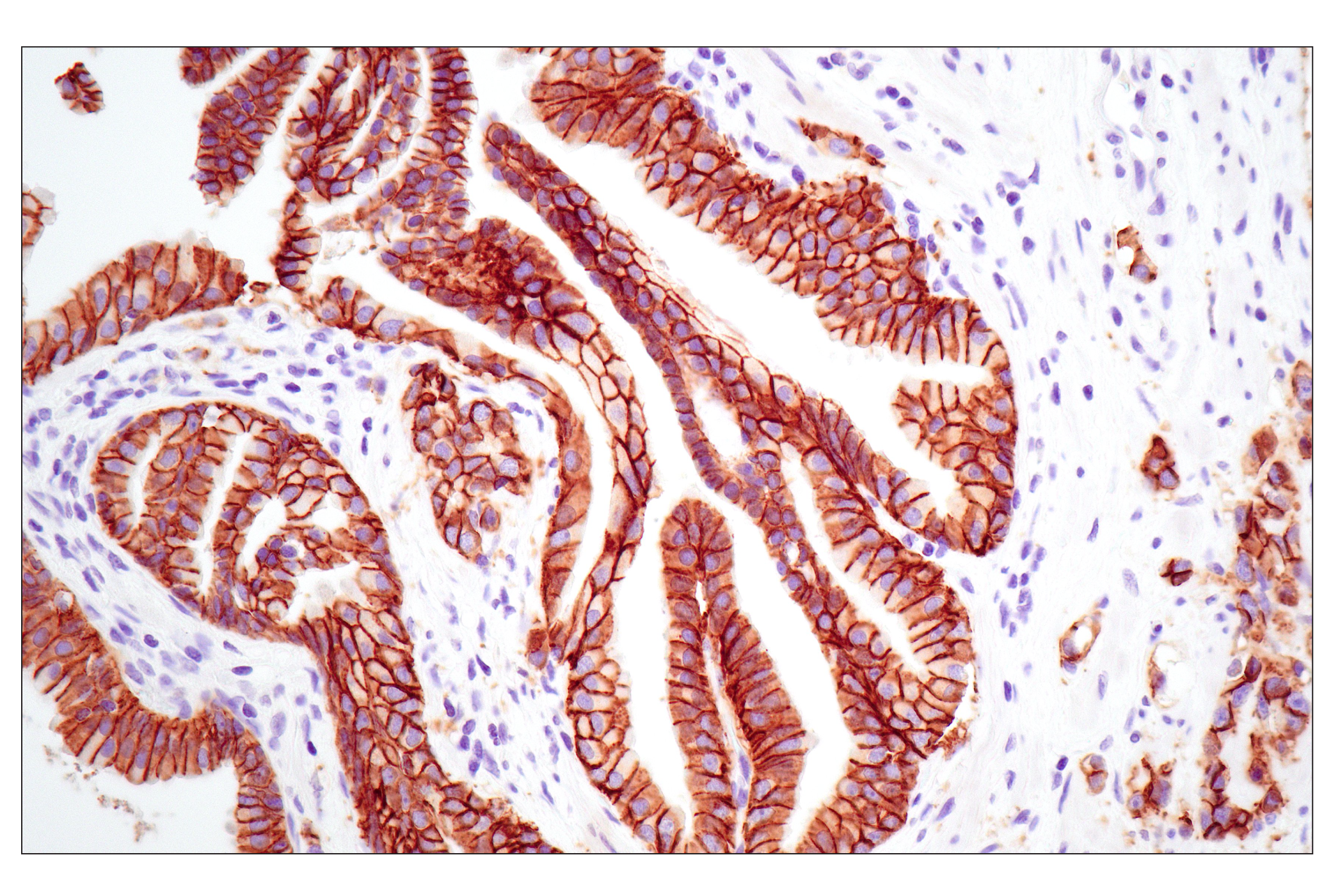
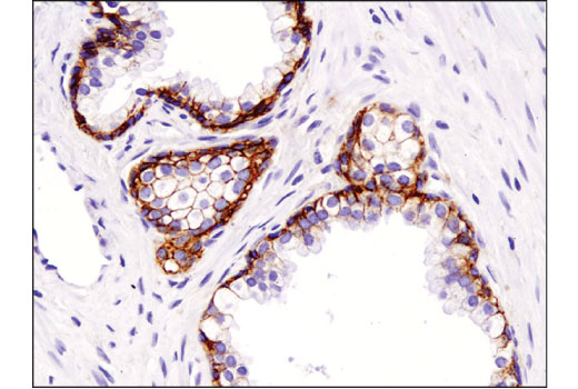
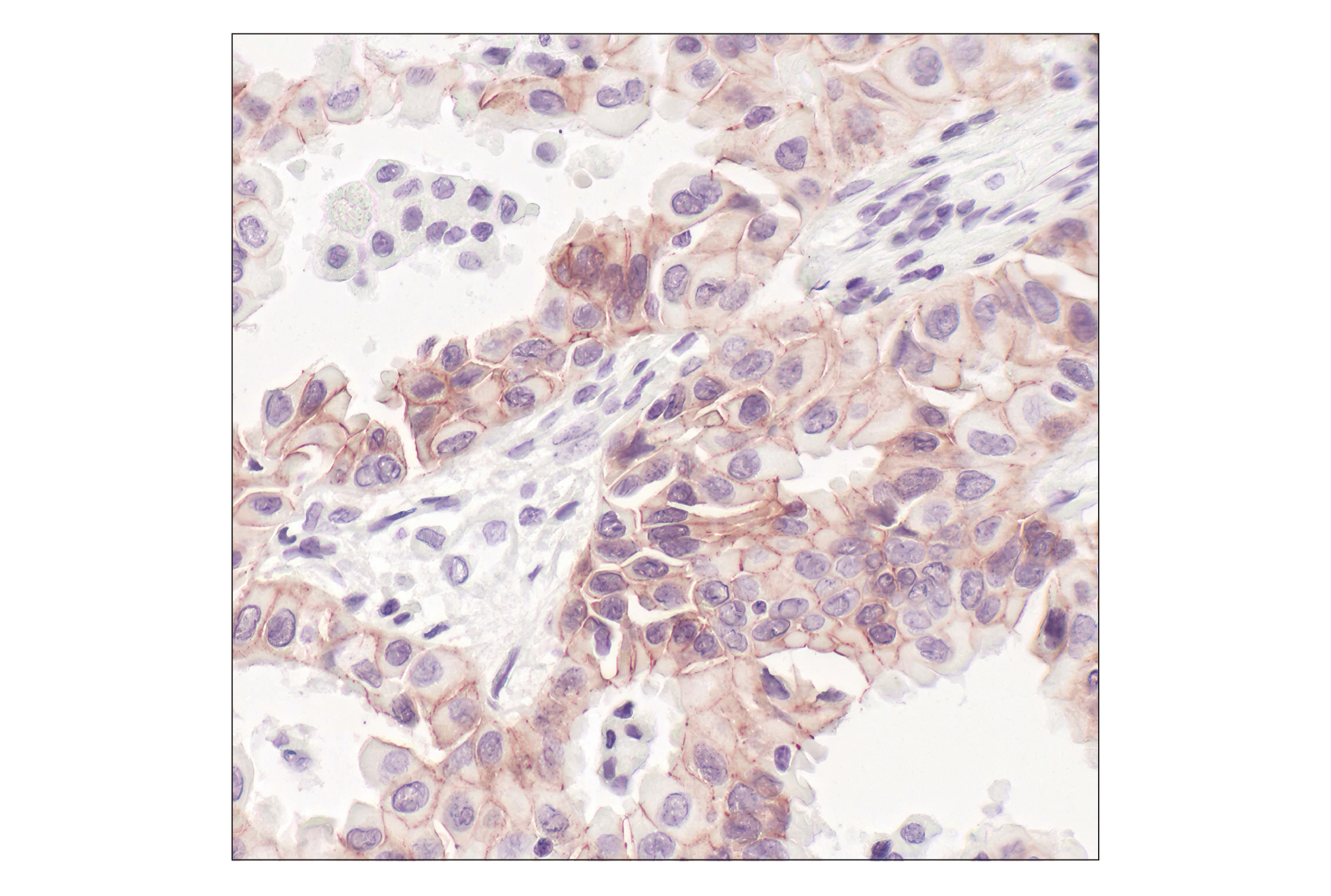
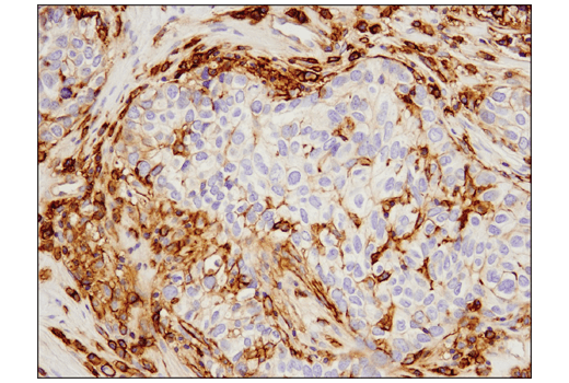
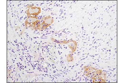
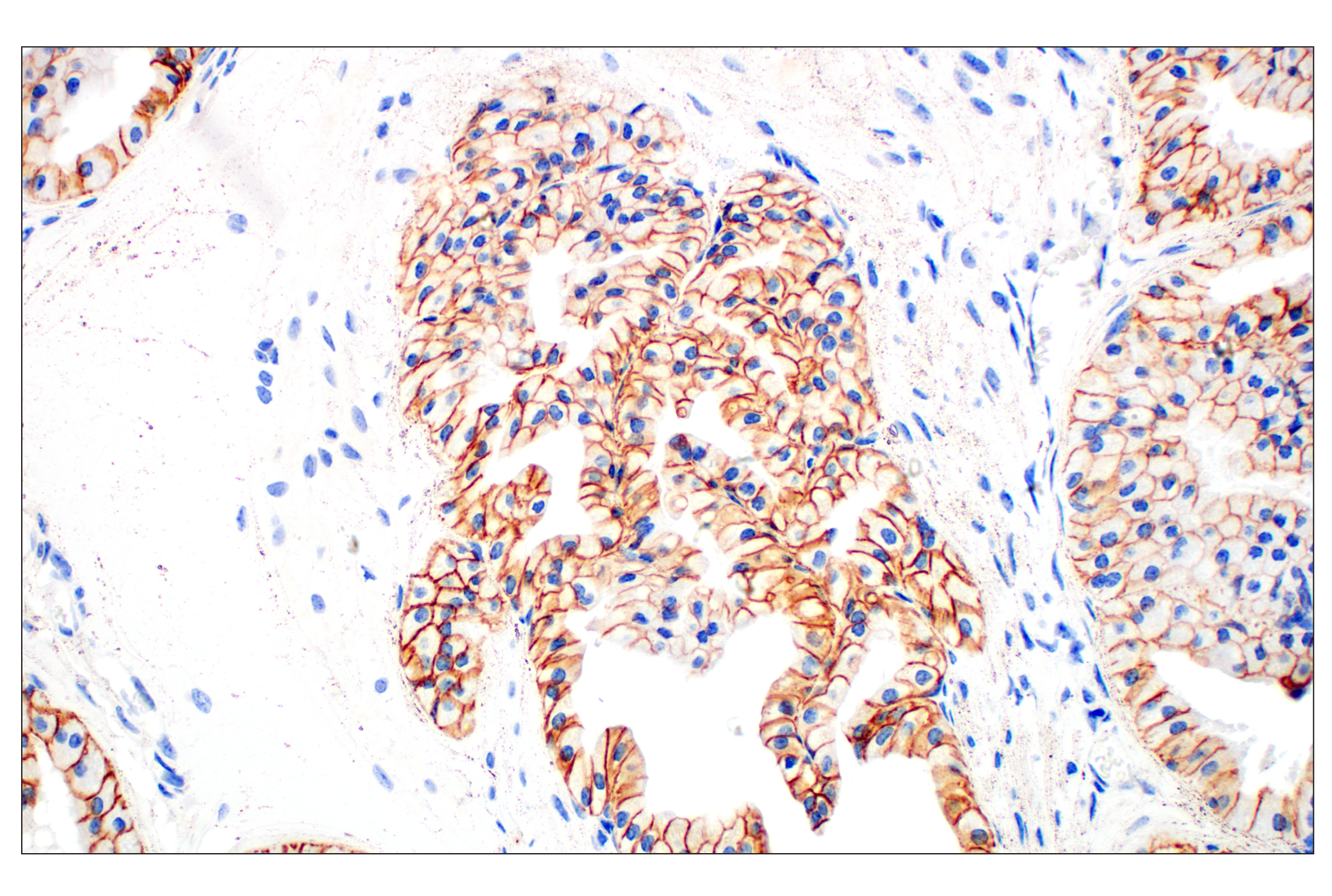
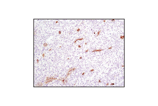
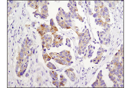
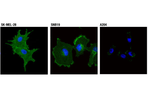
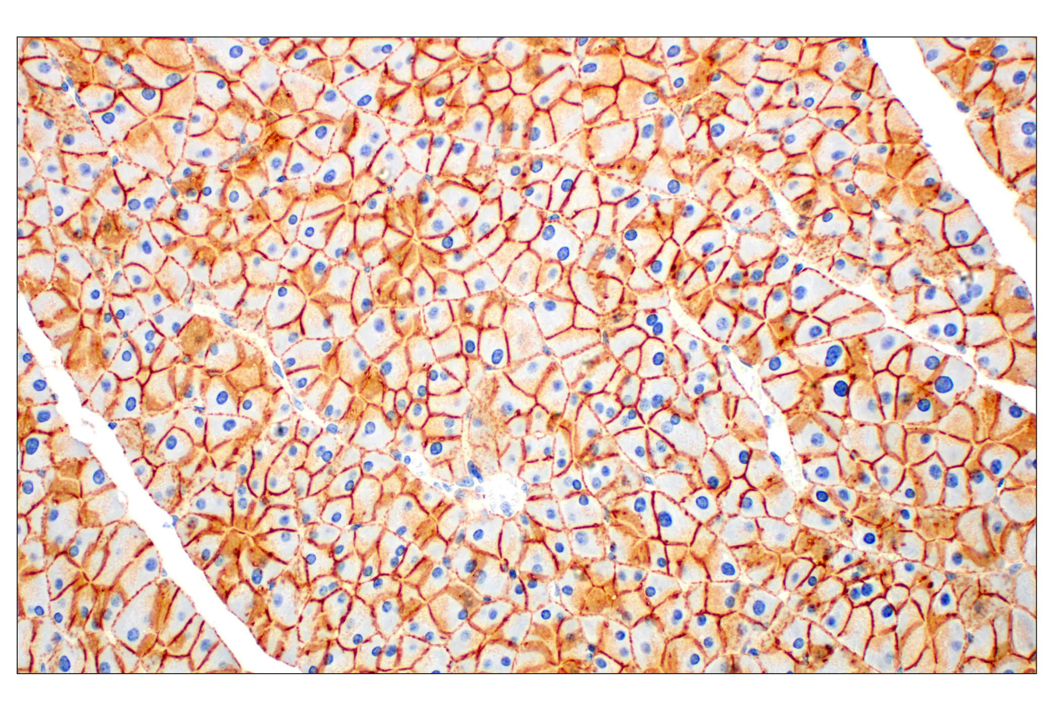
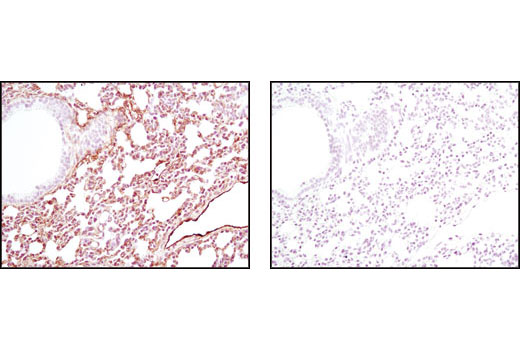
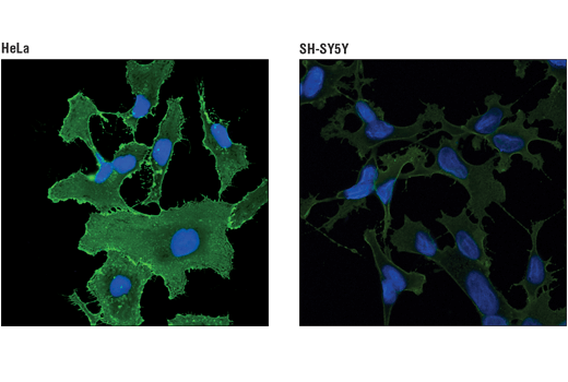
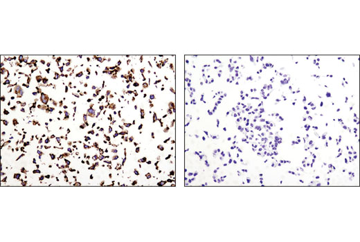
The Plasma Membrane Marker Antibody Sampler Kit provides an economical means of detecting plasma membrane markers in various cell types. The kit includes enough antibodies to perform two western blot experiments with each primary antibody.
Storage
Background
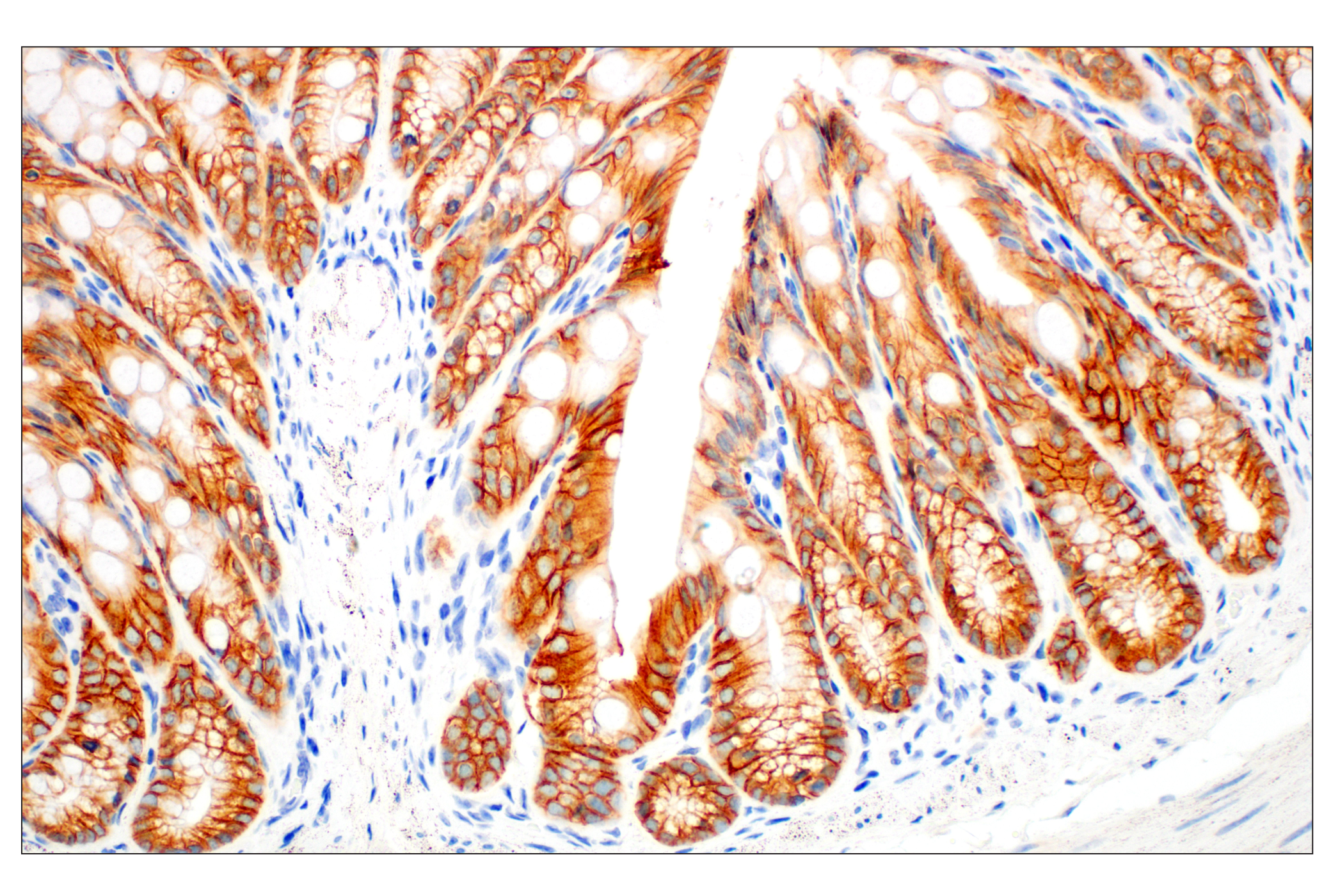
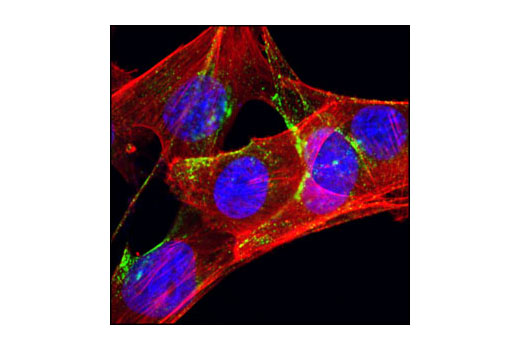
The Na,K-ATPase is an integral membrane heterodimer belonging to the P-type ATPase family. Phosphorylation of Na,K-ATPase at Tyr10 has been implicated in the regulation of enzyme activity in response to hormones and neurotransmitters (1). Cadherins are a superfamily of transmembrane glycoproteins that contain cadherin repeats of approximately 100 residues in their extracellular domain. Cadherins mediate calcium-dependent cell-cell adhesion and play critical roles in normal tissue development. The classic cadherin subfamily includes N-, P-, R-, B-, and E-cadherins, as well as about ten other members that are found in adherens junctions, a cellular structure near the apical surface of polarized epithelial cells (2). 4F2hc is a transmembrane protein that belongs to the solute carrier family. 4F2hc forms heterodimeric complexes with various amino acid transporters, such as LAT1 and LAT2, and regulates uptake of amino acids (3). The 21-24 kDa integral proteins, caveolins, are the principal structural components of the cholesterol/sphingolipid-enriched plasma membrane microdomain caveolae. Three members of the caveolin family (caveolin-1, -2, and -3) have been identified with different tissue distributions (4). Ectonucleotide pyrophosphatase-phosphodiesterase 1 (ENPP1) is a single-pass, type II transmembrane protein primarily involved in ATP hydrolysis at the plasma membrane. ENPP1 plays important roles in bone mineralization and soft tissue calcification (5).
- Therien, A.G. and Blostein, R. (2000) Am J Physiol Cell Physiol 279, C541-66.
- Wheelock, M.J. and Johnson, K.R. (2003) Annu Rev Cell Dev Biol 19, 207-35.
- Kanai, Y. et al. (1998) J Biol Chem 273, 23629-32.
- Okamoto, T. et al. (1998) J Biol Chem 273, 5419-22.
- Harmey, D. et al. (2004) Am J Pathol 164, 1199-209.
Background References
Trademarks and Patents
Limited Uses
Except as otherwise expressly agreed in a writing signed by a legally authorized representative of CST, the following terms apply to Products provided by CST, its affiliates or its distributors. Any Customer's terms and conditions that are in addition to, or different from, those contained herein, unless separately accepted in writing by a legally authorized representative of CST, are rejected and are of no force or effect.
Products are labeled with For Research Use Only or a similar labeling statement and have not been approved, cleared, or licensed by the FDA or other regulatory foreign or domestic entity, for any purpose. Customer shall not use any Product for any diagnostic or therapeutic purpose, or otherwise in any manner that conflicts with its labeling statement. Products sold or licensed by CST are provided for Customer as the end-user and solely for research and development uses. Any use of Product for diagnostic, prophylactic or therapeutic purposes, or any purchase of Product for resale (alone or as a component) or other commercial purpose, requires a separate license from CST. Customer shall (a) not sell, license, loan, donate or otherwise transfer or make available any Product to any third party, whether alone or in combination with other materials, or use the Products to manufacture any commercial products, (b) not copy, modify, reverse engineer, decompile, disassemble or otherwise attempt to discover the underlying structure or technology of the Products, or use the Products for the purpose of developing any products or services that would compete with CST products or services, (c) not alter or remove from the Products any trademarks, trade names, logos, patent or copyright notices or markings, (d) use the Products solely in accordance with CST Product Terms of Sale and any applicable documentation, and (e) comply with any license, terms of service or similar agreement with respect to any third party products or services used by Customer in connection with the Products.