| Product Includes | Product # | Quantity | Mol. Wt | Isotype/Source |
|---|---|---|---|---|
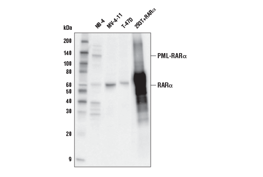
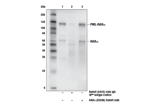
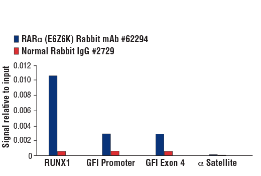
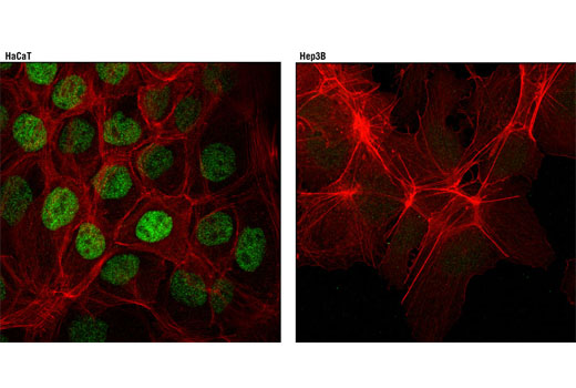
| RARα (E6Z6K) Rabbit mAb | 62294 | 20 µl | 60 kDa | Rabbit IgG |
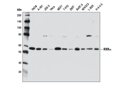
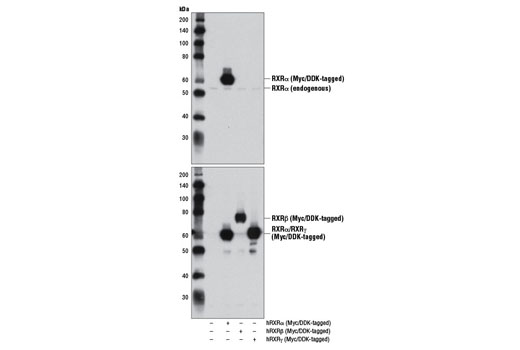
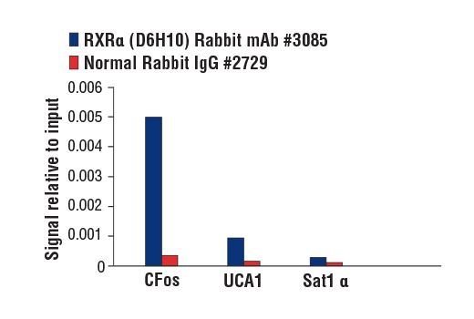
| RXRα (D6H10) Rabbit mAb | 3085 | 20 µl | 53 kDa | Rabbit IgG |
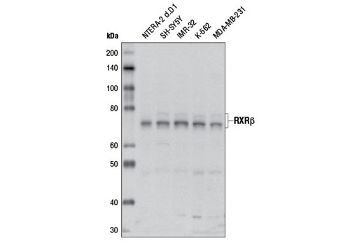
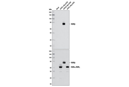
| RXRβ Antibody | 8715 | 20 µl | 70-72 kDa | Rabbit |
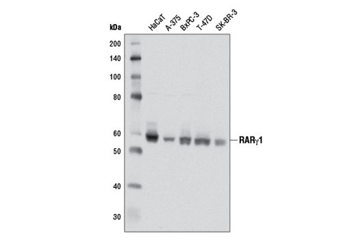
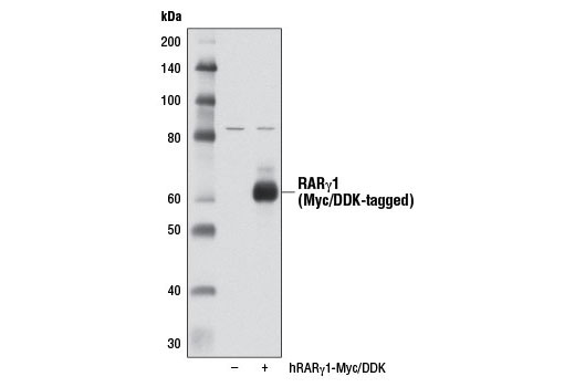
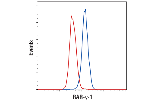
| RARγ1 (D3A4) XP® Rabbit mAb | 8965 | 20 µl | 58 kDa | Rabbit IgG |
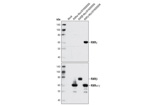
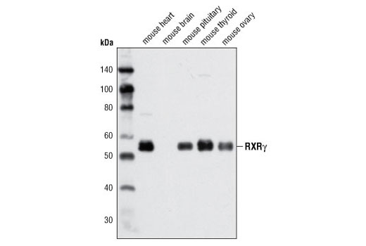
| RXRγ Antibody | 5629 | 20 µl | 55 kDa | Rabbit |
| Anti-rabbit IgG, HRP-linked Antibody | 7074 | 100 µl | Goat |
Please visit cellsignal.com for individual component applications, species cross-reactivity, dilutions, protocols, and additional product information.
Description
The Retinoic Acid and Retinoid X Receptors Antibody Sampler Kit provides an economical means to investigate the expression of various subtypes of retinoic acid and retinoid X receptors. The kit contains enough primary antibody to perform two western blot experiments per primary.
Storage
Background
Nuclear retinoic acid (RA) receptors (RARs) consist of three subtypes encoded by separate genes: α (NR1B1), β (NR1B2), and γ (NR1B3). For each subtype, there are at least two isoforms, which are generated by differential promoter usage and alternative splicing and differ only in their N-terminal regions. Retinoids, which are metabolites of vitamin A, serve as ligands for RARs (1). RARs function as ligand-dependent transcriptional regulators and are found to be heterodimerized with retinoid X receptors (RXRs). These transcriptionally active dimers regulate the expression of genes involved in cellular differentiation, proliferation, and apoptosis (2,3). Consequently, RARs play critical roles in a variety of biological processes, including development, reproduction, immunity, and organogenesis (4-6). RAR mutations, fusion proteins, altered expression levels, or aberrant post-translational modifications result in multiple diseases due to altered RAR function and disruption of homeostasis.
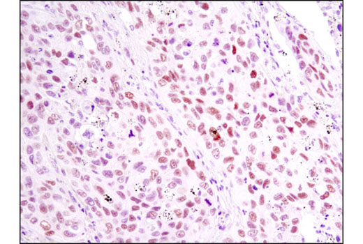
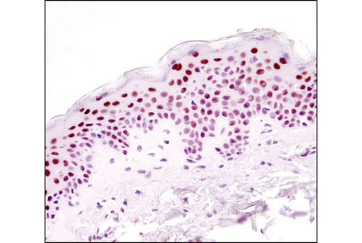
In contrast to the ubiquitously expressed RARα subtype, RARγ displays a complex tissue-specific expression pattern (7). The hematopoietic system expresses significant levels of RARγ, and a recent study identified a role for RARγ in hematopoietic stem cell maintenance (8). RARγ is the predominant subtype in human and mouse epidermis, representing 90% of the RARs in this tissue (9-11). Given the high level of RARγ expression in the skin, it has been suggested that this nuclear receptor participates in a transcriptional program that governs maintenance and differentiation of normal epidermis and skin appendages. The transcriptional activity of RARγ is under stringent control, in part, through retinoic acid-induced phosphorylation and proteasomal degradation (12).
The human retinoid X receptors (RXRs) are encoded by three distinct genes (RXRα, RXRβ, and RXRγ) and bind selectively and with high affinity to the vitamin A derivative, 9-cis-retinoic acid. RXRs are type-II nuclear hormone receptors that are largely localized to the nuclear compartment independent of ligand binding. Nuclear RXRs form heterodimers with nuclear hormone receptor subfamily 1 proteins, including thyroid hormone receptor, retinoic acid receptors, vitamin D receptor, peroxisome proliferator-activated receptors, liver X receptors, and farnesoid X receptor (13). Since RXRs heterodimerize with multiple nuclear hormone receptors, they play a central role in transcriptional control of numerous hormonal signaling pathways by binding to cis-acting response elements in the promoter/enhancer region of target genes (14).
- Rochette-Egly, C. and Germain, P. (2009) Nucl Recept Signal 7, e005.
- Delacroix, L. et al. (2010) Mol Cell Biol 30, 231-44.
- Eifert, C. et al. (2006) Mol Reprod Dev 73, 796-824.
- Mark, M. et al. (2006) Annu Rev Pharmacol Toxicol 46, 451-80.
- Niederreither, K. and Dollé, P. (2008) Nat Rev Genet 9, 541-53.
- Mark, M. et al. (2009) Nucl Recept Signal 7, e002.
- Dollé, P. (2009) Nucl Recept Signal 7, e006.
- Purton, L.E. et al. (2006) J Exp Med 203, 1283-93.
- Fisher, G.J. et al. (1994) J Biol Chem 269, 20629-35.
- Zelent, A. et al. (1989) Nature 339, 714-7.
- Elder, J.T. et al. (1991) J Invest Dermatol 96, 425-33.
- Giannì, M. et al. (2002) EMBO J 21, 3760-9.
- Gronemeyer, H. et al. (2004) Nat Rev Drug Discov 3, 950-64.
- Mangelsdorf, D.J. et al. (1992) Genes Dev 6, 329-44.
Background References
Trademarks and Patents
Limited Uses
Except as otherwise expressly agreed in a writing signed by a legally authorized representative of CST, the following terms apply to Products provided by CST, its affiliates or its distributors. Any Customer's terms and conditions that are in addition to, or different from, those contained herein, unless separately accepted in writing by a legally authorized representative of CST, are rejected and are of no force or effect.
Products are labeled with For Research Use Only or a similar labeling statement and have not been approved, cleared, or licensed by the FDA or other regulatory foreign or domestic entity, for any purpose. Customer shall not use any Product for any diagnostic or therapeutic purpose, or otherwise in any manner that conflicts with its labeling statement. Products sold or licensed by CST are provided for Customer as the end-user and solely for research and development uses. Any use of Product for diagnostic, prophylactic or therapeutic purposes, or any purchase of Product for resale (alone or as a component) or other commercial purpose, requires a separate license from CST. Customer shall (a) not sell, license, loan, donate or otherwise transfer or make available any Product to any third party, whether alone or in combination with other materials, or use the Products to manufacture any commercial products, (b) not copy, modify, reverse engineer, decompile, disassemble or otherwise attempt to discover the underlying structure or technology of the Products, or use the Products for the purpose of developing any products or services that would compete with CST products or services, (c) not alter or remove from the Products any trademarks, trade names, logos, patent or copyright notices or markings, (d) use the Products solely in accordance with CST Product Terms of Sale and any applicable documentation, and (e) comply with any license, terms of service or similar agreement with respect to any third party products or services used by Customer in connection with the Products.