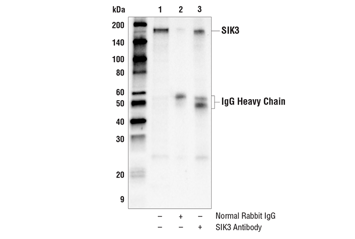
WB, IP
H
Endogenous
145
Rabbit
#Q9Y2K2
23387
Product Information
Product Usage Information
| Application | Dilution |
|---|---|
| Western Blotting | 1:1000 |
| Immunoprecipitation | 1:100 |
Storage
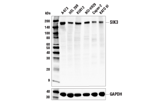
Specificity / Sensitivity
Species Reactivity:
Human
Source / Purification
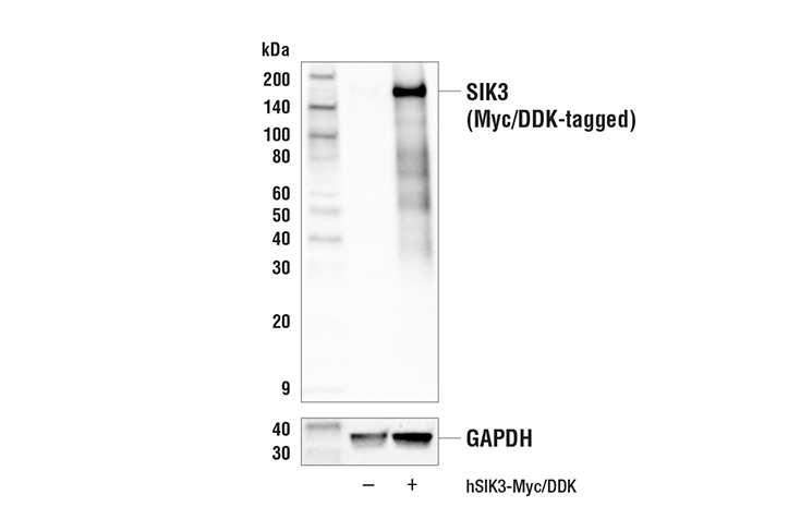
Polyclonal antibodies are produced by immunizing animals with a synthetic peptide corresponding to residues surrounding Gly1155 of human SIK3 protein. Antibodies are purified by protein A and peptide affinity chromatography.
Background
Salt-inducible kinase 1 (SIK1) was originally identified as a serine/threonine kinase from adrenocortical tissues of rats on a high salt diet (1). SIK1 is an SNF1/AMPK family kinase capable of autophosphorylation (1). SIK2 is an isoform of SIK1 and is specifically expressed in adipose tissues where it is induced during adipocyte differentiation (2). Studies suggest that SIK2 can phosphorylate human insulin receptor substrate 1 (IRS-1) at Ser794. Along with evidence that SIK2 expression and activity are increased in white adipocytes of diabetic mice, this finding suggests a possible role for SIK2 in regulating insulin signaling in adipocytes and in the development of insulin resistance (2,3). Insulin triggers Akt2-mediated phosphorylation of SIK2 at Ser358 and the resultant kinase activation during post-fasting feeding (4). The activated SIK2 then induces the phosphorylation of TORC2 at Ser171, resulting in translocation of this transcriptional coactivator from the nucleus to the cytoplasm, where it is degraded through the ubiquitin pathway, leading to inhibition of gluconeogenic gene expression (4).
SIK3 was identified in a screen for tumor-associated antigens that promoted cell proliferation and showed increased expression in ovarian cancer (5). Analysis of SIK3 knockout mice identified a key role in skeletal development via regulation of subcellular localization of HDAC4 (6). Additional studies have also found that SIK3 can regulate glucose and lipid metabolism in the liver (7). SIK1 and SIK3 were found to mediate the tumor suppressive activity of LKB1 in models of non-small cell lung cancer (8).
- Wang, Z. et al. (1999) FEBS Lett 453, 135-9.
- Horike, N. et al. (2003) J Biol Chem 278, 18440-7.
- Katoh, Y. et al. (2004) Mol Cell Endocrinol 217, 109-12.
- Dentin, R. et al. (2007) Nature 449, 366-9.
- Charoenfuprasert, S. et al. (2011) Oncogene 30, 3570-84.
- Sasagawa, S. et al. (2012) Development 139, 1153-63.
- Uebi, T. et al. (2012) PLoS One 7, e37803.
- Hollstein, P.E. et al. (2019) Cancer Discov 9, 1606-27.
Species Reactivity
Species reactivity is determined by testing in at least one approved application (e.g., western blot).
Western Blot Buffer
IMPORTANT: For western blots, incubate membrane with diluted primary antibody in 5% w/v BSA, 1X TBS, 0.1% Tween® 20 at 4°C with gentle shaking, overnight.
Applications Key
WB: Western Blotting IP: Immunoprecipitation
Cross-Reactivity Key
H: human M: mouse R: rat Hm: hamster Mk: monkey Vir: virus Mi: mink C: chicken Dm: D. melanogaster X: Xenopus Z: zebrafish B: bovine Dg: dog Pg: pig Sc: S. cerevisiae Ce: C. elegans Hr: horse GP: Guinea Pig Rab: rabbit All: all species expected
Trademarks and Patents
Limited Uses
Except as otherwise expressly agreed in a writing signed by a legally authorized representative of CST, the following terms apply to Products provided by CST, its affiliates or its distributors. Any Customer's terms and conditions that are in addition to, or different from, those contained herein, unless separately accepted in writing by a legally authorized representative of CST, are rejected and are of no force or effect.
Products are labeled with For Research Use Only or a similar labeling statement and have not been approved, cleared, or licensed by the FDA or other regulatory foreign or domestic entity, for any purpose. Customer shall not use any Product for any diagnostic or therapeutic purpose, or otherwise in any manner that conflicts with its labeling statement. Products sold or licensed by CST are provided for Customer as the end-user and solely for research and development uses. Any use of Product for diagnostic, prophylactic or therapeutic purposes, or any purchase of Product for resale (alone or as a component) or other commercial purpose, requires a separate license from CST. Customer shall (a) not sell, license, loan, donate or otherwise transfer or make available any Product to any third party, whether alone or in combination with other materials, or use the Products to manufacture any commercial products, (b) not copy, modify, reverse engineer, decompile, disassemble or otherwise attempt to discover the underlying structure or technology of the Products, or use the Products for the purpose of developing any products or services that would compete with CST products or services, (c) not alter or remove from the Products any trademarks, trade names, logos, patent or copyright notices or markings, (d) use the Products solely in accordance with CST Product Terms of Sale and any applicable documentation, and (e) comply with any license, terms of service or similar agreement with respect to any third party products or services used by Customer in connection with the Products.