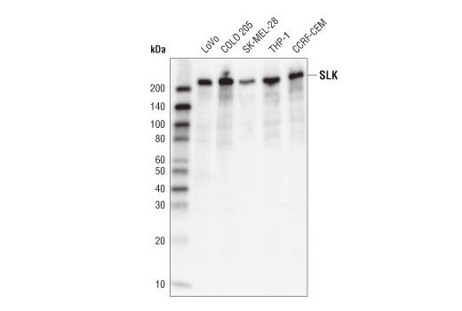
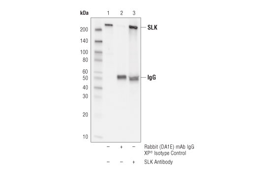
WB, IP
H
Endogenous
220
Rabbit
#Q9H2G2
9748
Product Information
Product Usage Information
| Application | Dilution |
|---|---|
| Western Blotting | 1:1000 |
| Immunoprecipitation | 1:50 |
Storage
Specificity / Sensitivity
Species Reactivity:
Human
Source / Purification
Polyclonal antibodies are produced by immunizing animals with a synthetic peptide corresponding to residues surrounding Glu431 of human SLK protein.
Background
SLK (Ste20-like Kinase) is a member of the germinal center kinase (GCK) family of proteins. SLK has a kinase domain located at the N terminus (1). The autophosphorylation of SLK at Thr183 and Ser189 is required for the upregulation of SLK kinase activity (1, 2). The protein also has a caspase cleavage site DXXD and a SH3 binding site PXXP located in the middle part of its sequence, and a regulatory C terminal coiled-coil domain for homodimerization and adaptor binding (1-4). SLK plays important roles in development, tissue regeneration and cancer cell migration by regulating several signaling pathways (5-7). SLK phosphorylates and activates ASK1 to induce downstream p38 phosphorylation and apoptosis (8,9). During cell cycle, SLK phosphorylates Polo-like kinase (PLK) at Thr210 to promote G2/M transition (10,11). SLK also promotes cell division by direct phosphorylation of ERMs and dynactin to activate microtubule reorganization and spindle orientation (12, 13). During focal adhesion and cell migration process, SLK is activated and colocalized to the focal adhesion complex where it promotes complex turnover by phosphorylating paxillin at Ser250 (14, 15).
- Al-Zahrani, K.N. et al. (2013) Cell Adh Migr 7, 1-10.
- Luhovy, A.Y. et al. (2012) J Biol Chem 287, 5446-58.
- Delarosa, S. et al. (2011) Am J Physiol Renal Physiol 301, F554-64.
- Baron, K.D. et al. (2015) Biochim Biophys Acta 1853, 1683-92.
- Al-Zahrani, K.N. et al. (2014) Dev Dyn 243, 640-51.
- Storbeck, C.J. et al. (2013) Skelet Muscle 3, 16.
- Roovers, K. et al. (2009) Oncogene 28, 2839-48.
- Hao, W. et al. (2006) J Biol Chem 281, 3075-84.
- Sabourin, L.A. et al. (2000) Mol Cell Biol 20, 684-96.
- Ellinger-Ziegelbauer, H. et al. (2000) Genes Cells 5, 491-8.
- Johnson, T.M. et al. (2008) Biochemistry 47, 3688-96.
- Zhapparova, O.N. et al. (2013) Mol Biol Cell 24, 3205-14.
- Machicoane, M. et al. (2014) J Cell Biol 205, 791-9.
- Wagner, S. et al. (2008) PLoS One 3, e1868.
- Quizi, J.L. et al. (2013) Oncogene 32, 4656-63.
Species Reactivity
Species reactivity is determined by testing in at least one approved application (e.g., western blot).
Western Blot Buffer
IMPORTANT: For western blots, incubate membrane with diluted primary antibody in 5% w/v BSA, 1X TBS, 0.1% Tween® 20 at 4°C with gentle shaking, overnight.
Applications Key
WB: Western Blotting IP: Immunoprecipitation
Cross-Reactivity Key
H: human M: mouse R: rat Hm: hamster Mk: monkey Vir: virus Mi: mink C: chicken Dm: D. melanogaster X: Xenopus Z: zebrafish B: bovine Dg: dog Pg: pig Sc: S. cerevisiae Ce: C. elegans Hr: horse GP: Guinea Pig Rab: rabbit All: all species expected
Trademarks and Patents
Limited Uses
Except as otherwise expressly agreed in a writing signed by a legally authorized representative of CST, the following terms apply to Products provided by CST, its affiliates or its distributors. Any Customer's terms and conditions that are in addition to, or different from, those contained herein, unless separately accepted in writing by a legally authorized representative of CST, are rejected and are of no force or effect.
Products are labeled with For Research Use Only or a similar labeling statement and have not been approved, cleared, or licensed by the FDA or other regulatory foreign or domestic entity, for any purpose. Customer shall not use any Product for any diagnostic or therapeutic purpose, or otherwise in any manner that conflicts with its labeling statement. Products sold or licensed by CST are provided for Customer as the end-user and solely for research and development uses. Any use of Product for diagnostic, prophylactic or therapeutic purposes, or any purchase of Product for resale (alone or as a component) or other commercial purpose, requires a separate license from CST. Customer shall (a) not sell, license, loan, donate or otherwise transfer or make available any Product to any third party, whether alone or in combination with other materials, or use the Products to manufacture any commercial products, (b) not copy, modify, reverse engineer, decompile, disassemble or otherwise attempt to discover the underlying structure or technology of the Products, or use the Products for the purpose of developing any products or services that would compete with CST products or services, (c) not alter or remove from the Products any trademarks, trade names, logos, patent or copyright notices or markings, (d) use the Products solely in accordance with CST Product Terms of Sale and any applicable documentation, and (e) comply with any license, terms of service or similar agreement with respect to any third party products or services used by Customer in connection with the Products.