| Product Includes | Product # | Quantity | Mol. Wt | Isotype/Source |
|---|---|---|---|---|
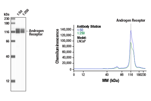
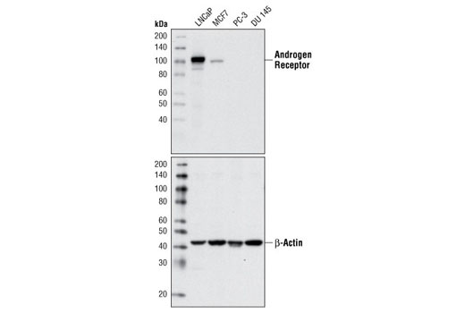
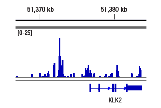
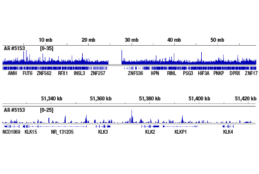
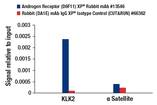
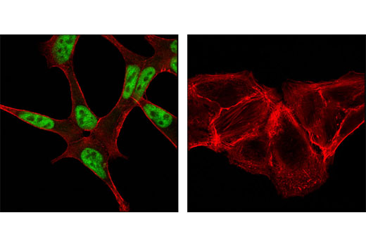
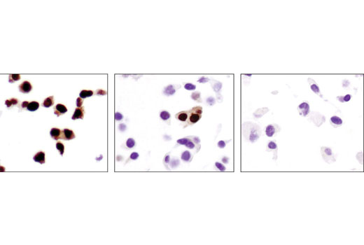
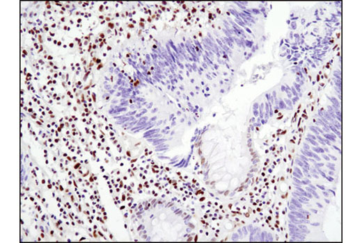
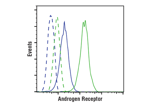
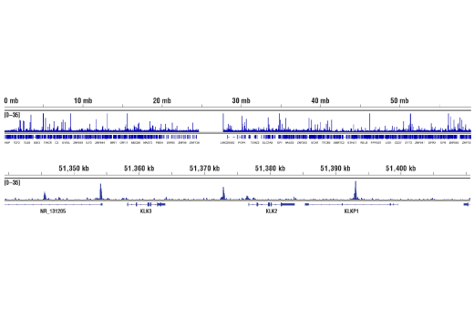
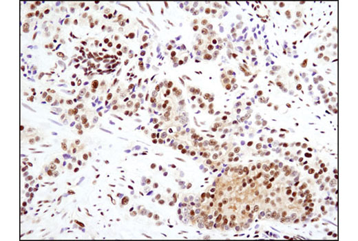
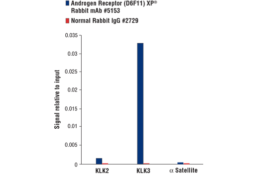
| Androgen Receptor (D6F11) XP® Rabbit mAb | 5153 | 20 µl | 110 kDa | Rabbit IgG |
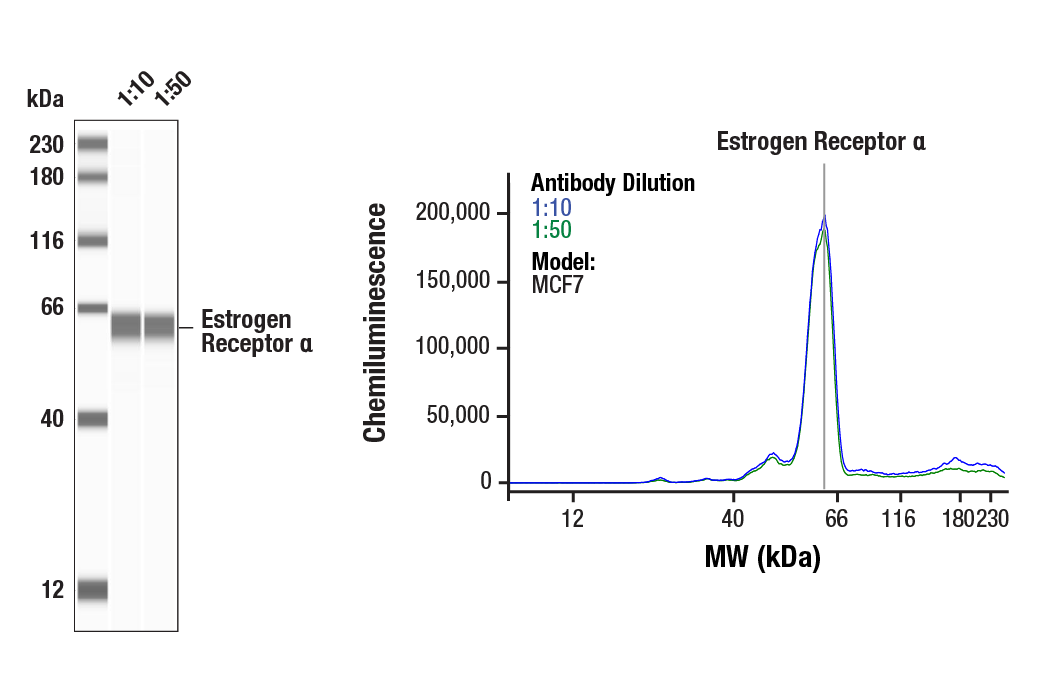
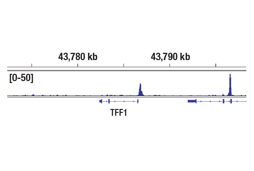
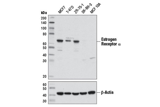
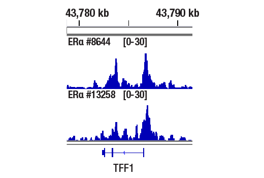
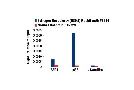
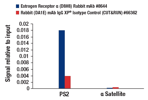
| Estrogen Receptor α (D8H8) Rabbit mAb | 8644 | 20 µl | 66 kDa | Rabbit IgG |
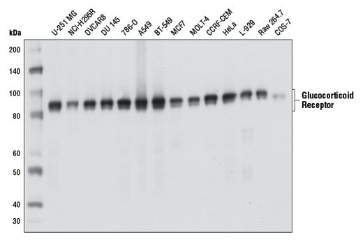
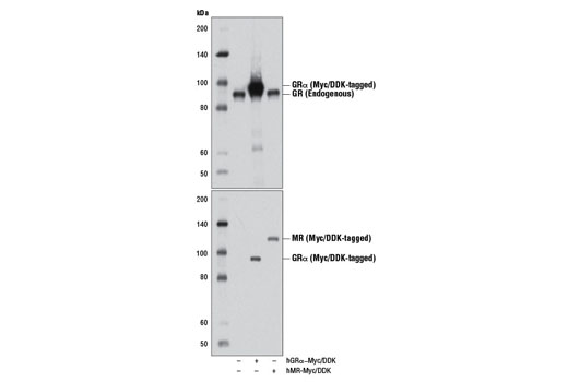
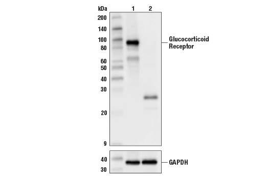
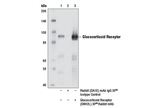
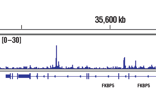
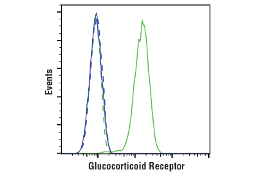
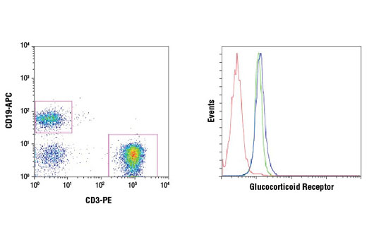
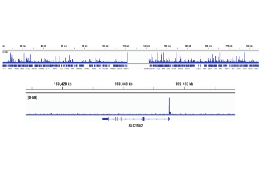
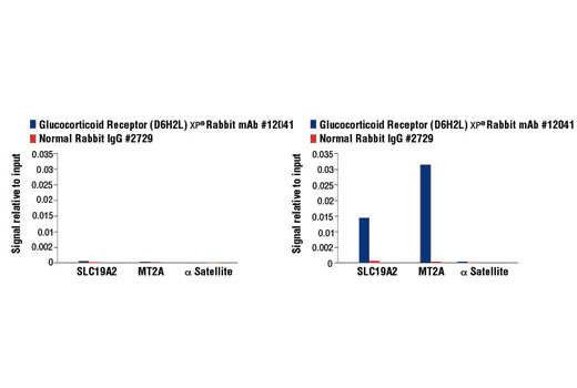
| Glucocorticoid Receptor (D6H2L) XP® Rabbit mAb | 12041 | 20 µl | 94, 91 kDa | Rabbit IgG |
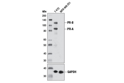
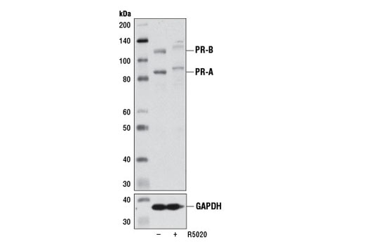
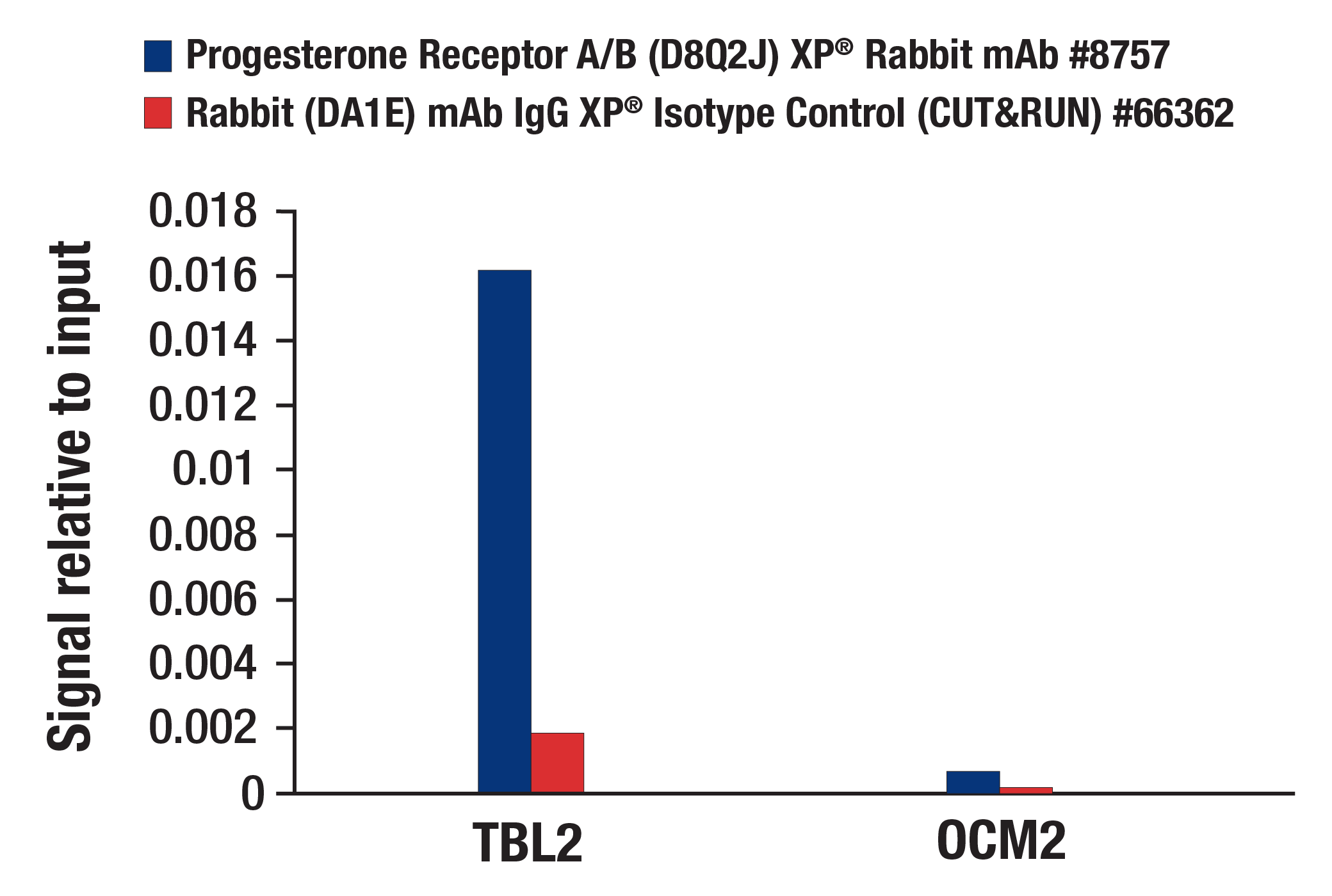
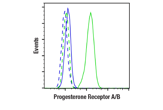
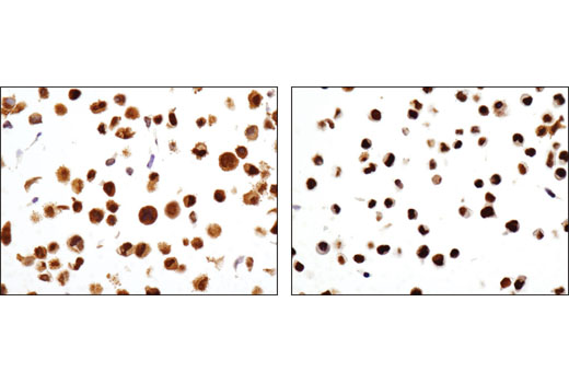
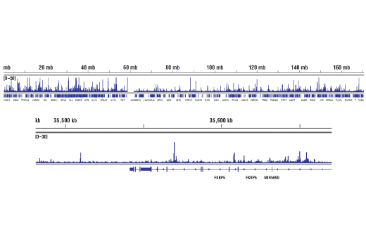
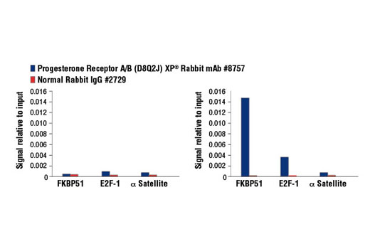
| Progesterone Receptor A/B (D8Q2J) XP® Rabbit mAb | 8757 | 20 µl | 90 (PR-A), 118 (PR-B) kDa | Rabbit IgG |
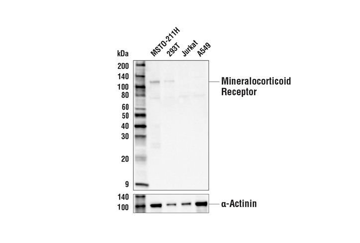
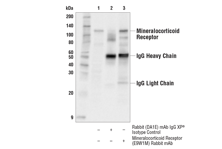
| Mineralocorticoid Receptor (E9W1M) Rabbit mAb | 58883 | 20 µl | 120 kDa | Rabbit IgG |
| Anti-rabbit IgG, HRP-linked Antibody | 7074 | 100 µl | Goat |
Please visit cellsignal.com for individual component applications, species cross-reactivity, dilutions, protocols, and additional product information.
Description
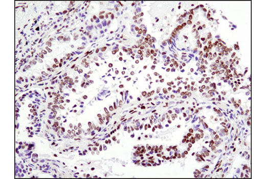
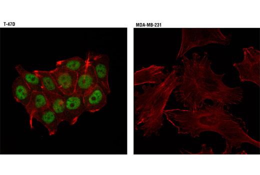
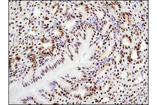
The Steroid Hormone Receptor Antibody Sampler Kit provides an economical means of detecting levels of steroid hormone nuclear receptors. The kit includes enough antibodies to perform two western blot experiments with each primary antibody.
Storage
Background
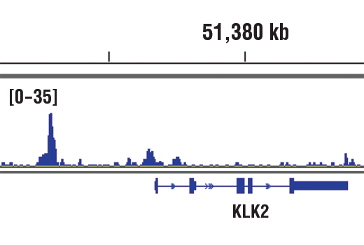
Androgen receptor (AR), a zinc finger transcription factor belonging to the nuclear receptor superfamily, is activated by phosphorylation and dimerization upon ligand binding (1). This promotes nuclear localization and binding of AR to androgen response elements in androgen target genes. Research studies have shown that AR plays a crucial role in several stages of male development and the progression of prostate cancer (2,3). Estrogen receptor α (ERα), a member of the steroid receptor superfamily, contains highly conserved DNA-binding and ligand-binding domains (4). Through its estrogen-independent and estrogen-dependent activation domains (AF-1 and AF-2, respectively), ERα regulates transcription by recruiting coactivator proteins and interacting with general transcriptional machinery (5). Human progesterone receptor (PR) is expressed as two forms: the full length PR-B and the short form PR-A. PR-A lacks the first 164 amino acid residues of PR-B (6,7). Both PR-A and PR-B are ligand activated, but differ in their relative ability to activate target gene transcription (8,9). Glucocorticoid hormones control cellular proliferation, inflammation, and metabolism through their association with the glucocorticoid receptor (GR)/NR3C1, a member of the nuclear hormone receptor superfamily of transcription factors (10). GR is composed of several conserved structural elements, including a carboxy-terminal ligand-binding domain (which also contains residues critical for receptor dimerization and hormone-dependent gene transactivation), a neighboring hinge region containing nuclear localization signals, a central zinc-finger-containing DNA-binding domain, and an amino-terminal variable region that participates in ligand-independent gene transcription. In the absence of hormone, a significant population of GR is localized to the cytoplasm in an inactive form via its association with regulatory chaperone proteins, such as HSP90, HSP70, and FKBP52. On hormone binding, GR is released from the chaperone complex and translocates to the nucleus as a dimer to associate with specific DNA sequences termed glucocorticoid response elements (GREs), thereby enhancing or repressing transcription of specific target genes (11). Mineralocorticoid receptor (MR) is a steroid hormone receptor with structural and functional similarities to GR. MR binds with high affinity to aldosterone and other mineralocorticoids as well as glucocorticoids (12-14). Upon ligand binding, MR undergoes conformational changes and enters the nucleus to bind to target mineralocorticoid response elements (MREs) (4,15,16). MR is also able to heterodimerize with GR and bind to hormone response elements on DNA in cells that express both receptors (17-19).
- Li, J. and Al-Azzawi, F. (2009) Maturitas 63, 142-8.
- Avila, D.M. et al. (2001) J Steroid Biochem Mol Biol 76, 135-42.
- Montgomery, J.S. et al. (2001) J Pathol 195, 138-46.
- Mangelsdorf, D.J. et al. (1995) Cell 83, 835-9.
- Glass, C.K. and Rosenfeld, M.G. (2000) Genes Dev 14, 121-41.
- Evans, R.M. (1988) Science 240, 889-95.
- Kastner, P. et al. (1990) EMBO J 9, 1603-14.
- Giangrande, P.H. et al. (2000) Mol Cell Biol 20, 3102-15.
- Wen, D.X. et al. (1994) Mol Cell Biol 14, 8356-64.
- Yamamoto, K.R. (1985) Annu Rev Genet 19, 209-52.
- Necela, B.M. and Cidlowski, J.A. (2003) Trends Pharmacol Sci 24, 58-61.
- Arriza, J.L. et al. (1987) Science 237, 268-75.
- Giguère, V. et al. (1988) Nature 331, 91-4.
- Beato, M. et al. (1995) Cell 83, 851-7.
- Guiochon-Mantel, A. et al. (1996) J Steroid Biochem Mol Biol 56, 3-9.
- Liu, W. et al. (1995) Proc Natl Acad Sci U S A 92, 12480-4.
- Liu, W. et al. (1996) Mol Endocrinol 10, 1399-406.
- Trapp, T. et al. (1994) Neuron 13, 1457-62.
- Funder, J.W. (1995) J Steroid Biochem Mol Biol 53, 53-5.
Background References
Trademarks and Patents
Limited Uses
Except as otherwise expressly agreed in a writing signed by a legally authorized representative of CST, the following terms apply to Products provided by CST, its affiliates or its distributors. Any Customer's terms and conditions that are in addition to, or different from, those contained herein, unless separately accepted in writing by a legally authorized representative of CST, are rejected and are of no force or effect.
Products are labeled with For Research Use Only or a similar labeling statement and have not been approved, cleared, or licensed by the FDA or other regulatory foreign or domestic entity, for any purpose. Customer shall not use any Product for any diagnostic or therapeutic purpose, or otherwise in any manner that conflicts with its labeling statement. Products sold or licensed by CST are provided for Customer as the end-user and solely for research and development uses. Any use of Product for diagnostic, prophylactic or therapeutic purposes, or any purchase of Product for resale (alone or as a component) or other commercial purpose, requires a separate license from CST. Customer shall (a) not sell, license, loan, donate or otherwise transfer or make available any Product to any third party, whether alone or in combination with other materials, or use the Products to manufacture any commercial products, (b) not copy, modify, reverse engineer, decompile, disassemble or otherwise attempt to discover the underlying structure or technology of the Products, or use the Products for the purpose of developing any products or services that would compete with CST products or services, (c) not alter or remove from the Products any trademarks, trade names, logos, patent or copyright notices or markings, (d) use the Products solely in accordance with CST Product Terms of Sale and any applicable documentation, and (e) comply with any license, terms of service or similar agreement with respect to any third party products or services used by Customer in connection with the Products.