| Product Includes | Product # | Quantity | Mol. Wt | Isotype/Source |
|---|---|---|---|---|
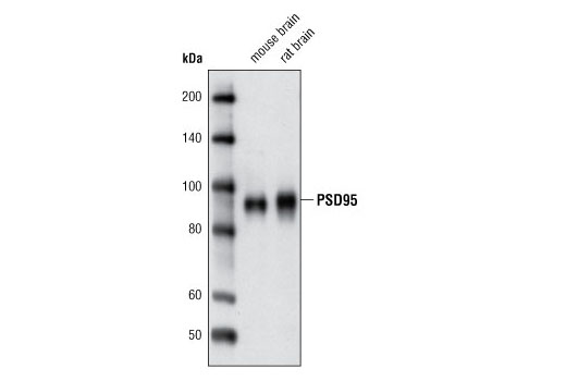
| PSD95 (D74D3) XP® Rabbit mAb | 3409 | 20 µl | 95 kDa | Rabbit IgG |
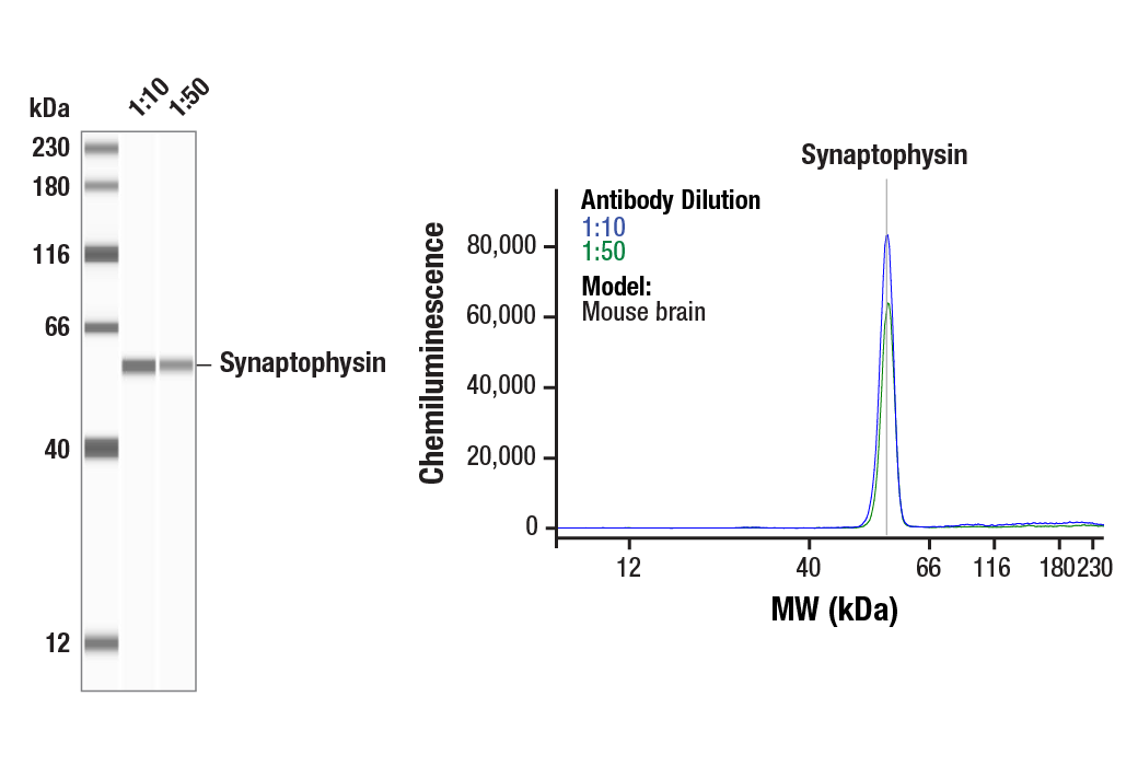
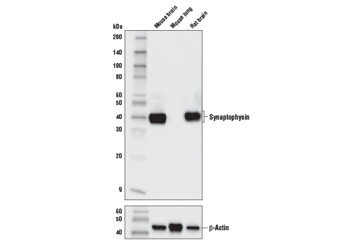
| Synaptophysin (D8F6H) XP® Rabbit mAb | 36406 | 20 µl | 38 kDa | Rabbit IgG |
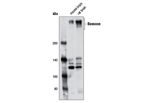
| Bassoon (D63B6) Rabbit mAb | 6897 | 20 µl | 420 kDa | Rabbit IgG |
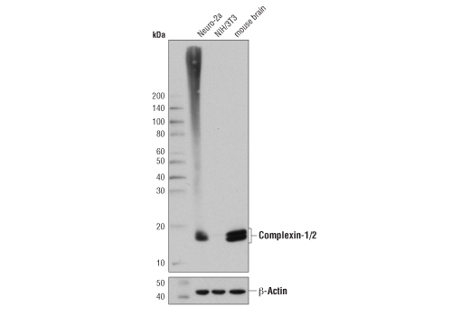
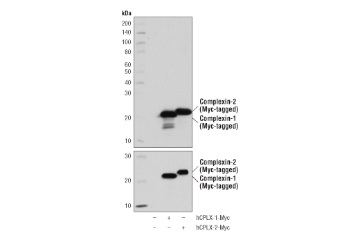
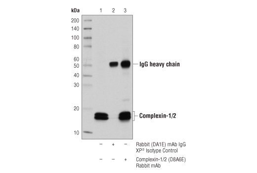
| Complexin-1/2 (D8A6E) Rabbit mAb | 28070 | 20 µl | 14-16 kDa | Rabbit IgG |
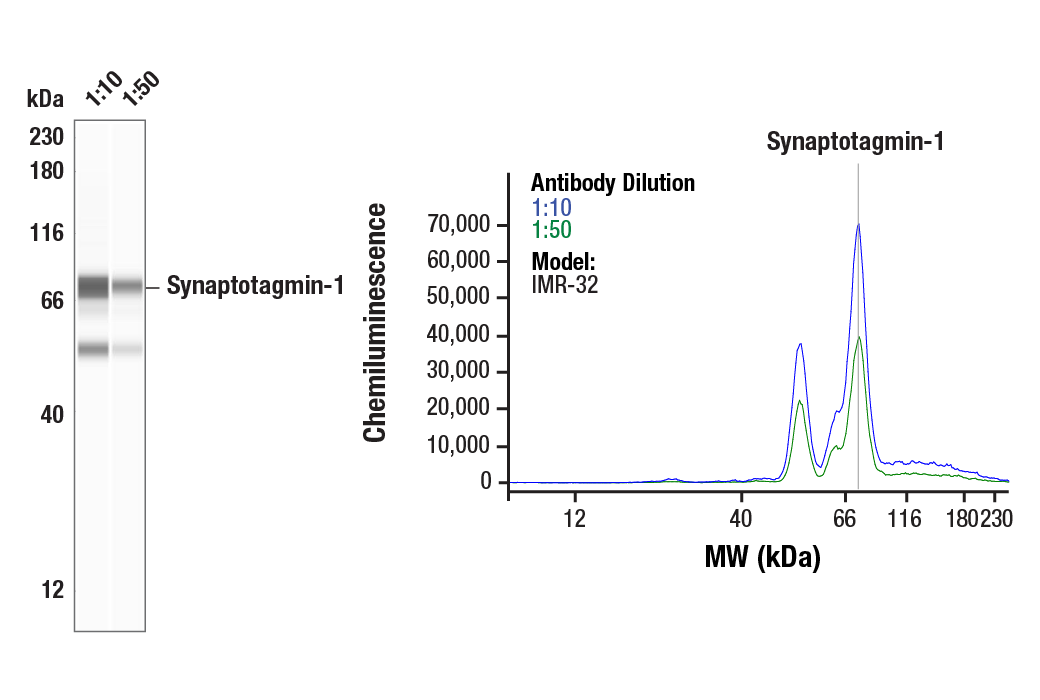
| Synaptotagmin-1 (D33B7) Rabbit mAb | 14558 | 20 µl | 60 kDa | Rabbit IgG |
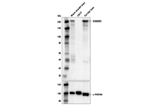
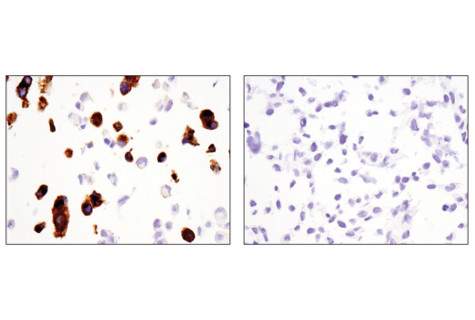
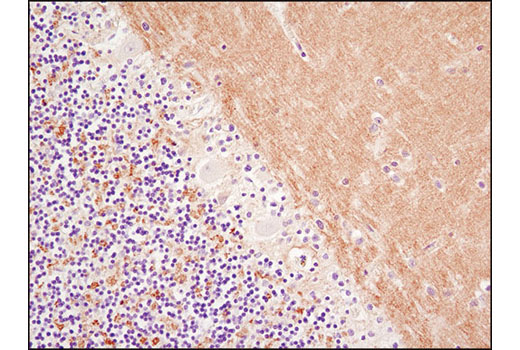
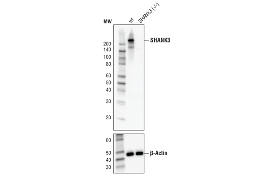
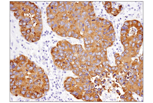
| SHANK3 (D5K6R) Rabbit mAb | 64555 | 20 µl | 220 kDa | Rabbit IgG |
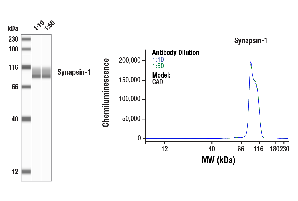
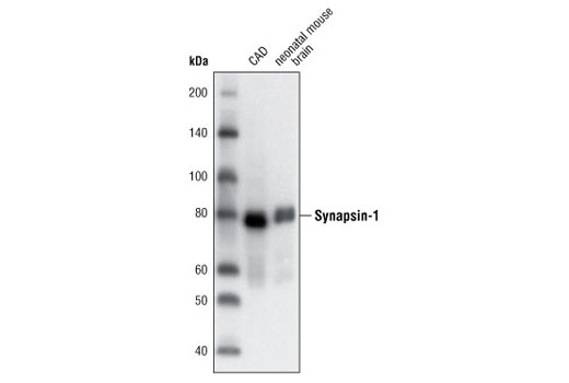
| Synapsin-1 (D12G5) XP® Rabbit mAb | 5297 | 20 µl | 77 kDa | Rabbit IgG |
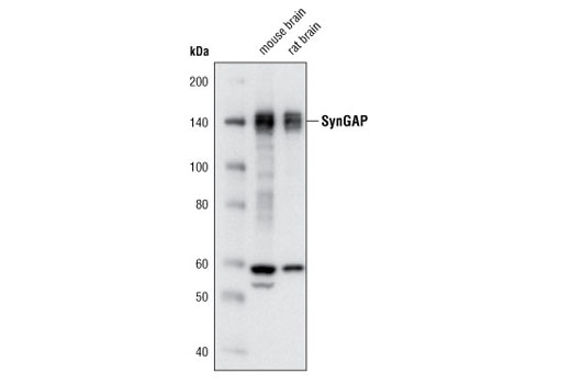
| SynGAP (D78B11) Rabbit mAb | 5540 | 20 µl | 140 kDa | Rabbit IgG |
| Anti-rabbit IgG, HRP-linked Antibody | 7074 | 100 µl | Goat |
Please visit cellsignal.com for individual component applications, species cross-reactivity, dilutions, protocols, and additional product information.
Description
The Synaptic Neuron Marker Antibody Sampler Kit provides an economical means of detecting presynaptic and postsynaptic proteins by western blot. This kit includes enough primary antibodies to perform at least two western blot experiments with each primary antibody.
Storage
Background
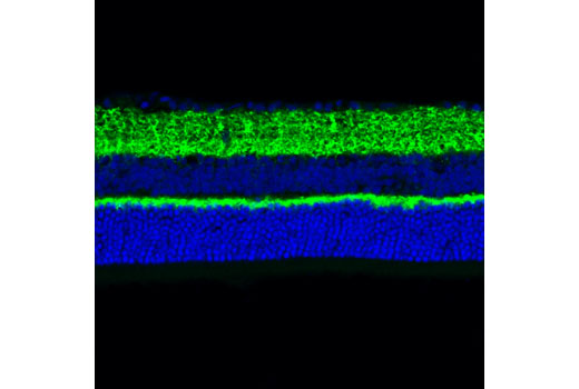
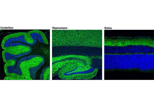
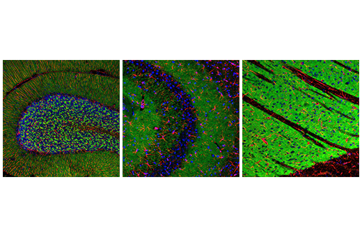
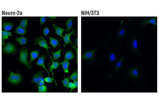
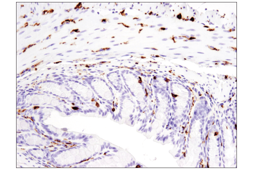
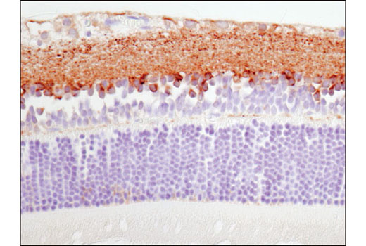
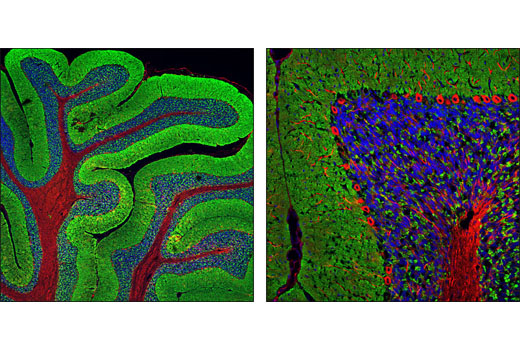
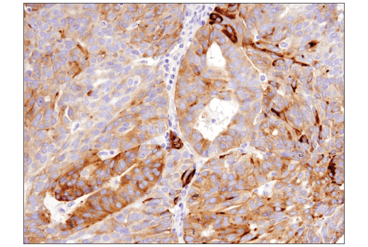
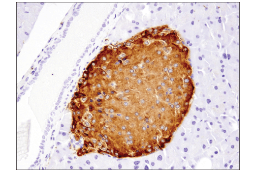
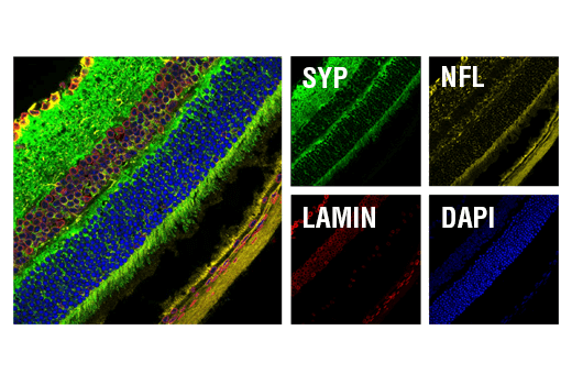
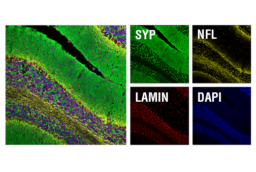
Synaptophysin (SYP) is a neuronal synaptic vesicle glycoprotein that is expressed in neuroendocrine cells and neoplasms (1). Synapsin-1 is a neuronal phospho-protein localized to presynaptic terminals. Synapsin-1 plays an important role in synapse formation, neurotransmitter regulation, and regulation of synaptic vesicle fusion and trafficking (2,3). Synaptotagmin-1 (SYT1) is an integral membrane protein found in synaptic vesicles thought to play a role in vesicle trafficking and exocytosis (4). Complexin isoforms 1 and 2 are small synaptic proteins that bind to SNARE complexes, responsible for regulating exocytosis and synaptic vesicle fusion (5). SynGAP is a synaptic GTPase-activating protein selectively expressed in the brain and found at higher concentrations specifically at excitatory synapses in the mammalian forebrain. SynGAP interacts with the PDZ domains of PSD95, a postsynaptic scaffolding protein that couples SynGAP to NMDA receptors (6). PSD95 is involved in the assembly and function of the postsynaptic density (PSD) complex (7,8). SHANK proteins act as scaffolds at the neuronal PSD (9), where they play a critical role in PSD assembly of excitatory synapses during development (10). Bassoon (BSN) is a scaffolding protein component of the synaptic ribbon and of the cytomatrix at the active zones of both excitatory and inhibitory synapses with a presumptive role in orchestrating events of the synaptic vesicle cycle (11-13). Together, these proteins can be used to measure presynaptic and postsynaptic proteins and synaptic development under normal and disease conditions.
- Wiedenmann, B. et al. (1986) Proc Natl Acad Sci U S A 83, 3500-4.
- Mirza, F.J. and Zahid, S. (2018) Neurosci Bull 34, 349-358.
- Takei, Y. et al. (1995) J Cell Biol 131, 1789-800.
- Fukuda, M. and Mikoshiba, K. (2001) Biochem Biophys Res Commun 281, 1226-33.
- Chang, S. et al. (2015) J Neurosci 35, 8272-90.
- Kim, J.H. et al. (1998) Neuron 20, 683-91.
- Cao, J. et al. (2005) J Cell Biol 168, 117-26.
- Chetkovich, D.M. et al. (2002) J Neurosci 22, 6415-25.
- Grabrucker, A.M. et al. (2011) Trends Cell Biol 21, 594-603.
- Boeckers, T.M. et al. (1999) J Neurosci 19, 6506-18.
- Winter, C. et al. (1999) Genomics 57, 389-97.
- Hallermann, S. et al. (2010) Neuron 68, 710-23.
- Frank, T. et al. (2010) Neuron 68, 724-38.
Background References
Trademarks and Patents
Limited Uses
Except as otherwise expressly agreed in a writing signed by a legally authorized representative of CST, the following terms apply to Products provided by CST, its affiliates or its distributors. Any Customer's terms and conditions that are in addition to, or different from, those contained herein, unless separately accepted in writing by a legally authorized representative of CST, are rejected and are of no force or effect.
Products are labeled with For Research Use Only or a similar labeling statement and have not been approved, cleared, or licensed by the FDA or other regulatory foreign or domestic entity, for any purpose. Customer shall not use any Product for any diagnostic or therapeutic purpose, or otherwise in any manner that conflicts with its labeling statement. Products sold or licensed by CST are provided for Customer as the end-user and solely for research and development uses. Any use of Product for diagnostic, prophylactic or therapeutic purposes, or any purchase of Product for resale (alone or as a component) or other commercial purpose, requires a separate license from CST. Customer shall (a) not sell, license, loan, donate or otherwise transfer or make available any Product to any third party, whether alone or in combination with other materials, or use the Products to manufacture any commercial products, (b) not copy, modify, reverse engineer, decompile, disassemble or otherwise attempt to discover the underlying structure or technology of the Products, or use the Products for the purpose of developing any products or services that would compete with CST products or services, (c) not alter or remove from the Products any trademarks, trade names, logos, patent or copyright notices or markings, (d) use the Products solely in accordance with CST Product Terms of Sale and any applicable documentation, and (e) comply with any license, terms of service or similar agreement with respect to any third party products or services used by Customer in connection with the Products.