| Product Includes | Product # | Quantity | Mol. Wt | Isotype/Source |
|---|---|---|---|---|
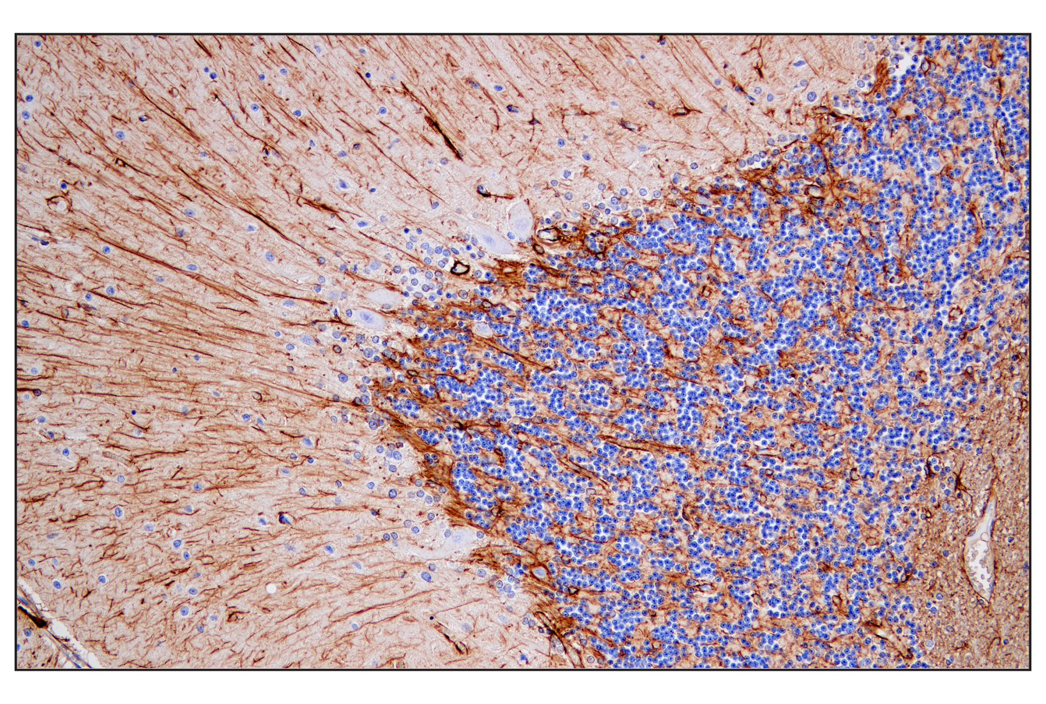
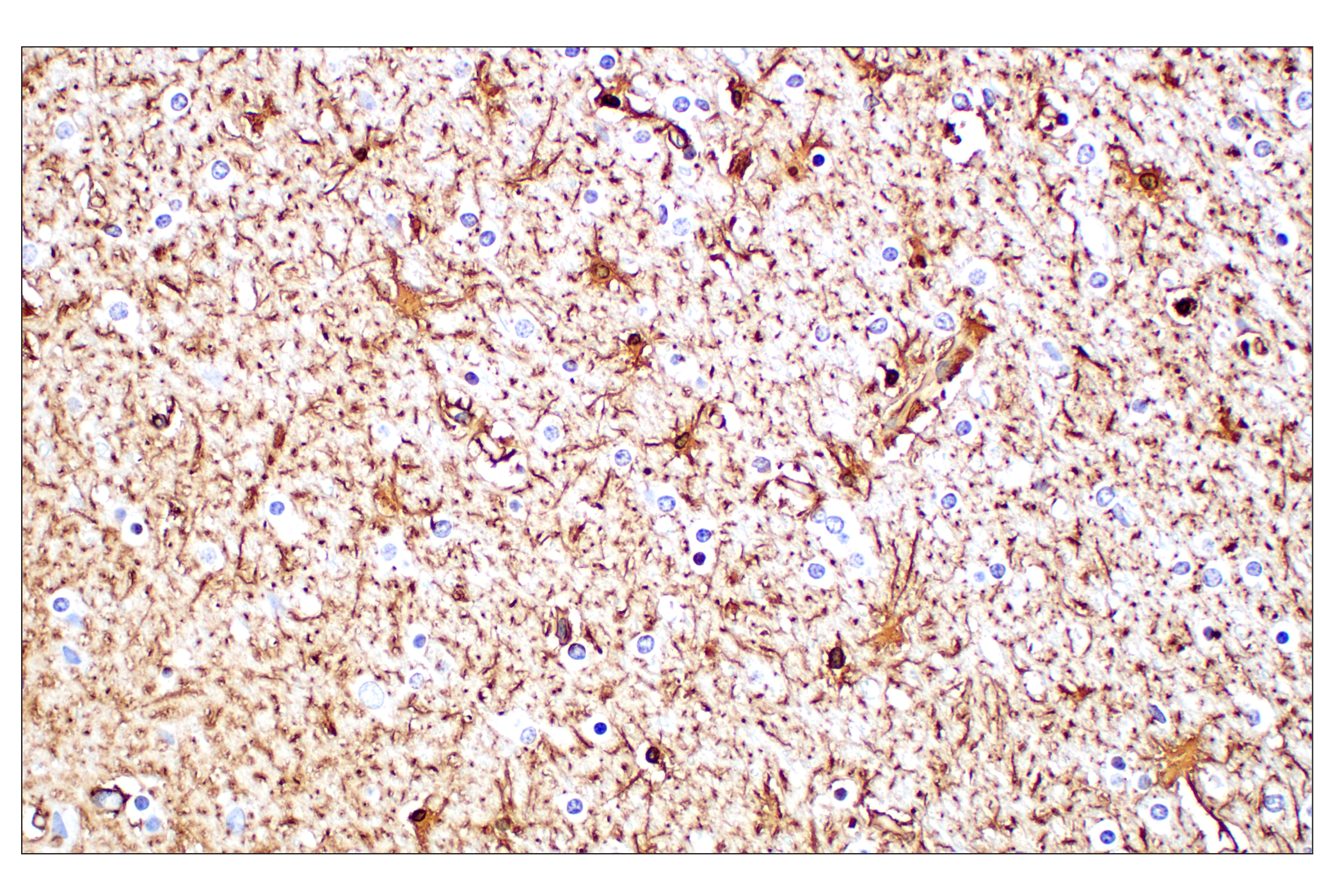
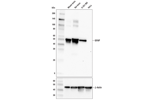
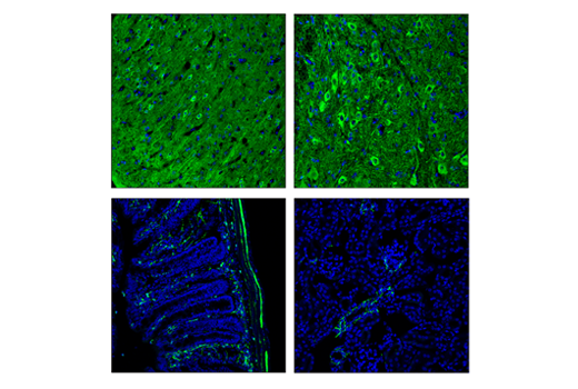
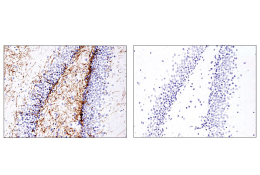
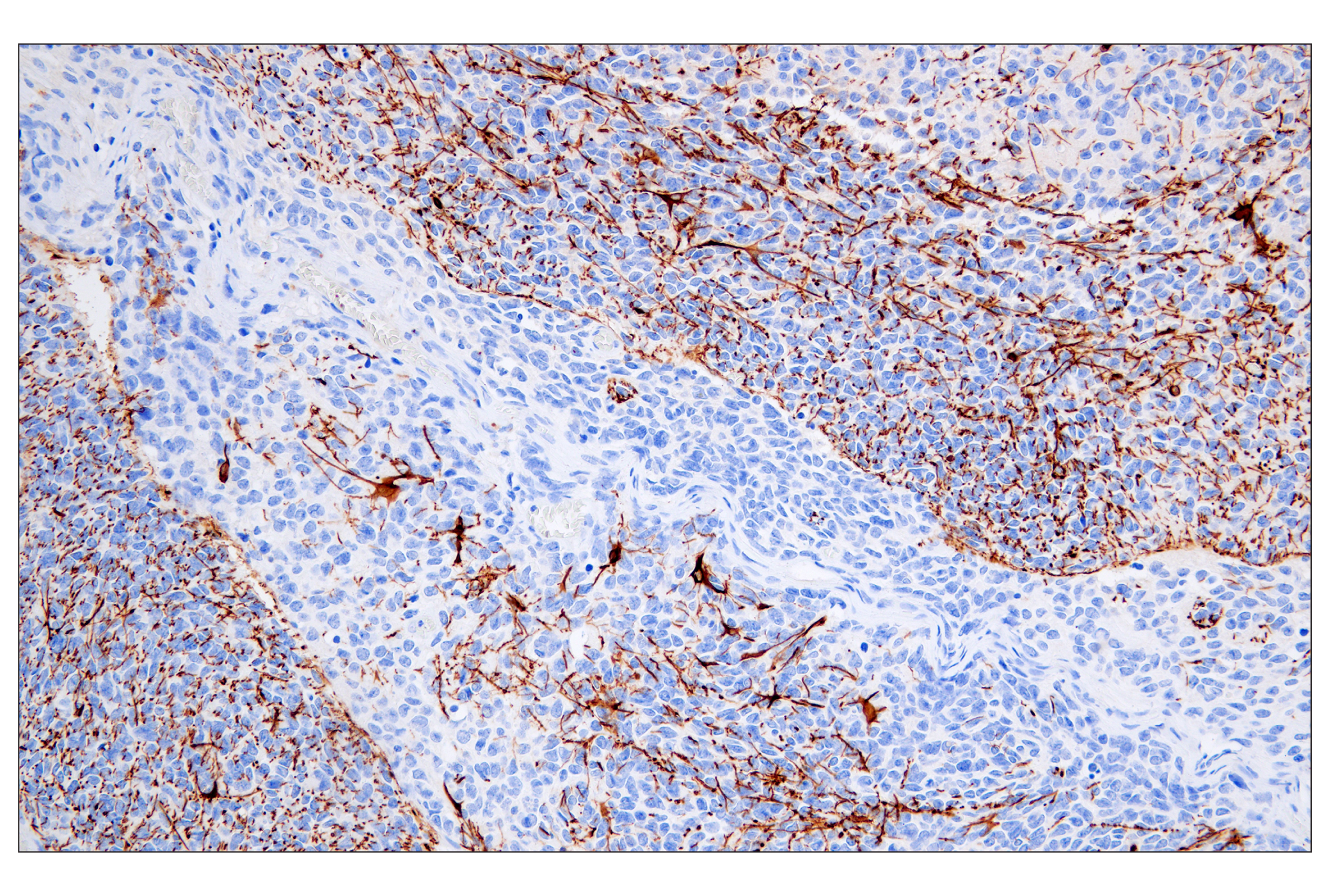
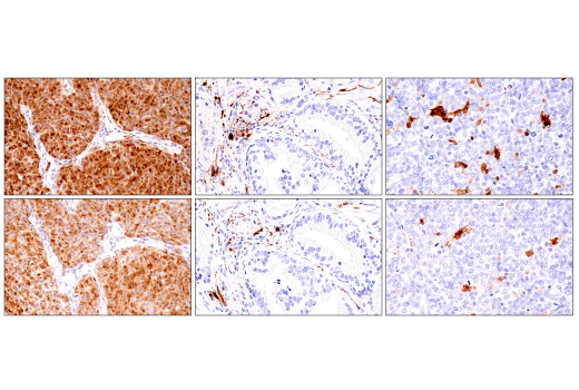
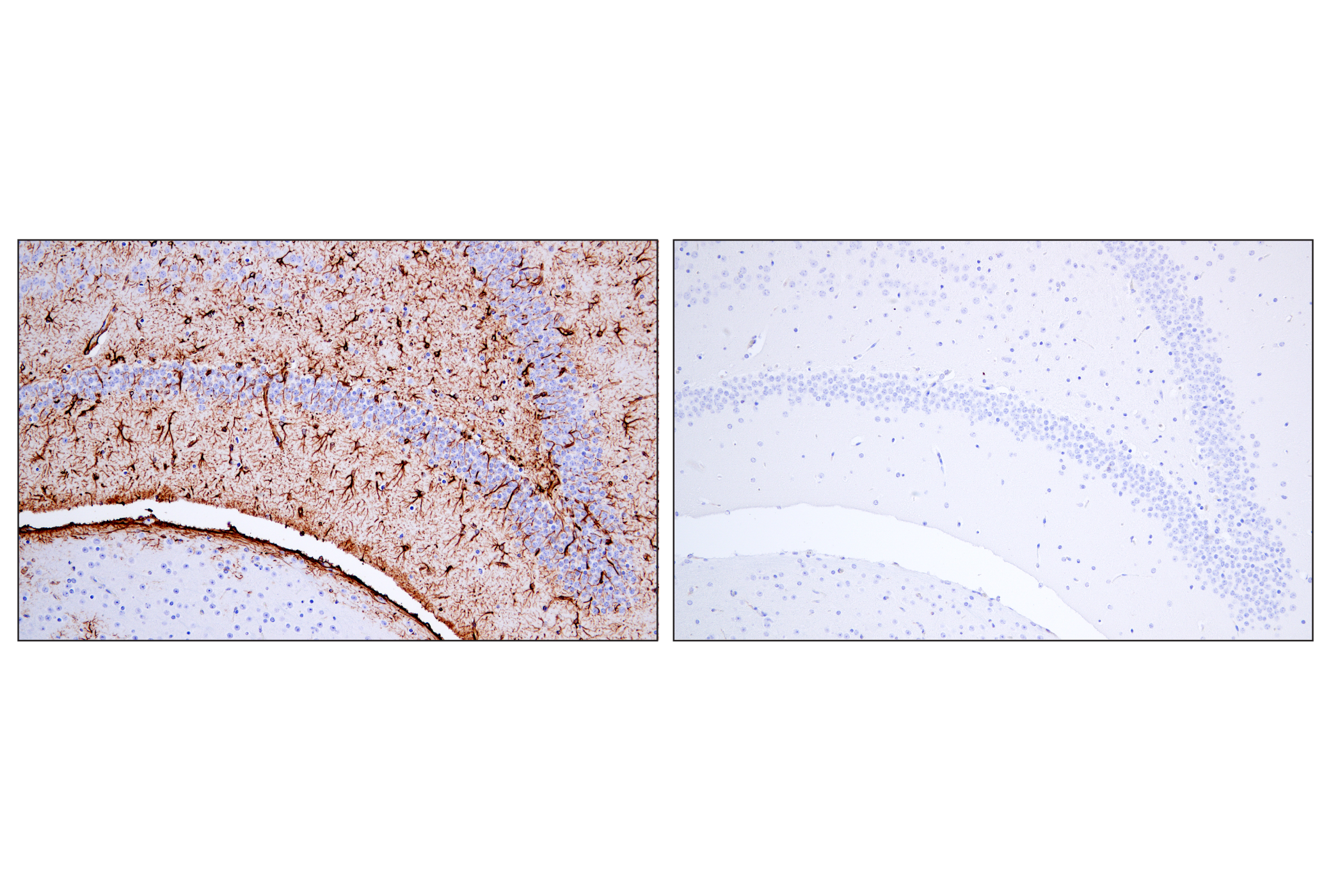
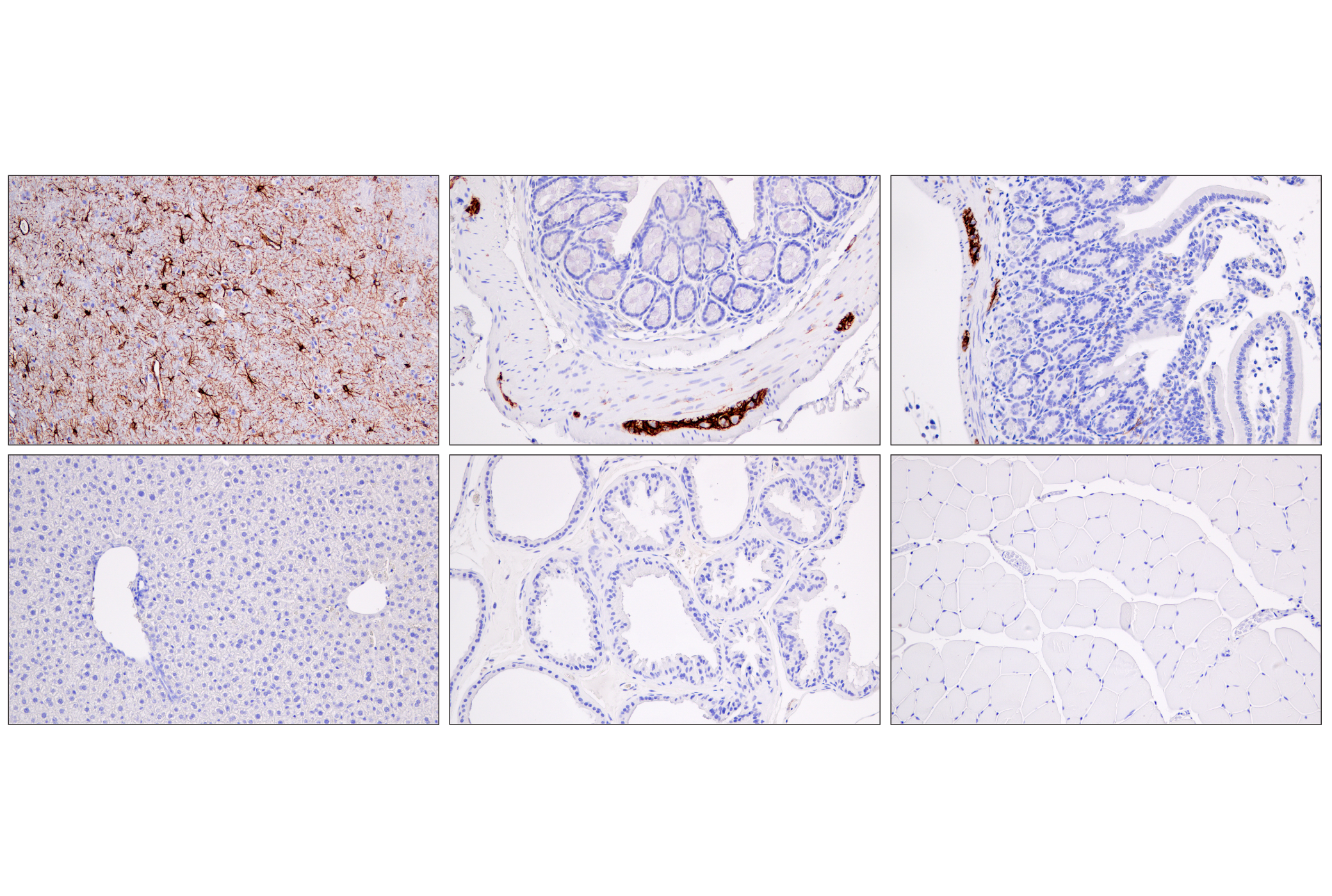
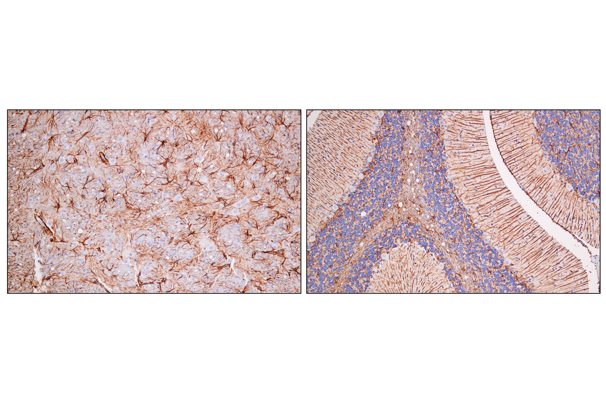
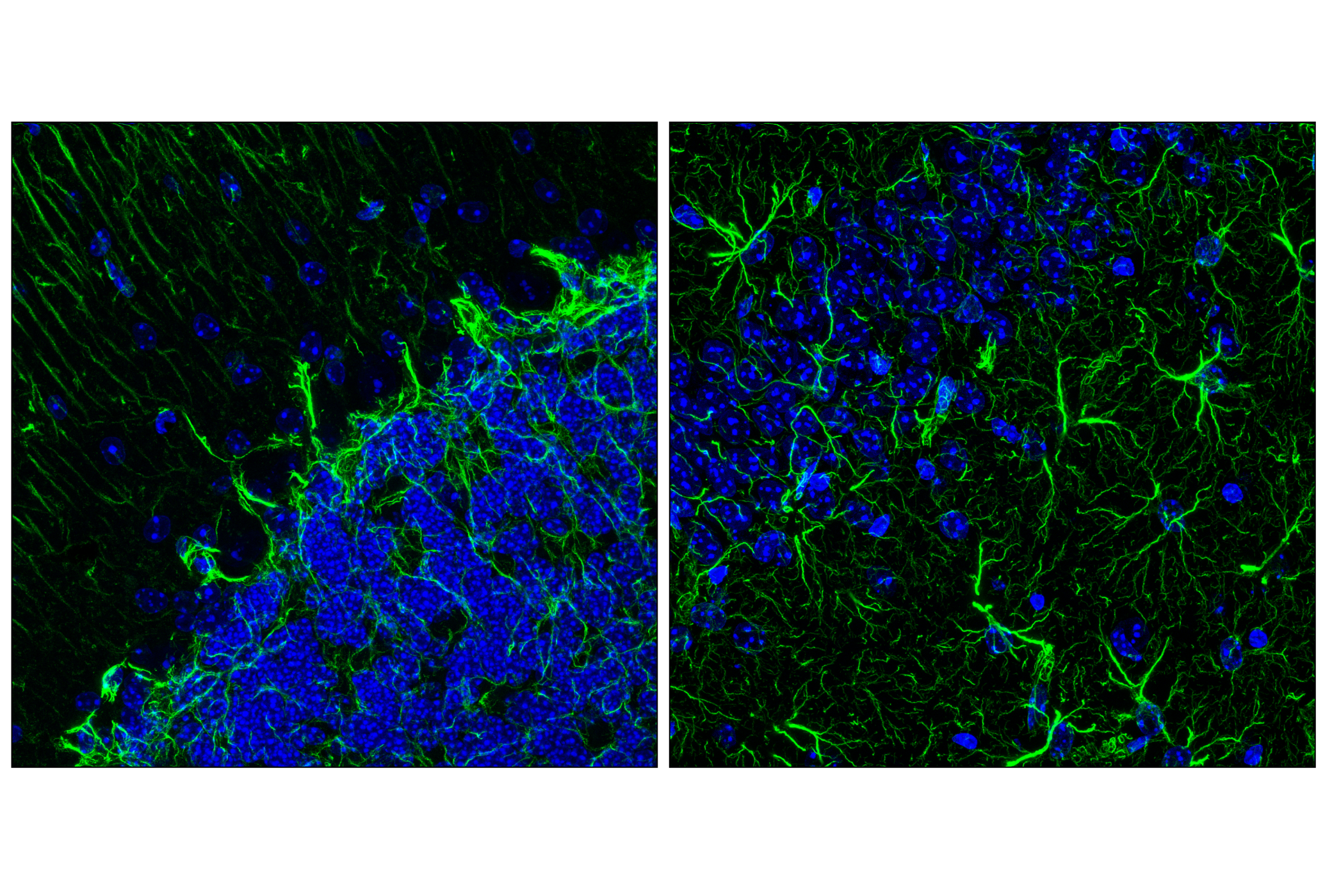
| GFAP (E4L7M) XP® Rabbit mAb | 80788 | 20 µl | 50 kDa | Rabbit IgG |
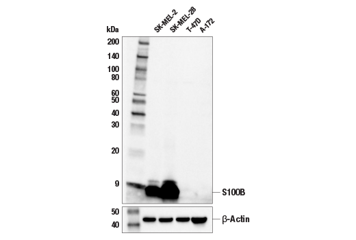
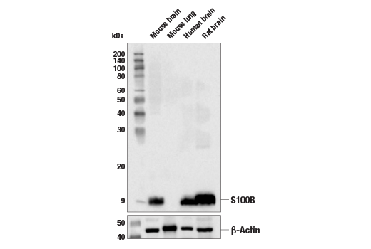
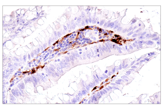
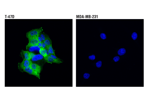
| S100B (E7C3A) Rabbit mAb | 90393 | 20 µl | 10 kDa | Rabbit IgG |
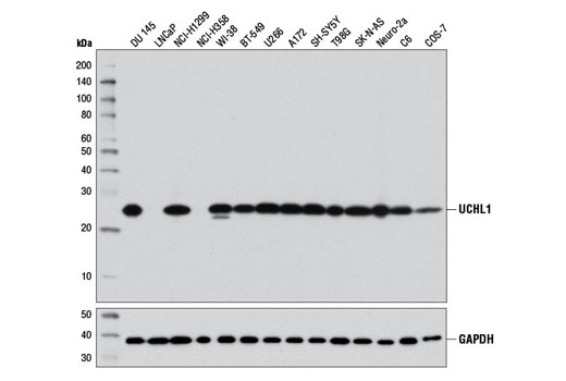
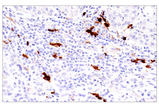
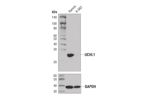
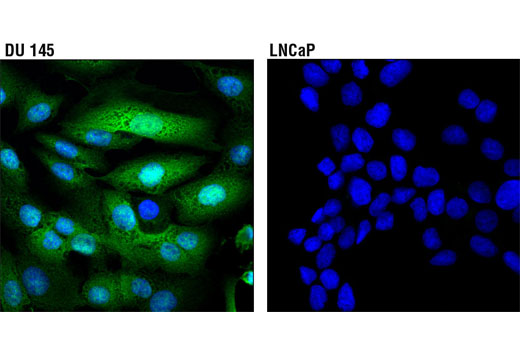
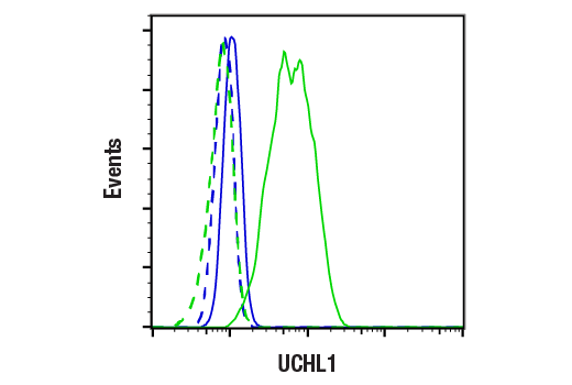
| UCHL1 (D3T2E) XP® Rabbit mAb | 13179 | 20 µl | 27 kDa | Rabbit IgG |
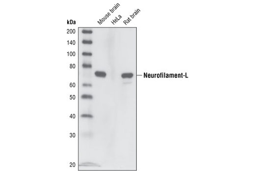
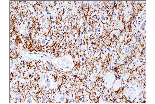
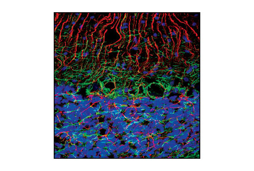
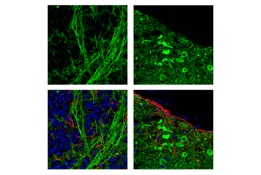
| Neurofilament-L (C28E10) Rabbit mAb | 2837 | 20 µl | 70 kDa | Rabbit IgG |
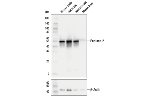
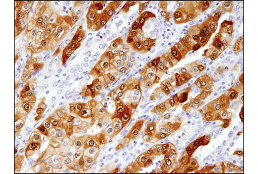
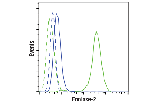
| Enolase-2 (E7D7I) Rabbit mAb | 65162 | 20 µl | 47 kDa | Rabbit IgG |
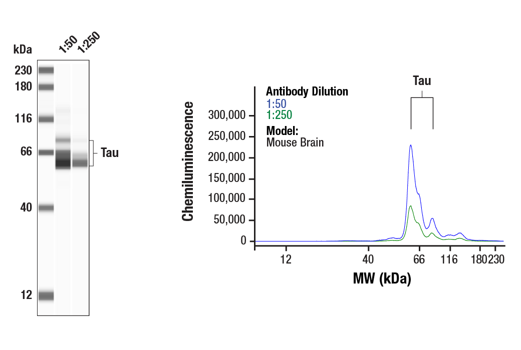
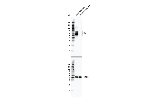
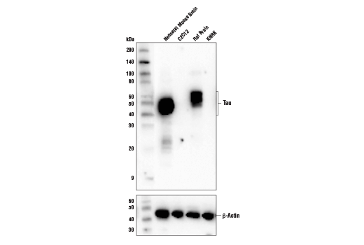
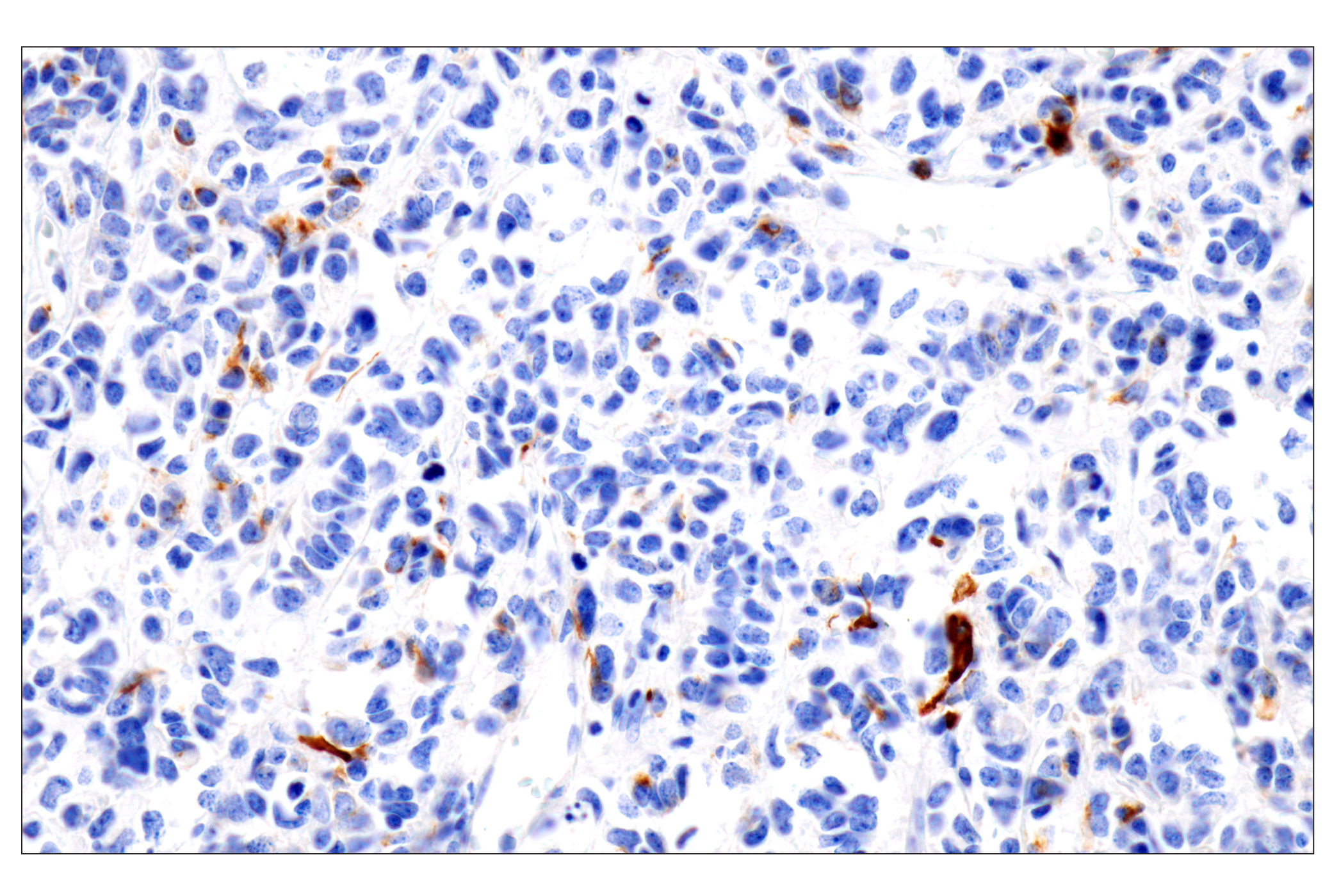
| Tau (D1M9X) XP® Rabbit mAb | 46687 | 20 µl | 50-80 kDa | Rabbit IgG |
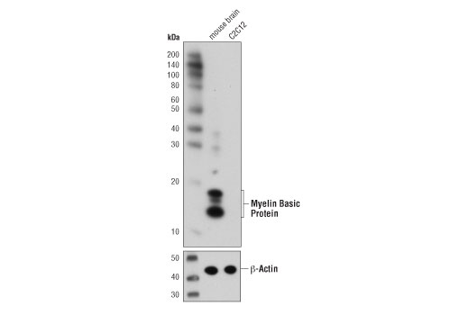
| Myelin Basic Protein (D8X4Q) XP® Rabbit mAb | 78896 | 20 µl | 12-18 kDa | Rabbit IgG |
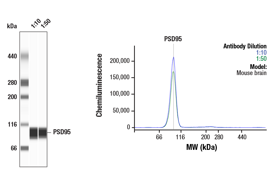
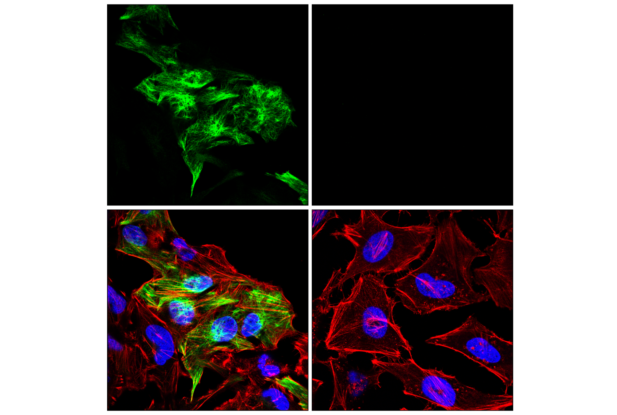
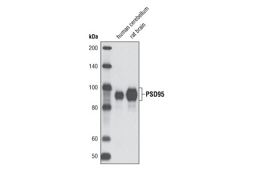
| PSD95 (D27E11) XP® Rabbit mAb | 3450 | 20 µl | 95 kDa | Rabbit IgG |
| Anti-rabbit IgG, HRP-linked Antibody | 7074 | 100 µl | Goat |
Please visit cellsignal.com for individual component applications, species cross-reactivity, dilutions, protocols, and additional product information.
Description
The Traumatic Brain Injury Biomarker Antibody Sampler Kit provides an economical means of detecting proteins involved in traumatic brain injury. The kit includes enough antibodies to perform two western blot experiments with each primary antibody.
Storage
Background
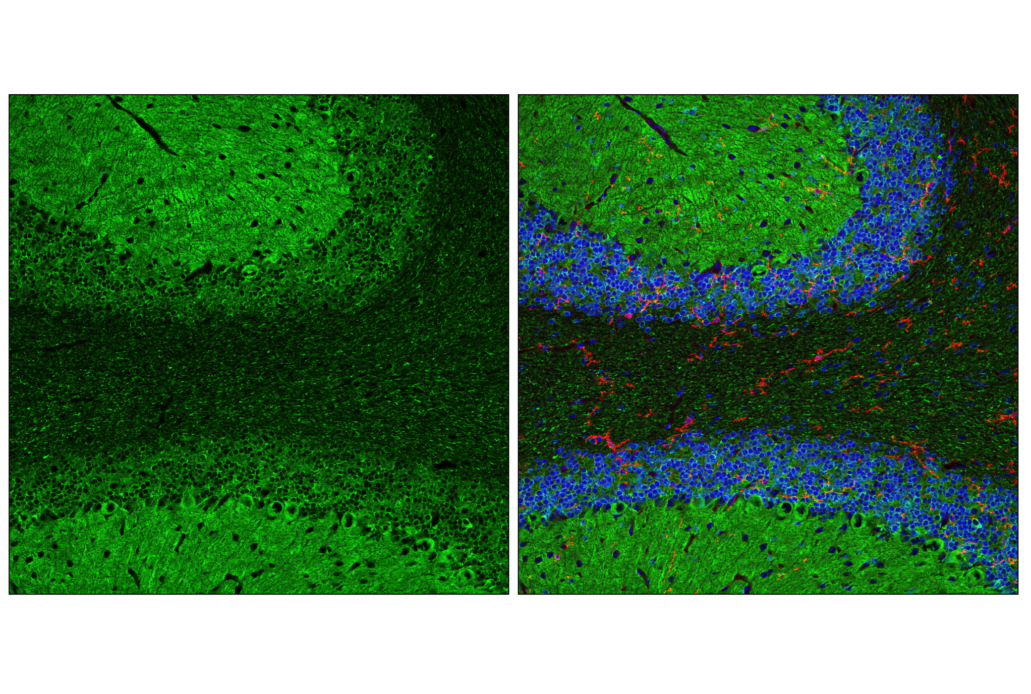
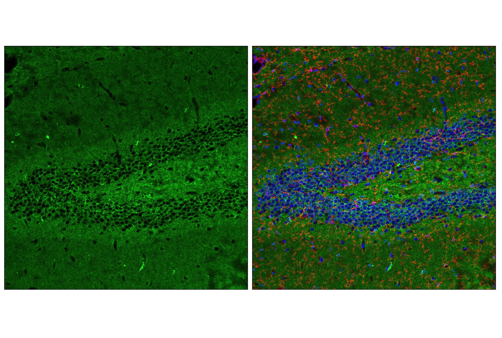
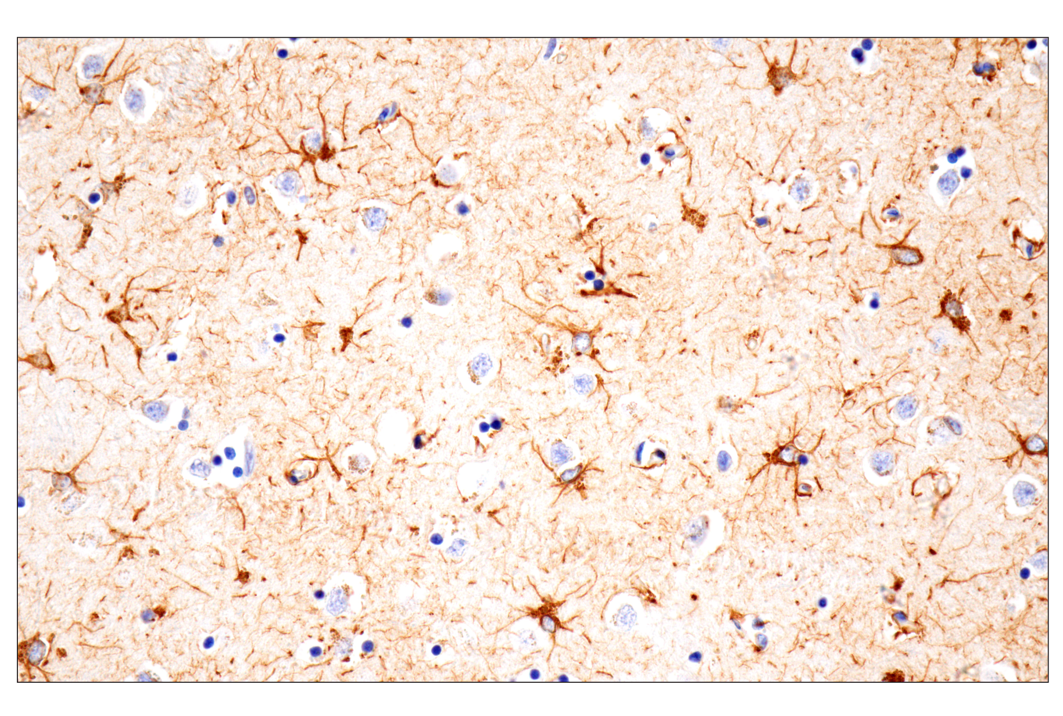
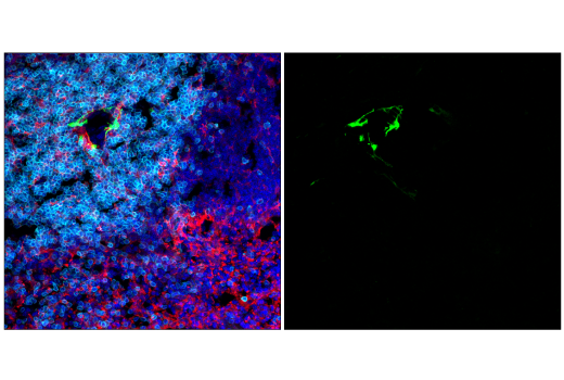
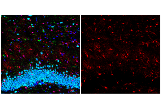
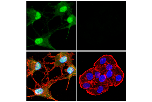
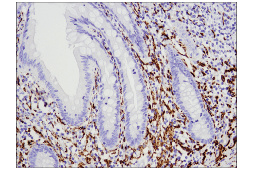
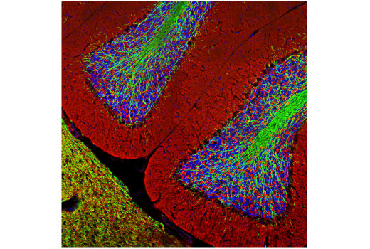
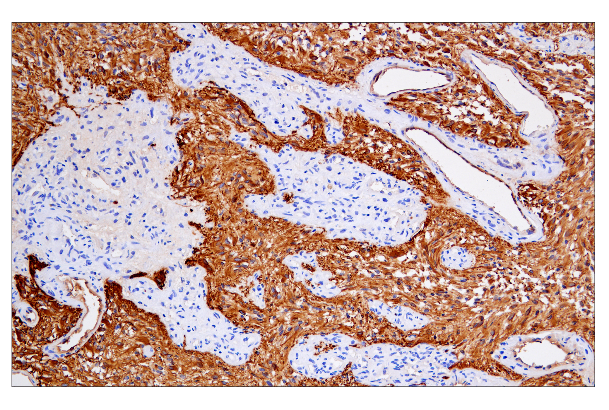
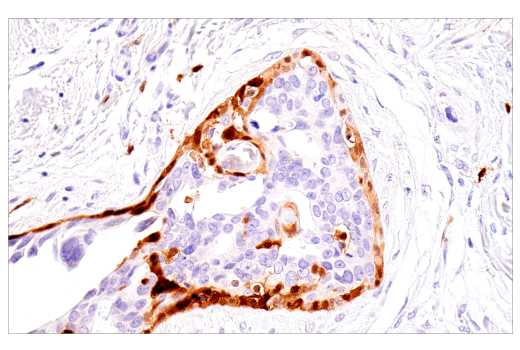
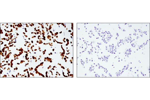
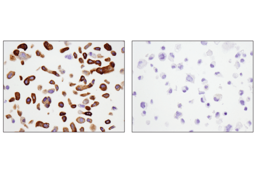
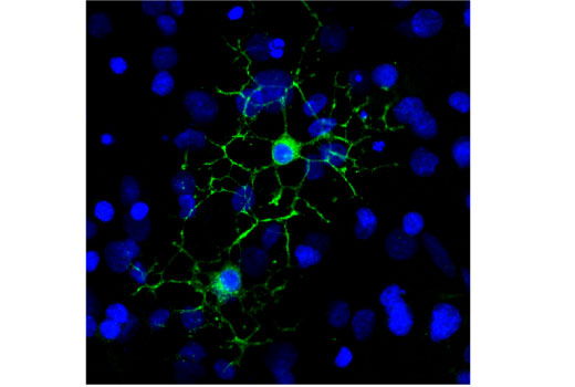
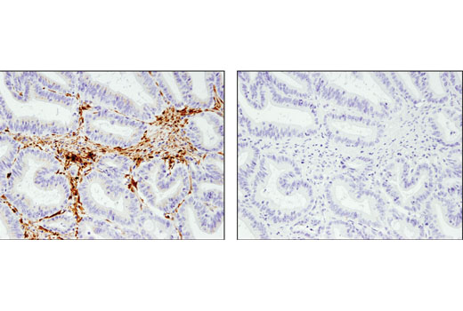
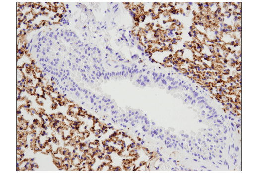
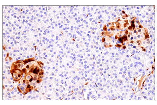
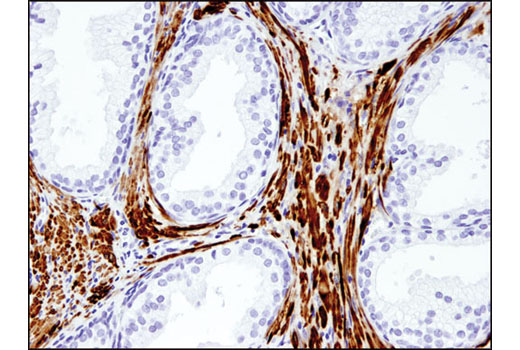
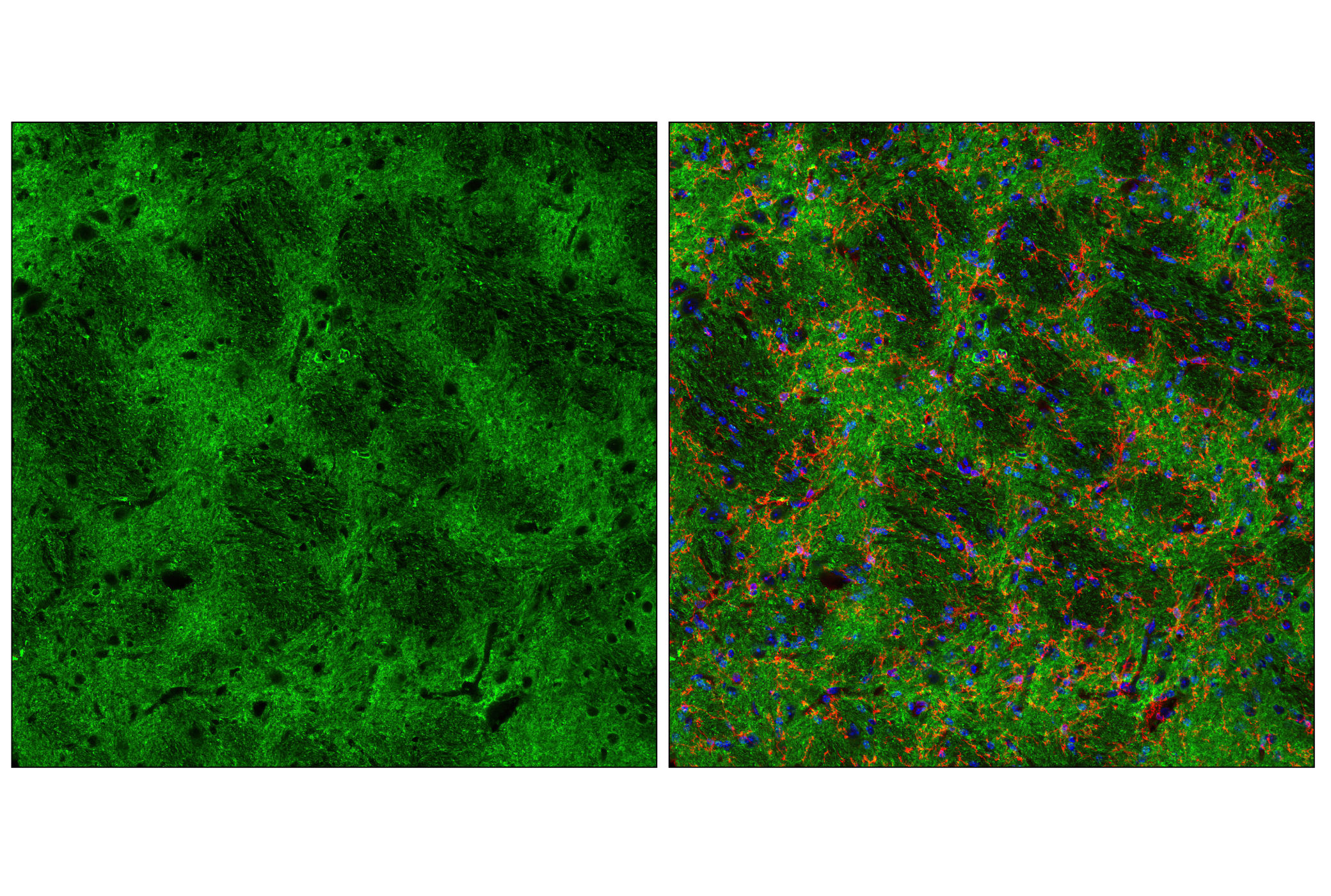
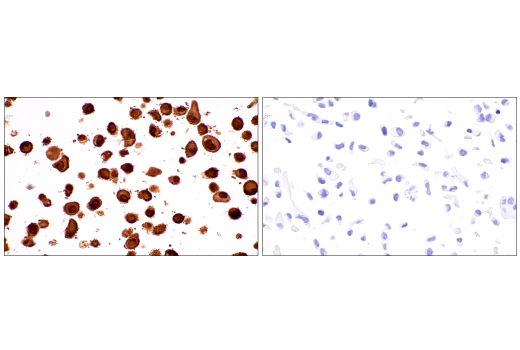
Traumatic brain injury (TBI) is a worldwide health issue that significantly affects the patient as well as their family. Annual total cost of nonfatal TBI in 2016 was $40.6 billion in the United States (1). In addition to acute brain injury, even mild cases, can lead to cognitive impairment and long-term psychiatric changes. More long term, TBI patients exhibit lower resilience to neurodegenerative disease-associated pathology (2). Treatment of TBI is made more difficult due to lack of reliable biomarkers to detect TBI (3). Several proteins are of interest, which are candidates for measurement in blood after TBI. Glial fibrillary acidic protein (GFAP) is an astrocytic intermediate filament protein. As a cytoskeletal protein, GFAP helps provide structural support to astrocytes, which provide metabolic support to neurons and maintains the blood brain barrier. The number and size of astrocytes, in a process called astrogliosis, is also positively correlated with brain injury (4). Also abundantly expressed in astrocytes, S100B is commonly used as an astrocytic marker and is positively correlated with TBI (5). Neurofilament-L (NfL) and tau are part of the neuronal cytoskeleton that provide structure to axons. Axons are covered by a multi-layered membrane called the myelin sheath. Myelin basic protein (MBP) is enriched in myelin and helps maintain its structure. UCHL1 and Enolase-2 are ubiquitin hydrolases and glycolytic enzymes, respectively, that are enriched in neurons. PSD95 is an adaptor protein enriched at postsynaptic sites in neurons. After brain injury, neuron-enriched proteins, as well as proteins that maintain neuronal/axonal integrity, can be measured in the blood, reflecting neuronal damage (6).
- Miller, G.F. et al. (2021) Med Care 59, 451-455.
- van Amerongen, S. et al. (2022) Dement Geriatr Cogn Dis Extra 12, 122-130.
- Alouani, A.T. and Elfouly, T. (2022) Biomedicines 10, 2472.
- Yang, Z. and Wang, K.K. (2015) Trends Neurosci 38, 364-74.
- Steinmüller, J.B. et al. (2022) Neurotrauma Rep 3, 447-455.
- Lippa, S.M. et al. (2022) Front Neurol 13, 816625.
Background References
Trademarks and Patents
Limited Uses
Except as otherwise expressly agreed in a writing signed by a legally authorized representative of CST, the following terms apply to Products provided by CST, its affiliates or its distributors. Any Customer's terms and conditions that are in addition to, or different from, those contained herein, unless separately accepted in writing by a legally authorized representative of CST, are rejected and are of no force or effect.
Products are labeled with For Research Use Only or a similar labeling statement and have not been approved, cleared, or licensed by the FDA or other regulatory foreign or domestic entity, for any purpose. Customer shall not use any Product for any diagnostic or therapeutic purpose, or otherwise in any manner that conflicts with its labeling statement. Products sold or licensed by CST are provided for Customer as the end-user and solely for research and development uses. Any use of Product for diagnostic, prophylactic or therapeutic purposes, or any purchase of Product for resale (alone or as a component) or other commercial purpose, requires a separate license from CST. Customer shall (a) not sell, license, loan, donate or otherwise transfer or make available any Product to any third party, whether alone or in combination with other materials, or use the Products to manufacture any commercial products, (b) not copy, modify, reverse engineer, decompile, disassemble or otherwise attempt to discover the underlying structure or technology of the Products, or use the Products for the purpose of developing any products or services that would compete with CST products or services, (c) not alter or remove from the Products any trademarks, trade names, logos, patent or copyright notices or markings, (d) use the Products solely in accordance with CST Product Terms of Sale and any applicable documentation, and (e) comply with any license, terms of service or similar agreement with respect to any third party products or services used by Customer in connection with the Products.