WB
H Mk
Endogenous
110
Rabbit IgG
#P41226
7318
Product Information
Product Usage Information
| Application | Dilution |
|---|---|
| Western Blotting | 1:1000 |
Storage
Specificity / Sensitivity
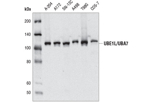
Species Reactivity:
Human, Monkey
Source / Purification
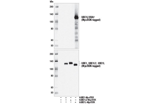
Monoclonal antibody is produced by immunizing animals with a synthetic peptide corresponding to residues near the carboxy terminus of human UBE1L/UBA7 protein.
Background
Interferon-stimulated 15 kDa protein (ISG15), also known as ubiquitin cross-reactive protein (UCRP), is a member of the ubiquitin-like protein family and functions in various biological pathways from pregnancy to innate immune responses (1). Expression of ISG15 is stimulated by cellular exposure to type 1 interferons α and β, in addition to infection with viruses such as influenza B (2,3). After exposure to type I interferons, both lymphocytes and monocytes, in addition to some fibroblasts and epithelial cells, release ISG15 into culture medium (1,4). ISG15 has been shown to function as a cytokine, stimulating interferon γ secretion by monocytes and macrophages, proliferation of natural killer cells, and chemotactic responses in neutrophils (4,5). ISG15 has also been shown to function intracellularly, being covalently conjugated to other proteins by E1 (Ube1L), E2 (UbcH8) and E3 ligases via a multi-step process analogous to ubiquitination (6,7). ISG15 is removed from proteins by the ubiquitin processing protease Ubp43 (8). ISG15-protein conjugation (ISGylation) is induced by type 1 interferons, and target proteins include the serine protease inhibitor Serpin 2A, PLCγ1, ERK1/2, Jak1 and Stat1 (9,10). Unlike ubiquitination, ISGylation does not target proteins for degradation, rather ISGylation increases Jak1 and Stat1 activity, enhancing the cellular response to interferons (11).
Ubiquitin-activating enzyme E1-like protein/Ubiquitin-activating enzyme 7 (UBE1L/UBA7) is the activating enzyme for ISG15. Research studies have suggested that loss of UBE1L/UBA7 expression contributes to the development of lung cancer due to compromised inhibition of cyclin D1 expression (12-15). UBE1L/UBA7 has also been implicated in the pathogenesis of acute promyelocytic leukemia through a mechanism in which UBE1L/UBA7 drives ISG15ylation of the oncogenic PML-RARα fusion protein to promote its degradation (16,17).
- Ritchie, K.J. and Zhang, D.E. (2004) Semin. Cell Dev. Biol. 15, 237-246.
- Korant, B.D. et al. (1984) J. Biol. Chem. 259, 14835-14839.
- Haas, A.L. et al. (1987) J. Biol. Chem. 262, 11315-11323.
- Knight, E. and Cordova, B. (1991) J. Immunol. 146, 2280-2284.
- D'Cunha, J. et al. (1996) Proc. Natl. Acad. Sci. USA 93, 211-215.
- Loeb, K.R. and Haas, A.L. (1992) J. Biol. Chem. 267, 7806-7813.
- Zhao, C. et al. (2005) Proc. Natl. Acad. Sci. USA 102, 10200-10205.
- Malakhov, M.P. et al. (2002) J. Biol. Chem. 277, 9976-9981.
- Malakhov, M.P. et al. (2003) J. Biol. Chem. 278, 16608-16613.
- Hamerman, J.A. et al. (2002) J. Immunol. 168, 2415-2423.
- Malakhova, O.A. et al. (2003) Genes Dev. 17, 455-460.
- McLaughlin, P.M. et al. (2000) Int J Cancer 85, 871-6.
- Kok, K. et al. (1995) Gene Expr 4, 163-75.
- Pitha-Rowe, I. et al. (2004) Cancer Res 64, 8109-15.
- Feng, Q. et al. (2008) Mol Cancer Ther 7, 3780-8.
- Shah, S.J. et al. (2008) Mol Cancer Ther 7, 905-14.
- Kitareewan, S. et al. (2002) Proc Natl Acad Sci U S A 99, 3806-11.
Species Reactivity
Species reactivity is determined by testing in at least one approved application (e.g., western blot).
Western Blot Buffer
IMPORTANT: For western blots, incubate membrane with diluted primary antibody in 5% w/v BSA, 1X TBS, 0.1% Tween® 20 at 4°C with gentle shaking, overnight.
Applications Key
WB: Western Blotting
Cross-Reactivity Key
H: human M: mouse R: rat Hm: hamster Mk: monkey Vir: virus Mi: mink C: chicken Dm: D. melanogaster X: Xenopus Z: zebrafish B: bovine Dg: dog Pg: pig Sc: S. cerevisiae Ce: C. elegans Hr: horse GP: Guinea Pig Rab: rabbit All: all species expected
Trademarks and Patents
Limited Uses
Except as otherwise expressly agreed in a writing signed by a legally authorized representative of CST, the following terms apply to Products provided by CST, its affiliates or its distributors. Any Customer's terms and conditions that are in addition to, or different from, those contained herein, unless separately accepted in writing by a legally authorized representative of CST, are rejected and are of no force or effect.
Products are labeled with For Research Use Only or a similar labeling statement and have not been approved, cleared, or licensed by the FDA or other regulatory foreign or domestic entity, for any purpose. Customer shall not use any Product for any diagnostic or therapeutic purpose, or otherwise in any manner that conflicts with its labeling statement. Products sold or licensed by CST are provided for Customer as the end-user and solely for research and development uses. Any use of Product for diagnostic, prophylactic or therapeutic purposes, or any purchase of Product for resale (alone or as a component) or other commercial purpose, requires a separate license from CST. Customer shall (a) not sell, license, loan, donate or otherwise transfer or make available any Product to any third party, whether alone or in combination with other materials, or use the Products to manufacture any commercial products, (b) not copy, modify, reverse engineer, decompile, disassemble or otherwise attempt to discover the underlying structure or technology of the Products, or use the Products for the purpose of developing any products or services that would compete with CST products or services, (c) not alter or remove from the Products any trademarks, trade names, logos, patent or copyright notices or markings, (d) use the Products solely in accordance with CST Product Terms of Sale and any applicable documentation, and (e) comply with any license, terms of service or similar agreement with respect to any third party products or services used by Customer in connection with the Products.