| Cat. # | Size | Qty. | Price |
|---|---|---|---|
| 9768T | 1 Kit (8 x 20 microliters) |
|
| Product Includes | Quantity | Applications | Reactivity | MW(kDa) | Isotype |
|---|---|---|---|---|---|
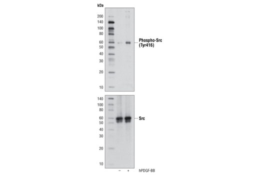
| Phospho-Src Family (Tyr416) (D49G4) Rabbit mAb 6943 | 20 µl |
|
H M R Mk | 60 | Rabbit IgG |
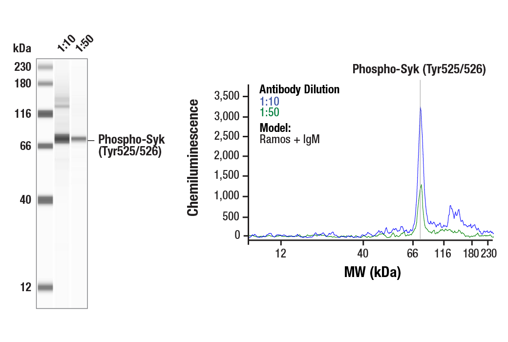
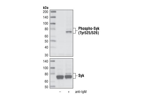
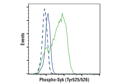
| Phospho-Syk (Tyr525/526) (C87C1) Rabbit mAb 2710 | 20 µl |
|
H | 72 | Rabbit IgG |
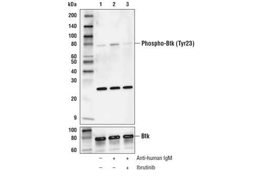
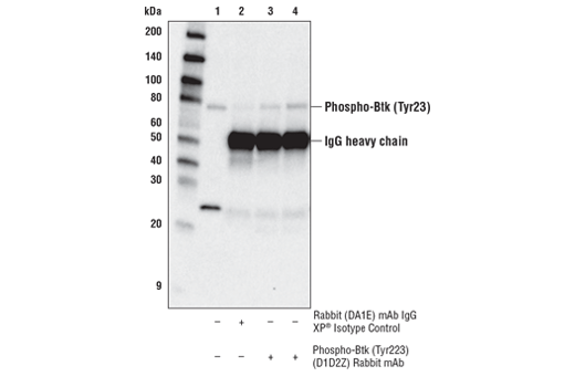
| Phospho-Btk (Tyr223) (D1D2Z) Rabbit mAb 87457 | 20 µl |
|
H M | 78 | Rabbit IgG |
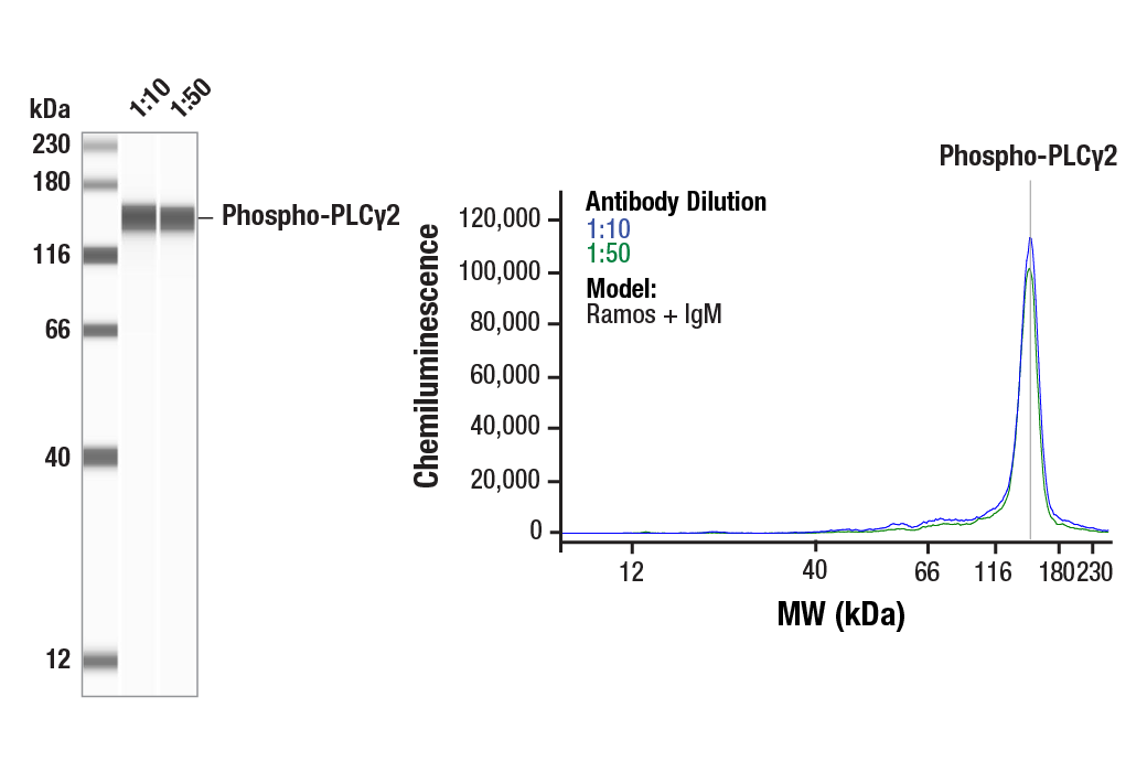
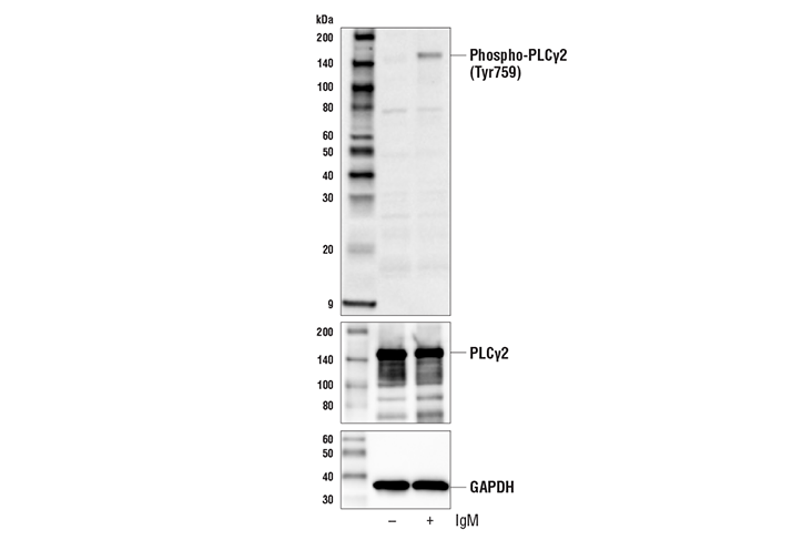
| Phospho-PLCγ2 (Tyr759) (E9E9Y) Rabbit mAb 50535 | 20 µl |
|
H | 150 | Rabbit IgG |
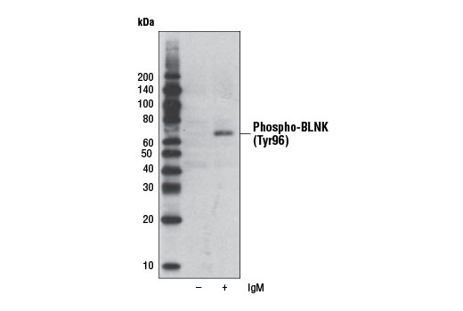
| Phospho-BLNK (Tyr96) Antibody 3601 | 20 µl |
|
H | 68, 70 | Rabbit |
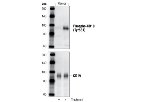
| Phospho-CD19 (Tyr531) Antibody 3571 | 20 µl |
|
H | 95 | Rabbit |
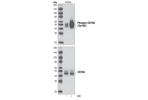
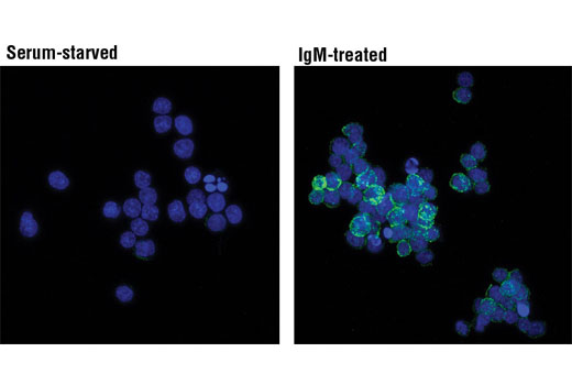
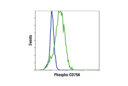
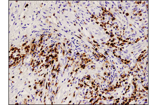
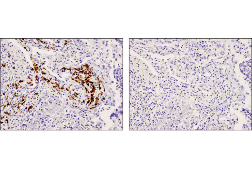
| Phospho-CD79A (Tyr182) Antibody 5173 | 20 µl |
|
H | 45-55 | Rabbit |
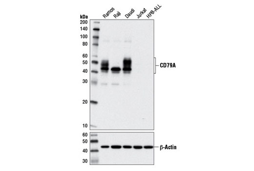
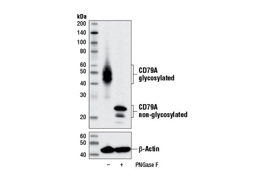
| CD79A (D1X5C) XP® Rabbit mAb 13333 | 20 µl |
|
H | 45-55 | Rabbit IgG |
| Anti-rabbit IgG, HRP-linked Antibody 7074 | 100 µl |
|
Goat |
Product Information
Polyclonal antibodies are produced by immunizing animals with a synthetic peptide and are purified by protein A and peptide affinity chromatography. Monoclonal antibodies are produced by immunizing animals with recombinant human proteins or synthetic peptides.
Antigen receptors found on the surface of B cells contain a heterodimeric signaling component composed of CD79A and CD79B, also known as Ig α and Ig ß, respectively. Presence of this receptor complex is essential for B-cell development and function. Antigen binding precedes formation of the CD79A and CD79B heterodimer and subsequent activation of receptor associated kinases. Tyr182 of mouse CD79A (corresponding to Tyr188 of human CD79A) is one of two key tyrosine residues in the immunoreceptor tyrosine-based activation motif (ITAM) of CD79A that are phosphorylated by Src family kinases (e.g., Lyn, Blk), and play a critical role in modulating signal transduction following immune receptor activation.
Syk is a protein tyrosine kinase that plays an important role in intracellular signal transduction in hematopoietic cells (1-3). Syk interacts with immunoreceptor tyrosine-based activation motifs (ITAMs) located in the cytoplasmic domains of immune receptors (4). It couples the activated immunoreceptors to downstream signaling events that mediate diverse cellular responses, including proliferation, differentiation, and phagocytosis (4). There is also evidence that Syk plays a role in nonimmune cells; Syk is a potential tumor suppressor in human breast carcinomas (5). Tyrosine 525 and 526 are located in the activation loop of the Syk kinase domain, and phosphorylation of Tyr525/526 of human Syk (equivalent to the Tyr519/520 of mouse Syk) is essential for Syk function (6).
Lyn, one of the Src family members, is predominantly expressed in hematopoietic cells (7). Two tyrosine residues have been reported to play a crucial role in the regulation of protein tyrosine kinases of the Src family. Autophosphorylation of Tyr396 (equivalent to Tyr416 of Src), located in the catalytic domain, correlates with enzyme activation. Csk-mediated phosphorylation of the carboxy-terminal Tyr507 (equivalent to Tyr527 of Src) inactivates the kinase. Tyrosine phosphorylation and activation of Lyn occurs upon association with cell surface receptors such as the B cell Ag receptor (BCR) and CD40 (8-10).
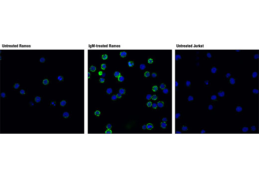
Bruton's tyrosine kinase (Btk) is a member of the Btk/Tec family of cytoplasmic tyrosine kinases. Btk plays an important role in B cell development (11,12). Activation of B cells by various ligands is accompanied by Btk membrane translocation mediated by its PH domain binding to phosphatidylinositol-3,4,5-trisphosphate (13-15). The membrane-located Btk is active and associated with transient phosphorylation of two tyrosine residues, Tyr551 and Tyr223. Tyr551 in the activation loop is transphosphorylated by the Src family tyrosine kinase, leading to autophosphorylation at Tyr223 within the SH3 domain, which is necessary for full activation (16,17).
CD19 is a 95 kDa coreceptor that amplifies the signaling cascade in B cells (18). On the B cell surface, CD19 associates with CD21, CD81, and Leu-13 to exert its function. The cytoplasmic tail of CD19 has nine conserved tyrosine residues playing critical roles in CD19-mediated function by coupling signaling molecules to the receptor (18). After BCR or CD19 ligation, Tyr531 and Tyr500 of CD19 are progressively phosphorylated. This phosphorylation enables the coupling of PI3 kinase and Src family tyrosine kinase to CD19 and activates the PI3K and Src signaling pathways (19,20).
B cell linker protein (BLNK), also known as SLP-65 or BASH, is an adaptor molecule that plays key roles in B cell activation and B cell antigen receptor (BCR) engagement. BLNK acts at the interface between BCR-associated Syk and downstream signaling cascades
Phosphoinositide-specific phospholipase C (PLC) plays a significant role in transmembrane signaling. PLCgamma2 is engaged in antigen-dependent signaling in B cells. Phosphorylation by Btk or Lck at tyrosines 753, 759, 1197 and 1217 is correlated with PLCgamma2 activity.
Explore pathways related to this product.
STRING - Known and Predicted Protein-Protein Interactions.
Except as otherwise expressly agreed in a writing signed by a legally authorized representative of CST, the following terms apply to Products provided by CST, its affiliates or its distributors. Any Customer's terms and conditions that are in addition to, or different from, those contained herein, unless separately accepted in writing by a legally authorized representative of CST, are rejected and are of no force or effect.
Products are labeled with For Research Use Only or a similar labeling statement and have not been approved, cleared, or licensed by the FDA or other regulatory foreign or domestic entity, for any purpose. Customer shall not use any Product for any diagnostic or therapeutic purpose, or otherwise in any manner that conflicts with its labeling statement. Products sold or licensed by CST are provided for Customer as the end-user and solely for research and development uses. Any use of Product for diagnostic, prophylactic or therapeutic purposes, or any purchase of Product for resale (alone or as a component) or other commercial purpose, requires a separate license from CST. Customer shall (a) not sell, license, loan, donate or otherwise transfer or make available any Product to any third party, whether alone or in combination with other materials, or use the Products to manufacture any commercial products, (b) not copy, modify, reverse engineer, decompile, disassemble or otherwise attempt to discover the underlying structure or technology of the Products, or use the Products for the purpose of developing any products or services that would compete with CST products or services, (c) not alter or remove from the Products any trademarks, trade names, logos, patent or copyright notices or markings, (d) use the Products solely in accordance with CST Product Terms of Sale and any applicable documentation, and (e) comply with any license, terms of service or similar agreement with respect to any third party products or services used by Customer in connection with the Products.