

Simplifying Multiplex IHC Panel Design for Spatial Biology
In spatial biology, multiplexing in immunohistochemistry (IHC) enables the simultaneous detection of multiple biomarkers within a single tissue section. This unravels insights into how cellular interactions influence the tissue microenvironment, deepening our understanding of disease progression and therapeutic responses.
SignalStar® Multiplex IHC is a technology that uses antibodies, oligonucleotides, and fluorophores to interrogate cellular presence, location, function, and biomarker co-expression patterns. Multiple biomarkers can be amplified simultaneously in FFPE tissue with high sensitivity and specificity, providing results for up to eight biomarkers in two days.
You Don’t Need to Be a Spatial Expert
SignalStar mIHC panels combine CST scientific expertise, our high-quality antibodies, and rigorous validation into an easy-to-use solution that enables you to generate multiplex spatial images and deepen your understanding of biology, even when you’re not a spatial expert. So you can easily design experiments and get answers to your most pressing scientific questions—fast.
- Automated SignalStar Multiplex IHC Panel Builder eliminates all the guesswork of pairing antibodies to fluorophores during panel design, letting you focus on results instead.
- IHC-validated antibodies, validated protocols, and antibody panels require minimal optimization and work with your existing fluorescence imagers.
- Signal amplification accurately detects up to 8 proteins in FFPE tissue—even those with low levels of expression.
- If your research needs change, it’s easy to redesign panels.
You can design a SignalStar mIHC panel specific to your needs, or reference human-reactive and mouse-reactive multiplex IHC panel designs curated by CST scientists.
Human Reactive Panels
Human Tumor Immune Microenvironment Panel
Detect immune cell types in a human tumor microenvironment (TME).
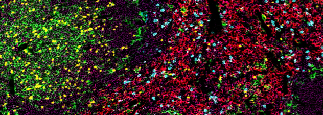
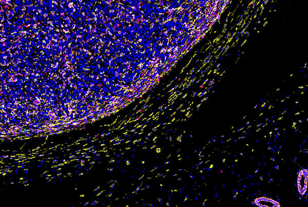
SignalStar® multiplex immunohistochemical analysis of paraffin-embedded human colorectal adenocarcinoma using CD11c (D3V1E) & CO-0017-488 SignalStar® Oligo-Antibody Pair #72146 (red), CD68 (D4B9C) & CO-0007-594 SignalStar® Oligo-Antibody Pair #77318 (green), FoxP3 (D2W8E) Rabbit Monoclonal Antibody #98377 detected with Anti-rabbit IgG & CO-0055-647 SignalStar® Secondary Antibody Kit #76126 (cyan), CD3 epsilon (D7A6E) & CO-0001-750 SignalStar® Oligo-Antibody Pair #51754 (yellow), CD4 (MSVA-004R) & CO-0071-488 SignalStar® Oligo-Antibody Pair #48809 (orange), CD20 (E7B7T) & CO-0011-594 SignalStar® Oligo-Antibody Pair #54189 (pink), CD8 alpha (D8A8Y) & CO-0004-647 SignalStar® Oligo-Antibody Pair #66676 (white), Pan-Keratin (C11) & CO-0003-750 SignalStar® Oligo-Antibody Pair #97227 (magenta), and DAPI #4083 (blue). Staining was performed on the BOND RX autostainer by Leica Biosystems.
Biomarker | Relevance |
|---|---|
Pan-Keratin | Epithelium (identifies tumor cells) |
CD3 | All T cells |
CD4 | Helper T cells |
CD8 | Cytotoxic T cells |
FoxP3 | Regulatory T cells |
CD68 | Macrophages |
CD11c | Dendritic cells |
CD20 | B cells |
Human T Cell Exhaustion Panel
Gain a comprehensive understanding of T cell dynamics within the TME.
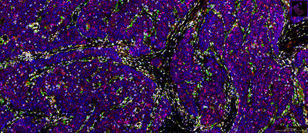
SignalStar® multiplex immunohistochemical analysis of paraffin-embedded human colorectal adenocarcinoma using Tox/Tox2 (E613Q) & CO-0016-488 SignalStar® Oligo-Antibody Pair #31189 (green), PD-1 (Intracellular Domain) (D4W2J) & CO-0008-594 SignalStar® Oligo-Antibody Pair #35347 (cyan), TIM-3 (D5D5R) & CO-0010-647 SignalStar® Oligo-Antibody Pair #15231 (red), CD3 epsilon (D7A6E) & CO-0001-750 SignalStar® Oligo-Antibody Pair #51754 (yellow), CD8 alpha (D8A8Y) & CO-0004-488 SignalStar® Oligo-Antibody Pair #45747 (magenta), TCF1/TCF7 (C63D9) & CO-0006-594 SignalStar® Oligo-Antibody Pair #34706 (white), LAG3 (D2G4O) & CO-0026-647 SignalStar® Oligo-Antibody Pair #40966 (orange), Granzyme B (D6E9W) & CO-0009-750 SignalStar® Oligo-Antibody Pair #60358 (pink), and DAPI #4083 (blue). Staining was performed on the BOND RX autostainer by Leica Biosystems.
Biomarker | Relevance |
|---|---|
Granzyme B | Cytotoxic potential of T cells |
CD3 | All T cells |
CD8 | Cytotoxic T cells |
Tox | Transcription factor that regulates T-cell exhaustion |
PD-1 | Stem-like T cells, exhausted T cells |
TIM3 | Exhausted T cells |
LAG3 | Exhausted T cells |
TCF1 | Stem-like T cells |
Learn More & View Key Features
Human Myeloid Cell Function Panel
Observe myeloid cells and relevant functional markers.
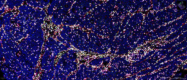
SignalStar® multiplex immunohistochemical analysis of paraffin-embedded human non-hodgkin’s lymphoma using LAMP3/CD208 (E6E5U) & CO-0218-488 SignalStar® Oligo-Antibody Pair #62662 (yellow), HLA-DRA (E9R2Q) & CO-0023-594 SignalStar® Oligo-Antibody Pair #89009 (cyan), XCR1 (D2F8T) & CO-0015-647 SignalStar® Oligo-Antibody Pair #82710 (white), CD11c (D3V1E) & CO-0017-750 SignalStar® Oligo-Antibody Pair #17879 (green), CD163 (D6U1J) & CO-0022-488 SignalStar® Oligo-Antibody Pair #35238 (pink), CD68 (D4B9C) & CO-0007-594 SignalStar® Oligo-Antibody Pair #77318 (orange), CD11b/ITGAM (D6X1N) & CO-0037-647 SignalStar® Oligo-Antibody Pair #29052 (red), CD86 (E2G8P) & CO-0038-750 SignalStar® Oligo-Antibody Pair #31722 (magenta), and DAPI #4083 (blue). Staining was performed on the BOND RX autostainer by Leica Biosystems.
Biomarker | Relevance |
|---|---|
CD11b | Myeloid cells |
CD11c | M1-polarized macrophage |
CD68 | Macrophages |
LAMP3 | mregsDCs |
CD163 | M2-polarized macrophage |
HLA-DRA | M1-polarized macrophage |
XCR1 | Conventional DC1 (CD8 T cell antigen) |
CD86 | M1-polarized macrophage |
Mouse Reactive Panels
Mouse Tumor Immune Microenvironment Panel
Gain detailed insights into immune cell infiltration versus immune exclusion within the TME.
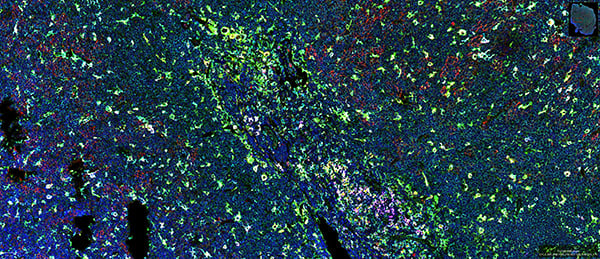
SignalStar® multiplex immunohistochemical analysis of paraffin-embedded GL261 syngeneic tumor using CD11c (D1V9Y) Rabbit Monoclonal Antibody #97585, detected with Anti-rabbit IgG & CO-0055-488 SignalStar® Secondary Antibody Kit #30599 (magenta), FoxP3 (D6O8R) & CO-0041-647 SignalStar® Oligo-Antibody Pair #45142 (green), CD8 alpha (D4W2Z) & CO-0040-750 SignalStar® Oligo-Antibody Pair #74502 (cyan), CD3 epsilon (E4T1B) & CO-0048-488 SignalStar® Oligo-Antibody Pair #92858 (red), F4/80 (D2S9R) & CO-0042-647 SignalStar® Oligo-Antibody Pair #35349 (yellow), Pan-Keratin (Type I) (E6S1S) & CO-0072-750 SignalStar® Oligo-Antibody Pair #53360 (orange), and DAPI #4083 (blue). Staining was performed on the BOND RX autostainer by Leica Biosystems.
Biomarker | Relevance |
|---|---|
Pan-Keratin | Epithelium (identifies tumor cells) |
CD3 | All T cells |
CD8 | Cytotoxic T cells |
FoxP3 | Regulatory T cells |
F4/80 | Macrophages |
CD11c | Dendritic cells |
Ly6G | Neutrophils and MDSCs (myeloid-derived suppressor cells) |
CD19 | B cells |
Learn More & View Key Features
Mouse Myeloid Cell Function Panel
Observe myeloid cells and relevant functional markers.
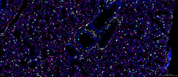
SignalStar® multiplex immunohistochemical analysis of paraffin-embedded mouse lung using CD11c (D1V9Y) Rabbit Monoclonal Antibody #97585 detected with Anti-rabbit IgG & CO-0055-488 SignalStar® Secondary Antibody Kit #30599 (cyan), Arginase-1 (D4E3M) & CO-0075-594 SignalStar® Oligo-Antibody Pair #66757 (white), CD86 (E5W6H) & CO-0051-674 SignalStar® Oligo-Antibody Pair #82598 (orange), F4/80 (D2S9R) & CO-0042-750 SignalStar® Oligo-Antibody Pair #51924 (green), Ly-6G (E6Z1T) & CO-0053-488 SignalStar® Oligo-Antibody Pair #69496 (pink), LAMP3/CD208 (E8T2T) & CO-0217-594 SignalStar® Oligo-Antibody Pair #60296 (red), CD11b/ITGAM (E4K8C) & CO-0083-647 SignalStar® Oligo-Antibody Pair #27353 (magenta), CD206/MRC1 (E6T5J) & CO-0032-750 SignalStar® Oligo-Antibody Pair #52089 (yellow), and DAPI #4083 (blue). Staining was performed on the BOND RX autostainer by Leica Biosystems.
Biomarker | Relevance |
|---|---|
CD11b | Myeloid cells |
CD11c | Dendritic cells |
F4/80 | Macrophages |
CD206 | M2-polarized macrophage |
CD86 | M1-polarized macrophage |
Arginase | M2-polarized macrophage |
Ly6G | Neutrophils and polymorphonuclear MDSCs |
LAMP3 | mregDCs |
Technical Support
Visit the Technical Support page to search for troubleshooting information and answers to technical questions.
Cell Signaling Technology, CST, and SignalStar are trademarks of Cell Signaling Technology, Inc. All other trademarks are the property of their respective owners. Visit cellsignal.com/trademarks for more information.
U.S. Patent No. 10,781,477, foreign equivalents, and child patents deriving therefrom.