Recombinant antibodies offer several key advantages compared to traditional antibodies. These include superior lot-to-lot consistency, continuous supply, and animal-free manufacturing. As such, recombinant antibodies are seeing increased use for scientific research, especially as a means of addressing the ongoing reproducibility crisis.

Traditional polyclonal and monoclonal antibodies are the product of normal B cell development and genetic recombination. They are generated by immunizing an animal with an antigen to elicit an immune response. While polyclonal antibodies are secreted by many different B cell clones and recognize multiple antigenic epitopes, monoclonals originate from a single B cell clone and are specific for just one epitope.
Recombinant antibodies are monoclonal, but their production involves in vitro genetic manipulation. After cloning the antibody genes into an expression vector, this is then transfected into an appropriate host cell line for antibody expression. Mammalian cell lines are most commonly used for recombinant antibody production, although cell lines of bacterial, yeast, or insect origin are also suitable.
Because recombinant antibody production involves sequencing the antibody light and heavy chains, it is a highly controlled and reliable process. In contrast, hybridoma-based systems for producing monoclonal antibodies are subject to genetic drift and instability, increasing the potential for lot-to-lot variability or loss of antibody expression. Recombinant antibodies are highly consistent from lot to lot, thereby ensuring reproducible experimental results.
In vitro methods for producing antibodies are amenable to large-scale production, meaning antibody availability is unlikely to become a limiting factor. Moreover, since the recombinant antibody sequence is known, continuity of supply is assured; in situations where an antibody will be used to support large, long-term studies, this can be an especially critical factor.
Unlike traditional methods for antibody production, recombinant approaches avoid the need to use animals. Where polyclonal antibodies are purified directly from the serum of the immunized host, and monoclonals are purified from either hybridoma-derived tissue culture supernatant or ascites, recombinant antibodies are instead purified from the tissue culture supernatants of transfected host cell lines. Regardless of whether an antibody is polyclonal, monoclonal or recombinant, it must always be properly validated in the intended application prior to experimental use. At CST, we adhere to the Hallmarks of Antibody Validation™, six complementary strategies for determining the specificity, sensitivity, and functionality of an antibody in any given assay. By carefully tailoring these strategies to each antibody product, we guarantee that CST antibodies will work as expected, to help you achieve results you can trust.

| Cat. # | Size | Qty. | Price |
|---|---|---|---|
| 72829SF | 100 µg |
|
| REACTIVITY | H M R |
| SENSITIVITY | Endogenous |
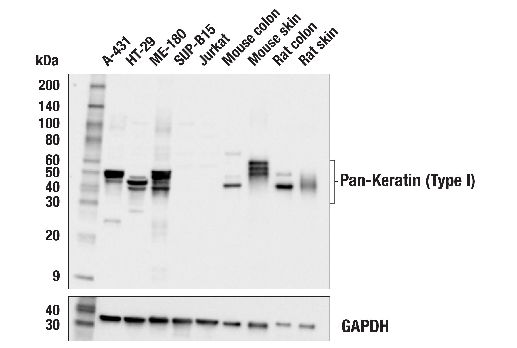
| MW (kDa) | 45-65 |
| Source/Isotype | Rabbit IgG |
Product Information
Human, Mouse, Rat
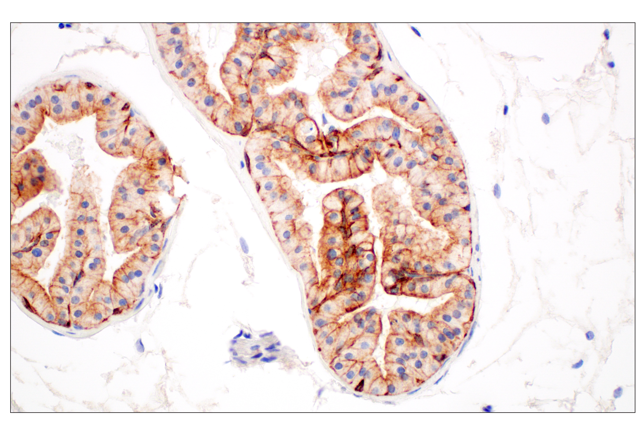
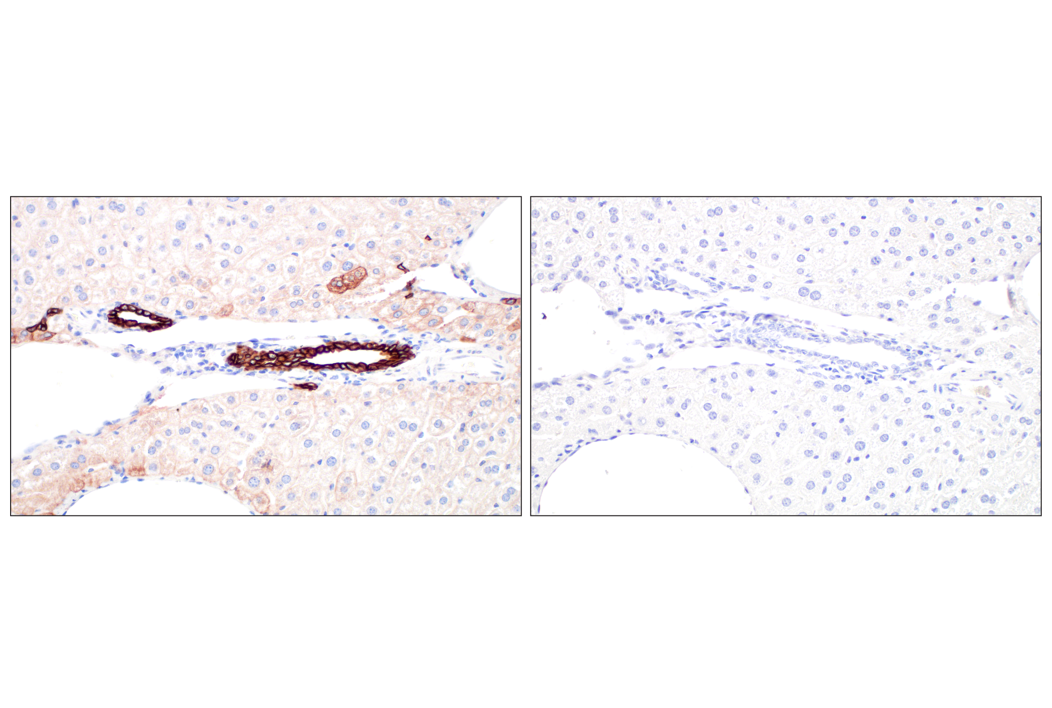
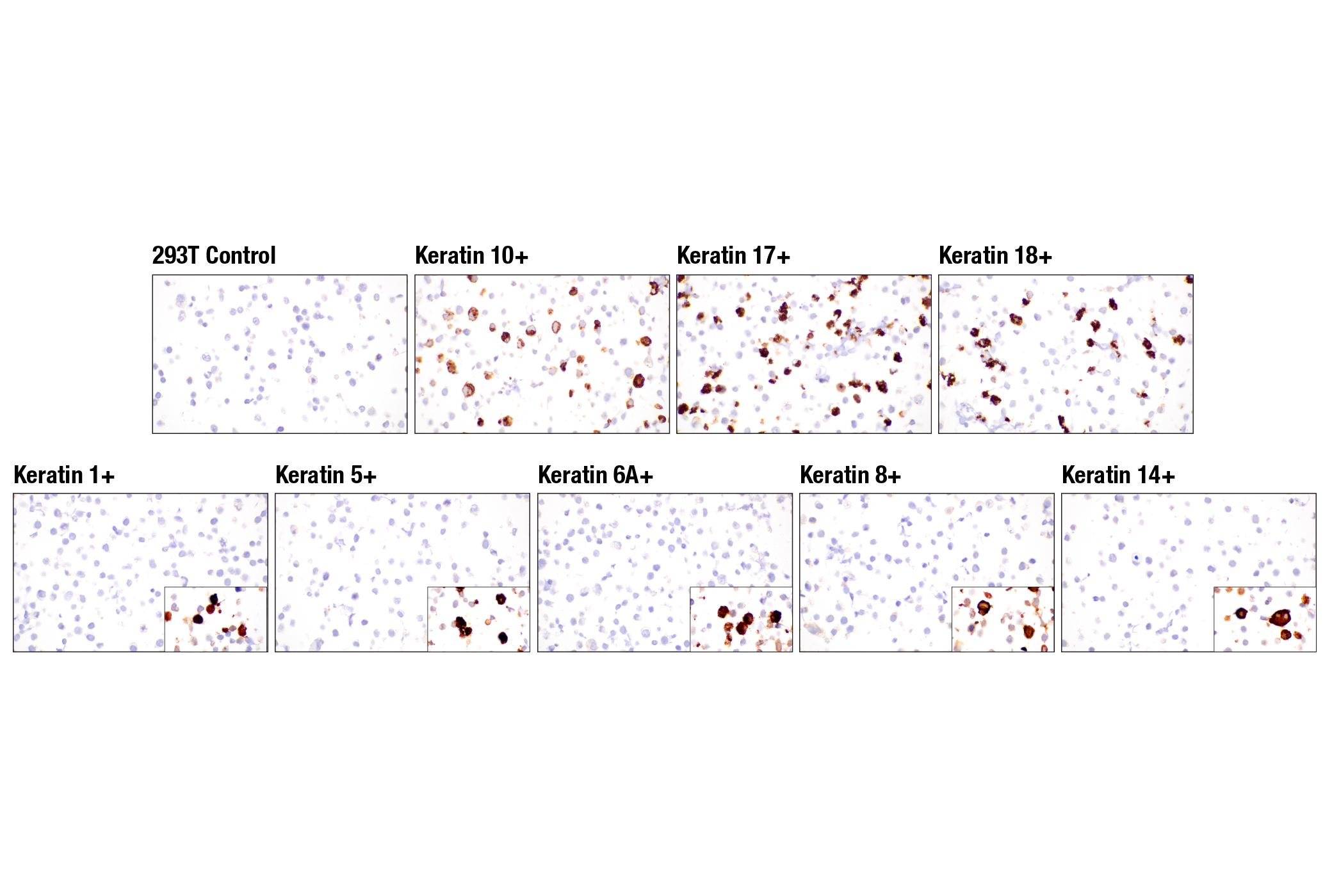
Monoclonal antibody is produced by immunizing animals with a mix of synthetic peptides corresponding to residues highly conserved among type I keratins.
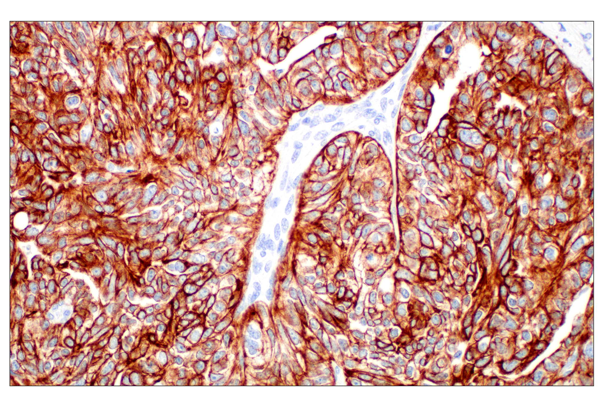
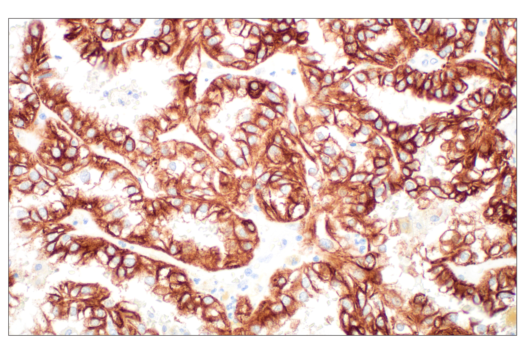
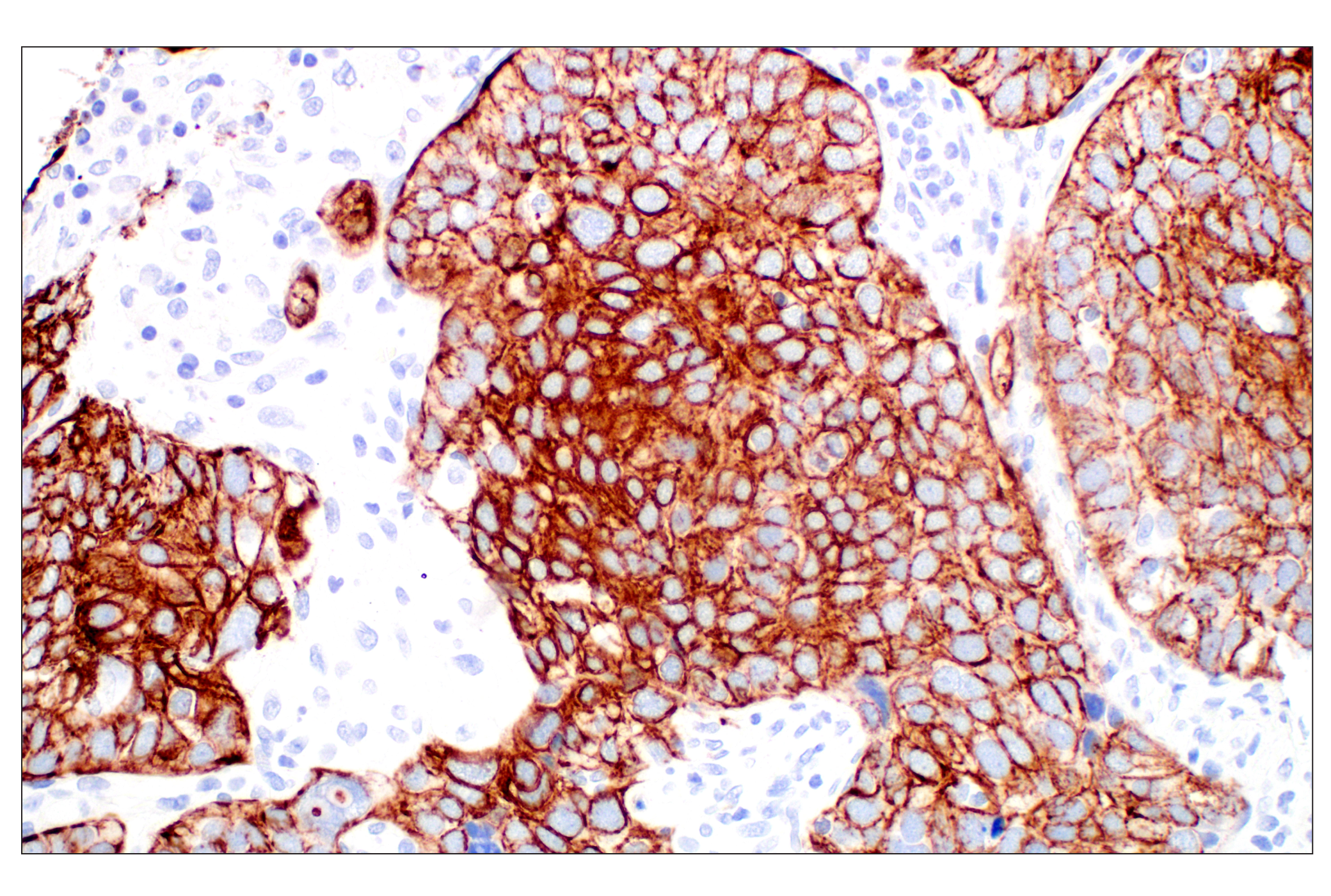
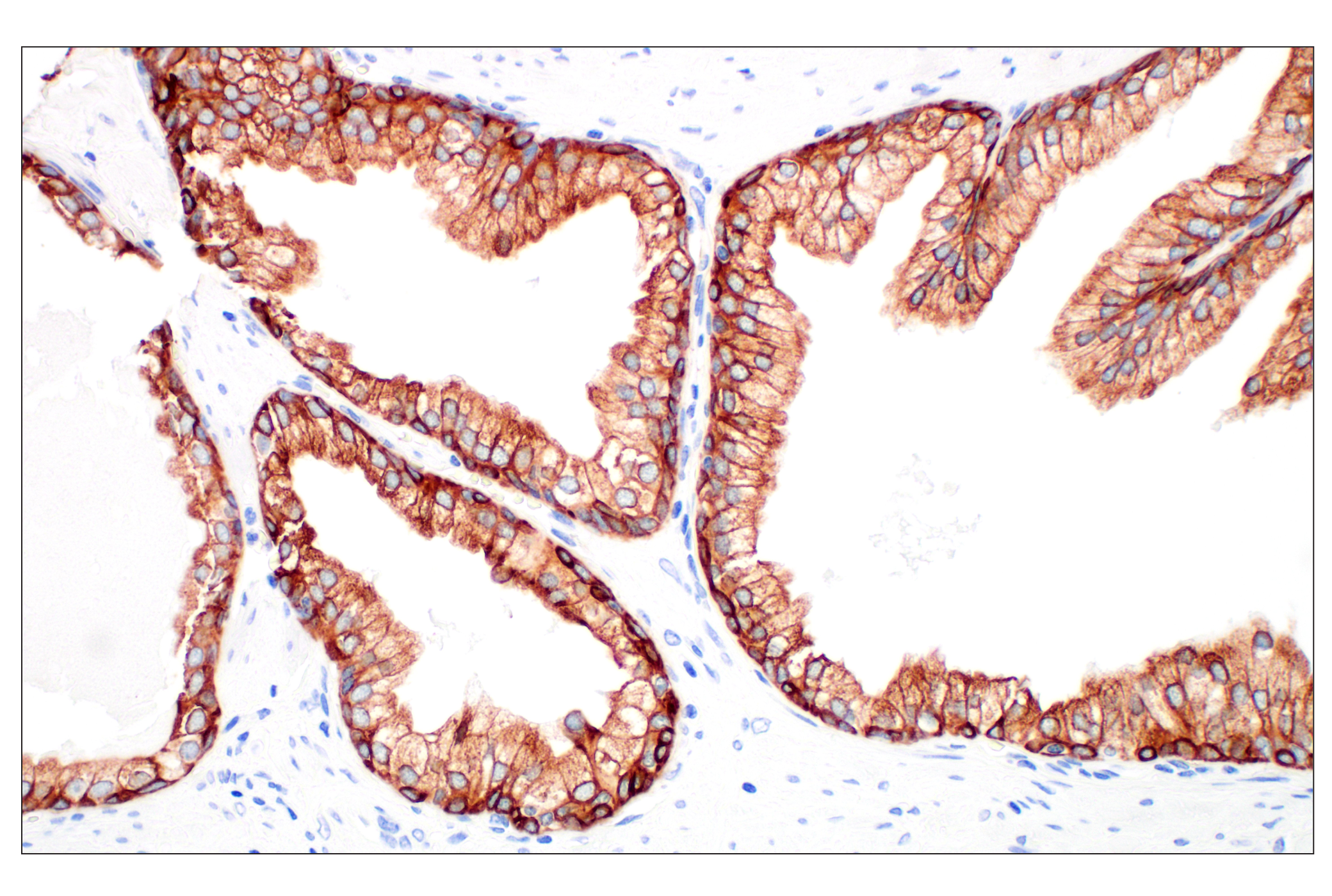
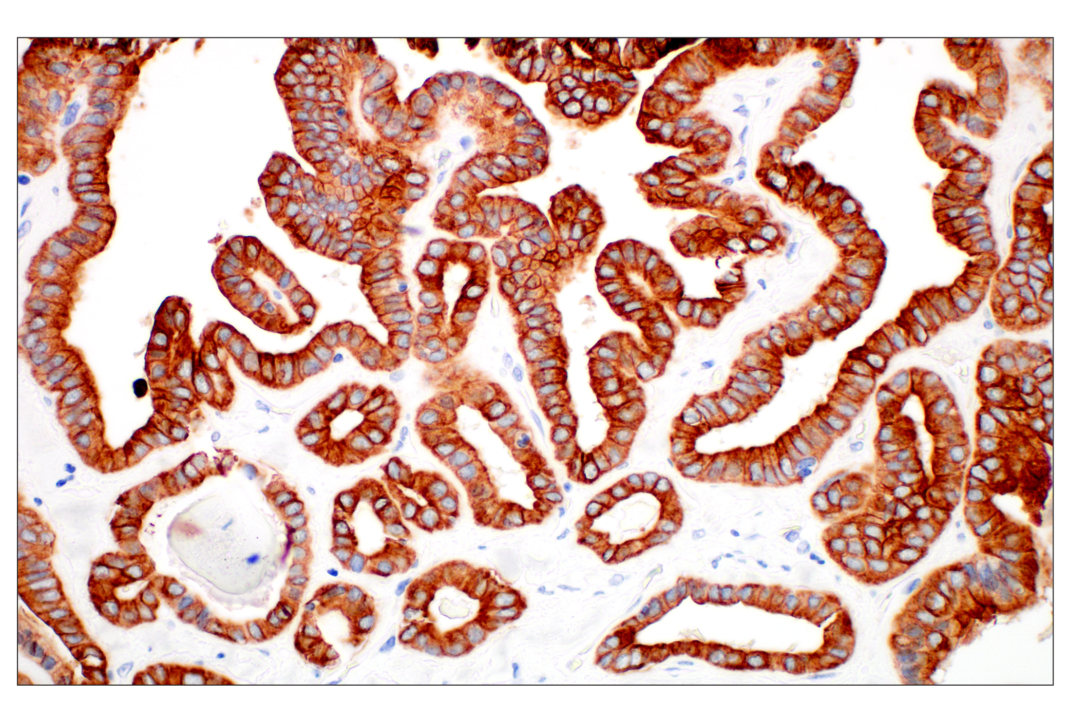
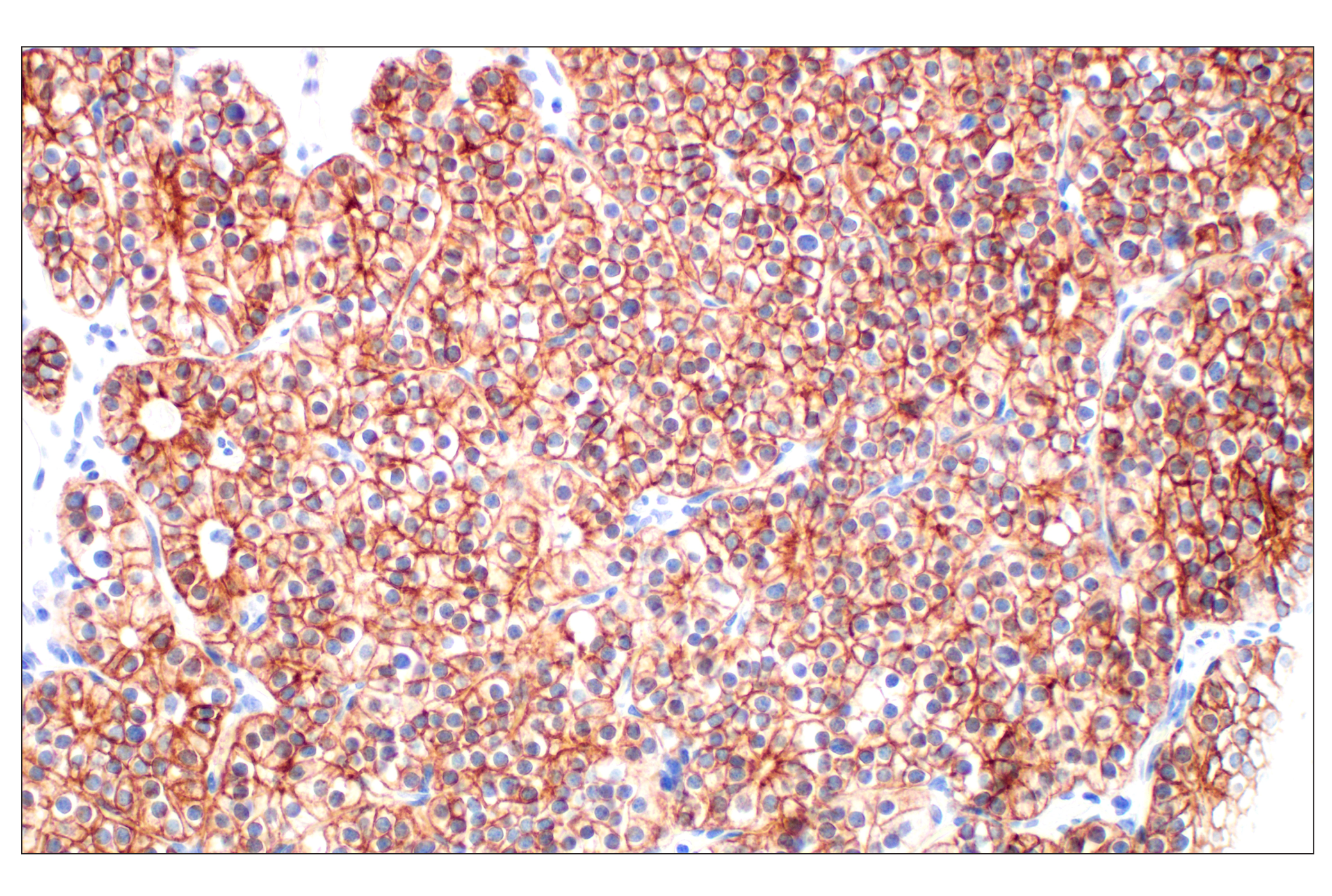
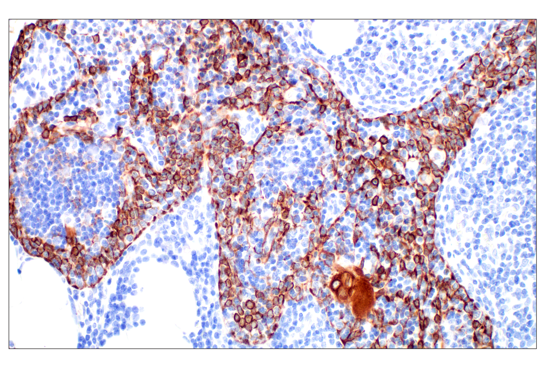
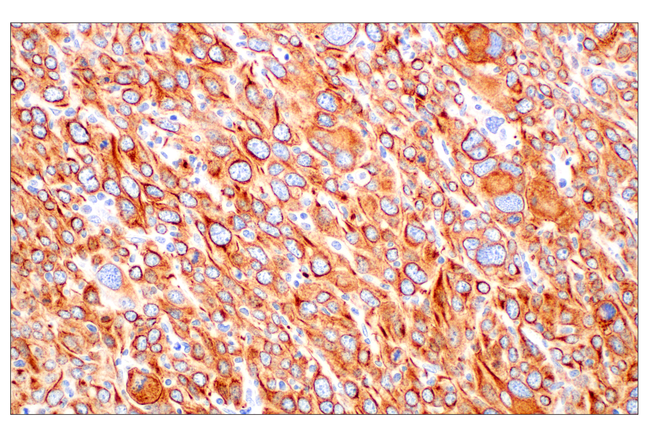
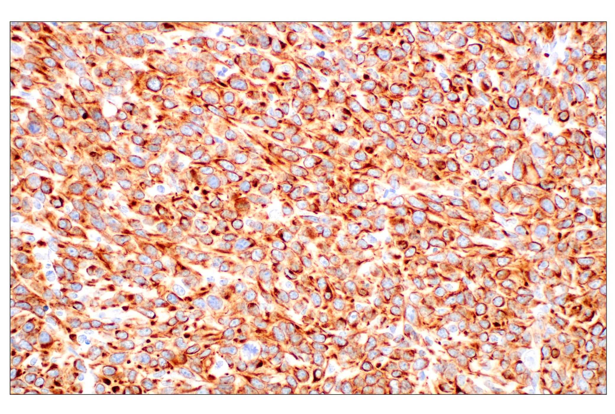
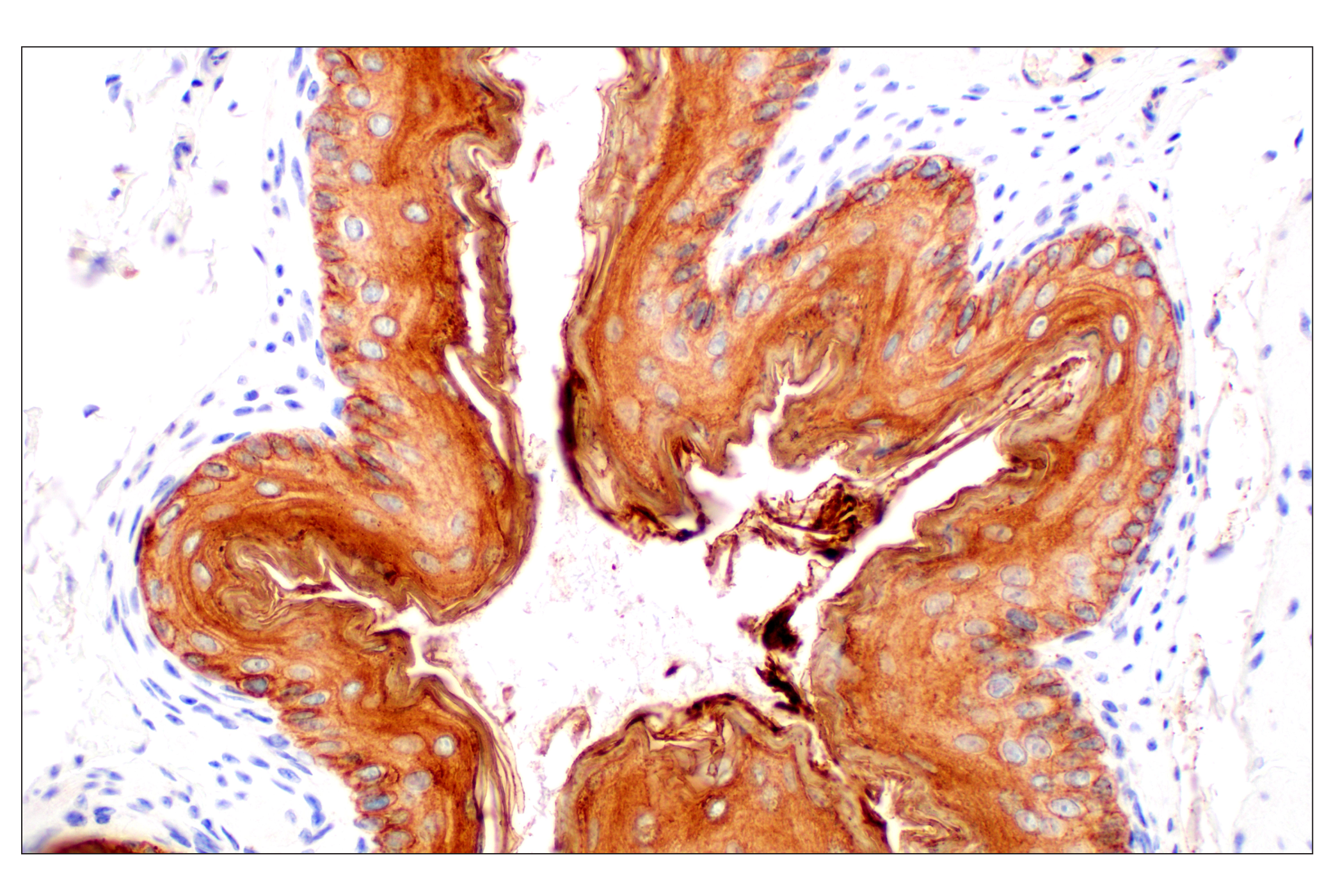
Keratins (cytokeratins) are intermediate filament proteins that are mainly expressed in epithelial cells. Keratin heterodimers composed of an acidic keratin (or type I keratin, keratins K9-K28) and a basic keratin (or type II keratin, keratins K1-K8 and K71-K80) assemble to form filaments. Keratin isoforms demonstrate tissue- and differentiation-specific profiles that make them useful as research and clinical biomarkers (1,2).
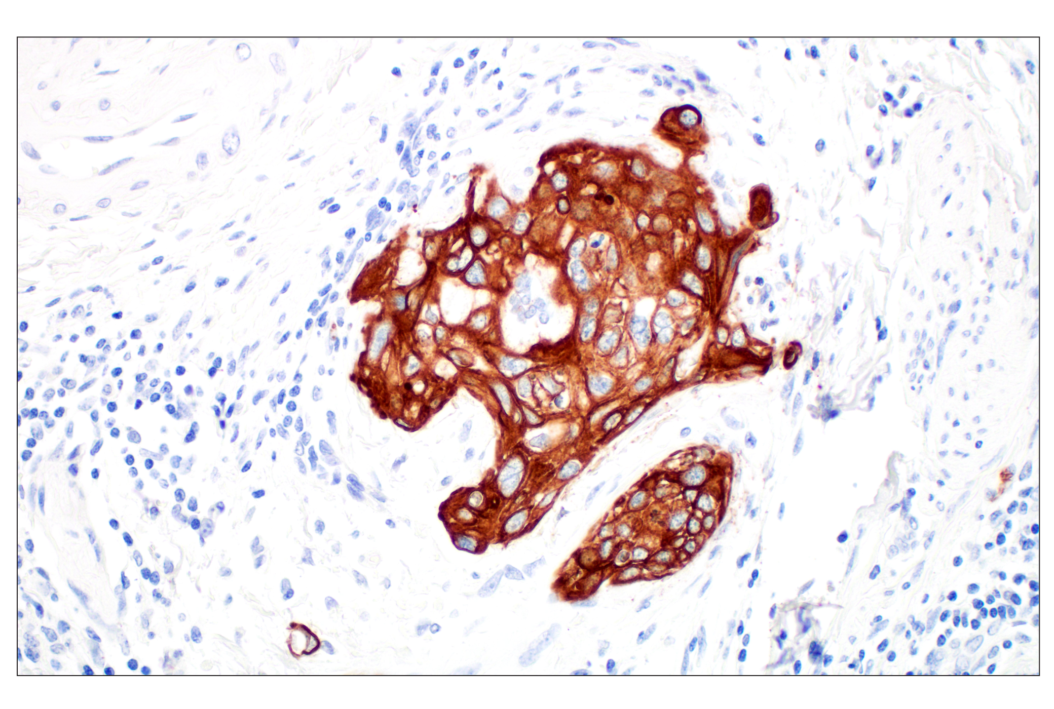
Dysregulation/mutations in keratin genes can lead to a variety of disorders affecting the skin, hair, nails, and other epithelial tissues (3). While expression of keratins can be variable, immunohistochemical staining of keratins is widely used to help in the identification and classification of epithelial tumors, and may also provide prognostic information.
Keratins 8 and 18 (K8/K18) are expressed in simple epithelia of normal tissue, as well as in adenocarcinomas of the breast, lung, ovary, and gastrointestinal tract. Keratin 17 is expressed in basal keratinocytes of stratified epithelia, hair follicles, and sebaceous glands. Onset of keratin 17 expression coincides with the definition of major epithelial lineages during skin development (4). Keratin 14 (K14) is expressed in basal cells of stratified epithelia, and in basal-like subtypes of breast cancer and squamous cell carcinomas. Keratin 19 (K19) is expressed in glandular epithelia, including the liver, gallbladder, and pancreas, as well as in adenocarcinomas of the breast, thyroid, and bile duct. Keratin 20 (K20) is expressed in gastrointestinal epithelium, urothelium, and Merkel cells in the skin, as well as in colorectal carcinomas and some urothelial carcinomas. Keratin 5/6 (K5/6) is expressed in basal cells of stratified epithelia, including the skin, prostate, and breast, as well as in basal-like breast cancers, squamous cell carcinomas, and some lung carcinomas. Keratin 7 (K7) is expressed in glandular epithelia, such as those in the lung, breast, and female reproductive tract, as well as in adenocarcinomas of the lung, breast, and ovary (5,6).
Keratins, particularly K8, K18, and K19, serve as biomarkers for identification of circulating tumor cells (CTCs) (5).
Post-translational modifications, including phosphorylation, acetylation, ubiquitylation, sumoylation, glycosylation, and transamidation, have been shown to affect the functions of keratins in normal and disease states (6). Understanding the molecular mechanisms underlying these PTMs may provide insights into cancer pathogenesis.
Except as otherwise expressly agreed in a writing signed by a legally authorized representative of CST, the following terms apply to Products provided by CST, its affiliates or its distributors. Any Customer's terms and conditions that are in addition to, or different from, those contained herein, unless separately accepted in writing by a legally authorized representative of CST, are rejected and are of no force or effect.
Products are labeled with For Research Use Only or a similar labeling statement and have not been approved, cleared, or licensed by the FDA or other regulatory foreign or domestic entity, for any purpose. Customer shall not use any Product for any diagnostic or therapeutic purpose, or otherwise in any manner that conflicts with its labeling statement. Products sold or licensed by CST are provided for Customer as the end-user and solely for research and development uses. Any use of Product for diagnostic, prophylactic or therapeutic purposes, or any purchase of Product for resale (alone or as a component) or other commercial purpose, requires a separate license from CST. Customer shall (a) not sell, license, loan, donate or otherwise transfer or make available any Product to any third party, whether alone or in combination with other materials, or use the Products to manufacture any commercial products, (b) not copy, modify, reverse engineer, decompile, disassemble or otherwise attempt to discover the underlying structure or technology of the Products, or use the Products for the purpose of developing any products or services that would compete with CST products or services, (c) not alter or remove from the Products any trademarks, trade names, logos, patent or copyright notices or markings, (d) use the Products solely in accordance with CST Product Terms of Sale and any applicable documentation, and (e) comply with any license, terms of service or similar agreement with respect to any third party products or services used by Customer in connection with the Products.