| Cat. # | Size | Qty. | Price |
|---|---|---|---|
| 83718T | 1 Kit (8 x 20 microliters) |
|
| Product Includes | Quantity | Applications | Reactivity | MW(kDa) | Isotype |
|---|---|---|---|---|---|
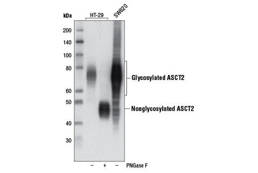
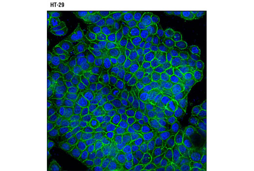
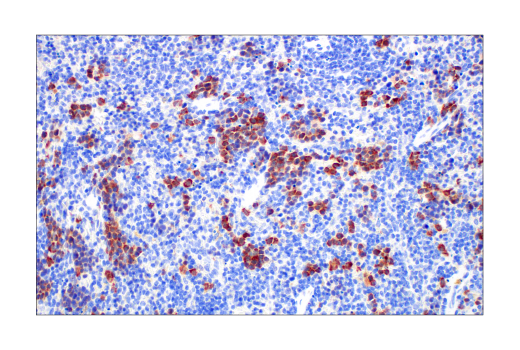
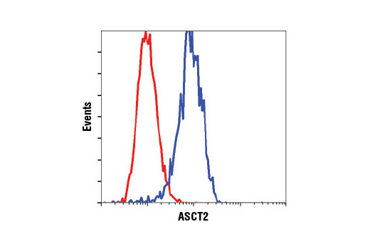
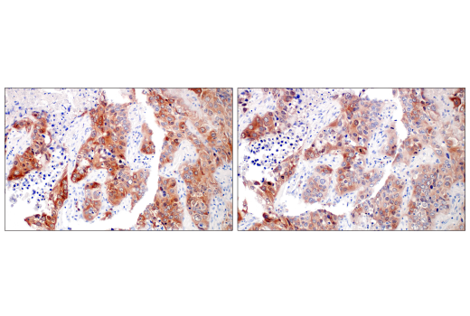
| ASCT2 (D7C12) Rabbit mAb 8057 | 20 µl |
|
H Mk | 49, 75 | Rabbit |
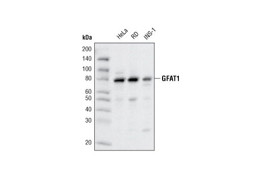
| GFAT1 (D12F4) Rabbit mAb 5322 | 20 µl |
|
H R | 80 | Rabbit IgG |
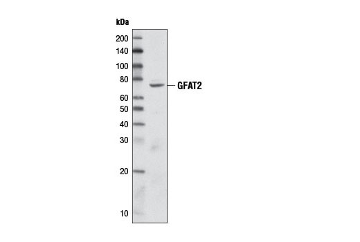
| GFAT2 (D40C7) Rabbit mAb 6917 | 20 µl |
|
H | 78 | Rabbit IgG |
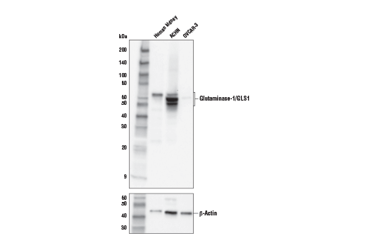
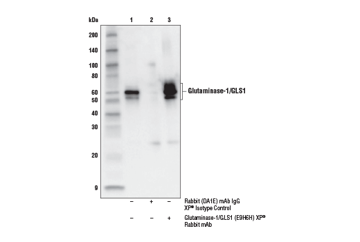
| Glutaminase-1/GLS1 (E9H6H) XP® Rabbit mAb 56750 | 20 µl |
|
H Mk | 55-65 | Rabbit IgG |
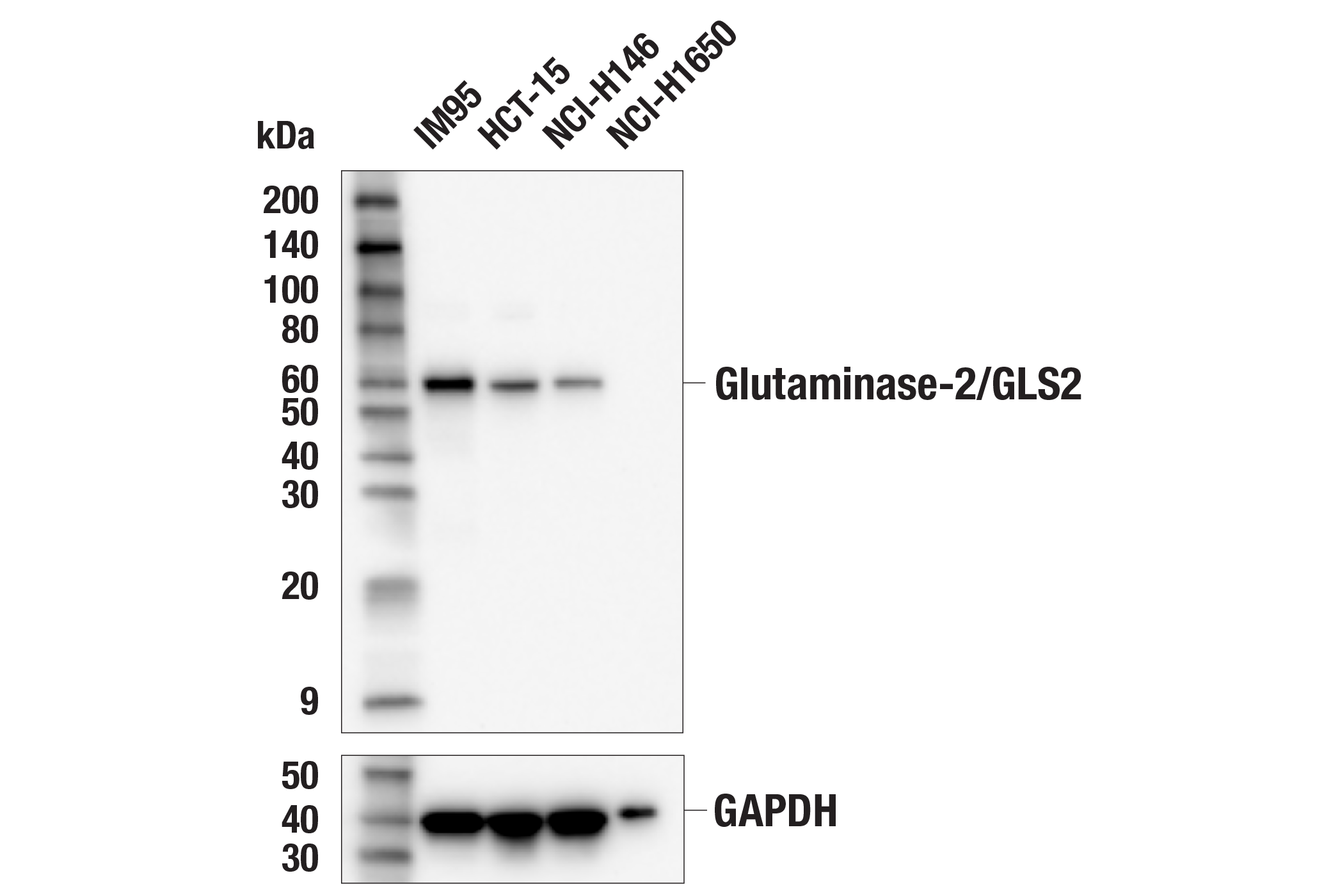
| Glutaminase-2/GLS2 (E9C7V) Rabbit mAb 85934 | 20 µl |
|
H | 60 | Rabbit IgG |
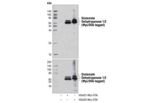
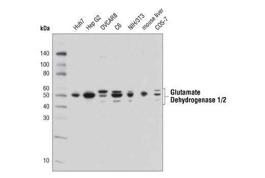
| Glutamate Dehydrogenase 1/2 (D9F7P) Rabbit mAb 12793 | 20 µl |
|
H M R Mk | 52 | Rabbit IgG |
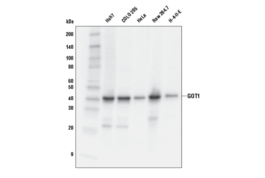
| GOT1 (E4A4O) Rabbit mAb 34423 | 20 µl |
|
H M R Mk | 41 | Rabbit IgG |
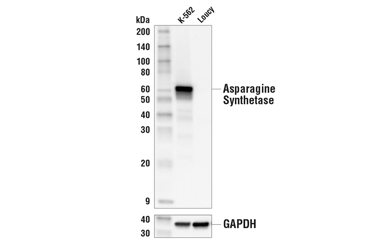
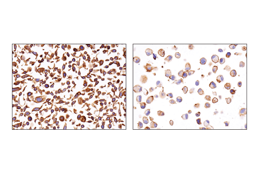
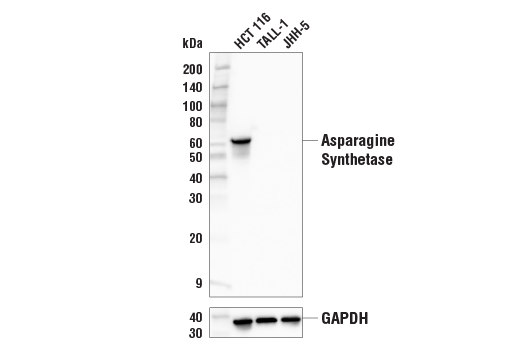
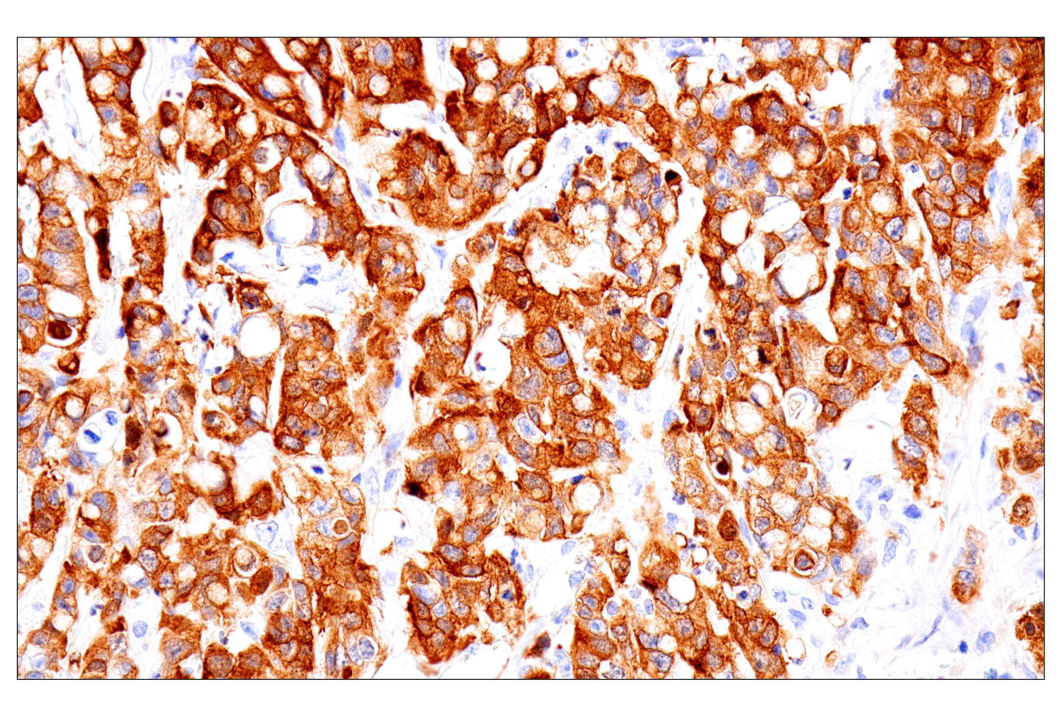
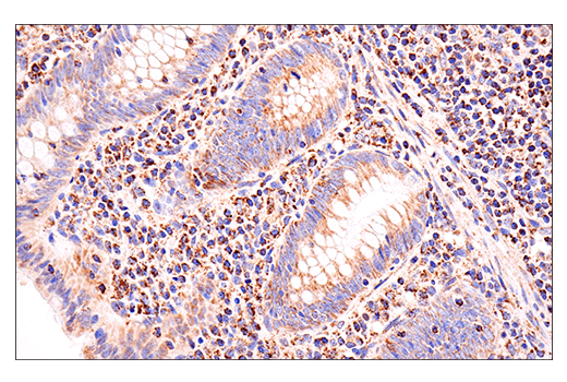
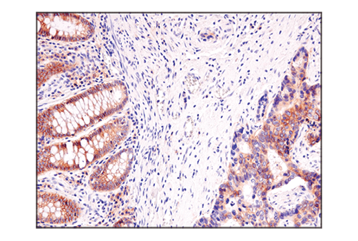
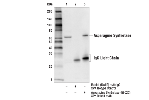
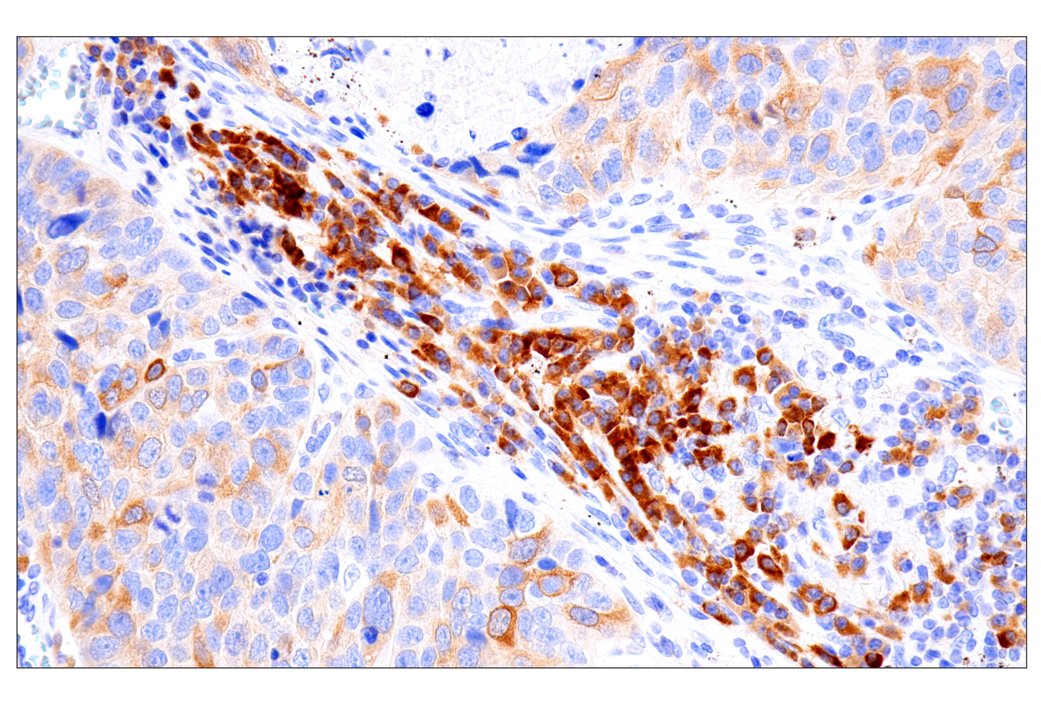
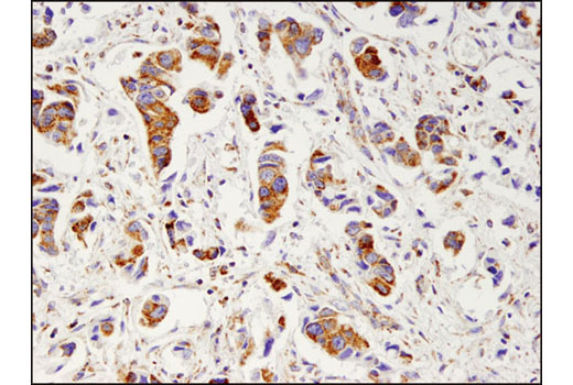
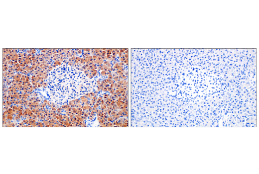
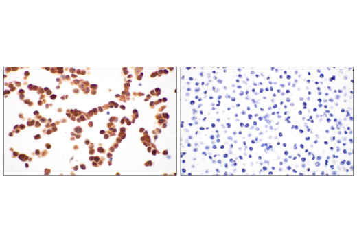
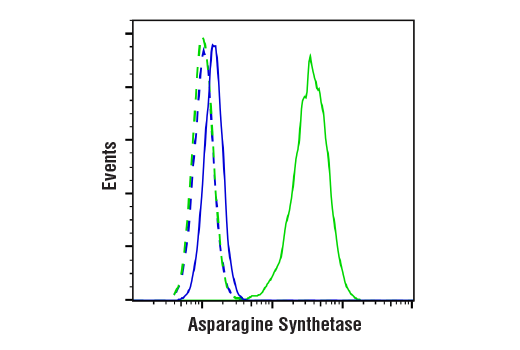
| Asparagine Synthetase (E6C2C) XP® Rabbit mAb 92479 | 20 µl |
|
H M R | 64 | Rabbit IgG |
| Anti-rabbit IgG, HRP-linked Antibody 7074 | 100 µl |
|
Goat |
Product Information
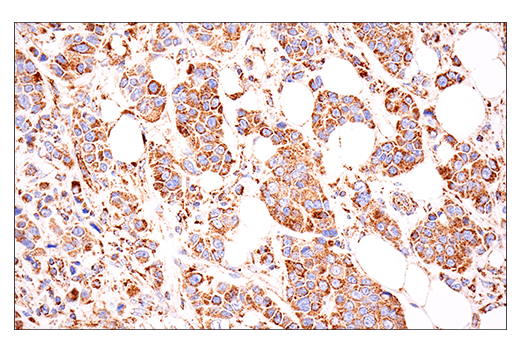
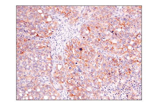
Sodium-dependent neutral amino acid transporter type 2 (ASCT2 or SLC1A5) is a neutral amino acid transporter that regulates the uptake of essential amino acids in conjunction with the SLC7A5 bilateral transporter (1,2). ASCT2 appears to be the major glutamine transporter in hepatoma cells and is thought to provide essential amino acids needed for tumor growth (3). Additional evidence suggests that ASCT2 plays a role in activating mTORC1 signaling and is required to suppress autophagy (4,5). Cell surface ASCT2 serves as a receptor for several mammalian interference retroviruses associated with cases of infectious immunodeficiency; variation in a small region of an extracellular loop (ECL2) may be responsible for species-specific differences in receptor function (6).
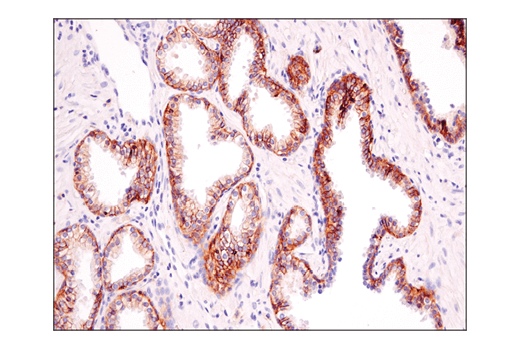
GFAT1, glutamine:fructose-6-phosphate aminotransferase 1, is the rate-limiting enzyme of the hexosamine biosynthesis pathway (7). This enzyme catalyzes the conversion of fructose-6-phosphate and glutamine to glucosamine-6-phosphate and glutamate (8). The hexosamine biosynthesis pathway generates the building blocks for protein and lipid glycosylation (8). Furthermore, studies suggest that increased activity of this pathway is a contributing factor to hyperglycemia-induced insulin resistance (7,8). GFAT1 is more active in non-insulin-dependent diabetes mellitus (NIDDM) patients (9). Transgenic mice overexpressing this enzyme in skeletal muscle and adipose tissue show an insulin resistance phenotype (10,11). GFAT2, an isoenzyme of GFAT1, was later identified (12,13). Studies show that the regulation of GFAT2 is different from that of GFAT1, suggesting differential regulation of the hexosamine pathway in different tissues (13).
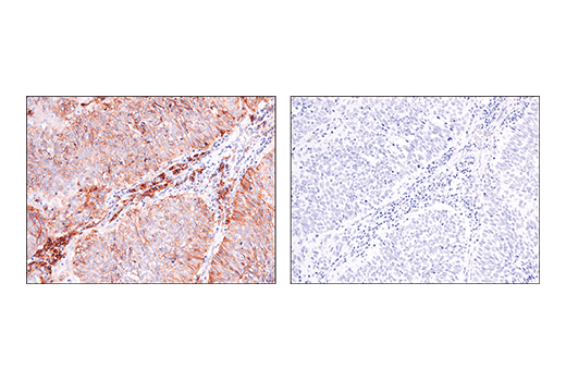
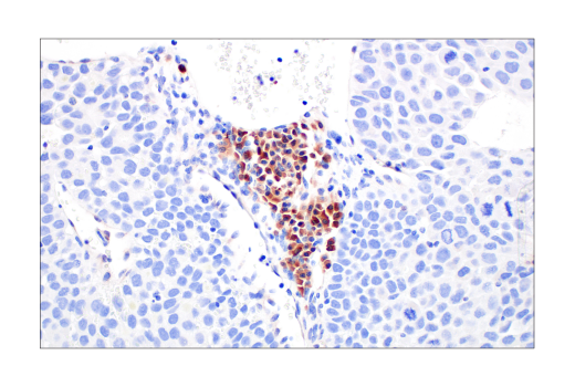
Glutaminase catalyzes the conversion of glutamine to glutamate, the first and rate-limiting step of glutaminolysis (14). Both kidney-type glutaminase (GLS1) and liver-type glutaminase (GLS2) are found in mammals (15). GLS1-mediated glutathione synthesis plays an essential role in redox homeostasis and contributes to increased survival of postimplantation bone cells preconditioned to the hypoxic and ischemic environment in the bone defect site (16). In addition, KEAP1–NRF2-mutant LUAD (KRAS-mutant lung adenocarcinoma) tumors are dependent on increased glutaminolysis (14). Furthermore, recent studies showed higher glutaminolysis and glucose production from glutamine in human primary hepatocytes with GLS2 gain-of-function missense mutations (17). These findings suggest GLS1 and GLS2 as potential targets in the therapy of bone regeneration and in the treatments of diseases such as cancer and hyperglycemia, respectively (14,16,17).
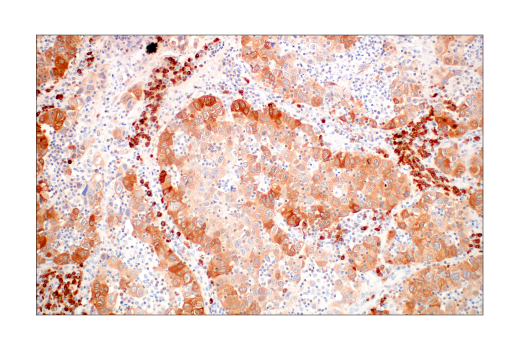
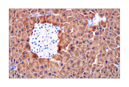
Glutamate dehydrogenase is a mitochondrial enzyme that catalyzes the oxidative deamination of glutamate to α-ketoglutarate through association with the cofactor nicotinamide adenine dinucleotide phosphate (18). Glutamate dehydrogenase is highly expressed in various tissues such as the liver, brain, kidney, heart, pancreas, ovaries, and testis. Two isoforms produced by two distinct genes are found in mammalian tissues. The GLUD1 gene is ubiquitously expressed (19), while the GLUD2 gene is specifically expressed in testicular tissues and astrocytes (20,21). Glutamate dehydrogenase links glutamate to the Krebs cycle, thereby playing a critical role in the regulation of energy homeostasis. Research studies have shown that changes in glutamate dehydrogenase activity in pancreatic β-cells can cause a hyperinsulinism syndrome (22).
Glutamate oxaloacetate transaminase 1 (GOT1) catalyzes the interconversion of aspartate and oxaloacetate (23).
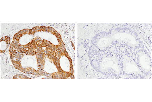
Asparagine synthetase (ASNS) catalyzes the synthesis of asparagine from aspartate and glutamine (24). In subsets of gastric and hepatic cancers, ASNS promoter hypermethylation correlates with low ASNS expression, sensitizing these cancers to the asparaginase treatment (25).
Explore pathways related to this product.
STRING - Known and Predicted Protein-Protein Interactions.
Except as otherwise expressly agreed in a writing signed by a legally authorized representative of CST, the following terms apply to Products provided by CST, its affiliates or its distributors. Any Customer's terms and conditions that are in addition to, or different from, those contained herein, unless separately accepted in writing by a legally authorized representative of CST, are rejected and are of no force or effect.
Products are labeled with For Research Use Only or a similar labeling statement and have not been approved, cleared, or licensed by the FDA or other regulatory foreign or domestic entity, for any purpose. Customer shall not use any Product for any diagnostic or therapeutic purpose, or otherwise in any manner that conflicts with its labeling statement. Products sold or licensed by CST are provided for Customer as the end-user and solely for research and development uses. Any use of Product for diagnostic, prophylactic or therapeutic purposes, or any purchase of Product for resale (alone or as a component) or other commercial purpose, requires a separate license from CST. Customer shall (a) not sell, license, loan, donate or otherwise transfer or make available any Product to any third party, whether alone or in combination with other materials, or use the Products to manufacture any commercial products, (b) not copy, modify, reverse engineer, decompile, disassemble or otherwise attempt to discover the underlying structure or technology of the Products, or use the Products for the purpose of developing any products or services that would compete with CST products or services, (c) not alter or remove from the Products any trademarks, trade names, logos, patent or copyright notices or markings, (d) use the Products solely in accordance with CST Product Terms of Sale and any applicable documentation, and (e) comply with any license, terms of service or similar agreement with respect to any third party products or services used by Customer in connection with the Products.