| Cat. # | Size | Qty. | Price |
|---|---|---|---|
| 84884T | 1 Kit (4 x 20 microliters) |
|
| Product Includes | Quantity | Applications | Reactivity | MW(kDa) | Isotype |
|---|---|---|---|---|---|
| TET1 (E5F1O) Rabbit mAb 40142 | 20 µl |
|
H M Mk | 300 | Rabbit IgG |
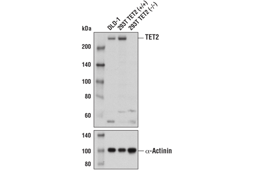
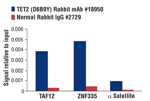
| TET2 (D6B9Y) Rabbit mAb 18950 | 20 µl |
|
H | 280 | Rabbit IgG |
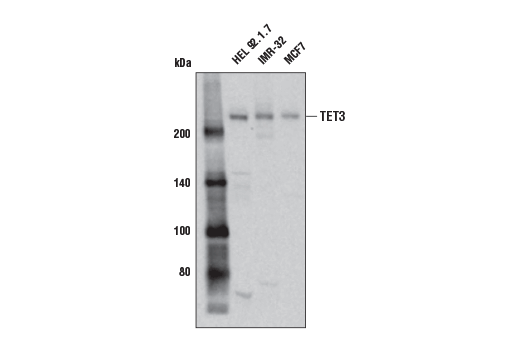
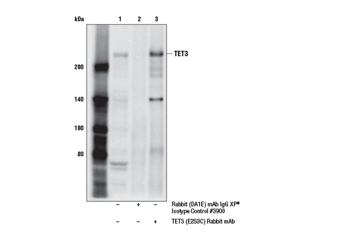
| TET3 (E2S3C) Rabbit mAb 85016 | 20 µl |
|
H Mk | 235 | Rabbit IgG |
| TDG (E5T5G) Rabbit mAb 99105 | 20 µl |
|
H M R Mk | 58, 60 | Rabbit IgG |
| Anti-rabbit IgG, HRP-linked Antibody 7074 | 100 µl |
|
Goat |
Product Information
Methylation of DNA at cytosine residues is a heritable, epigenetic modification that is critical for proper regulation of gene expression, genomic imprinting, and mammalian development (1,2). 5-methylcytosine is a repressive epigenetic mark established de novo by two enzymes, DNMT3a and DNMT3b, and is maintained by DNMT1 (3,4). 5-methylcytosine was originally thought to be passively depleted during DNA replication. However, subsequent studies have shown that Ten-Eleven Translocation (TET) proteins TET1, TET2, and TET3 can catalyze the oxidation of methylated cytosine to 5-hydroxymethylcytosine (5-hmC) (5). Additionally, TET proteins can further oxidize 5-hmC to form 5-formylcytosine (5-fC) and 5-carboxylcytosine (5-caC), both of which are excised by thymine-DNA glycosylase (TDG), effectively linking cytosine oxidation to the base excision repair pathway and supporting active cytosine demethylation (6,7). TET1 is highly expressed in embryonic stem cells and is essential for maintaining stem cell pluripotency (8). Aberrant TET1 expression has also been implicated in a variety of cancers, including hepatocellular carcinoma, T-cell acute lymphoblastic leukemia (T-ALL), and triple-negative breast cancer (TNBC), among others (9-11). TET2 is frequently mutated in myeloid dysplastic syndrome (MDS) and diffuse large B-cell lymphomas (12,13). TET2 protein expression is often reduced in solid tumors such as prostate cancer, melanoma, and oral squamous cell carcinoma (14-16). TET3 plays key roles in regulating early development and neonatal growth (17,18). TET2/TET3 deficiency can lead to myeloid cell, B cell, and invariant natural killer T (iNKT) cell malignancies. In Tregs, TET2/TET3 deficiency in mice leads to hyperproliferation and inflammatory disease (19,20). Knockout or catalytic inactivation of TDG leads to embryonic lethality (21,22). SUMOylation of TDG has been reported to help it dissociate from its abasic product, thereby increasing catalytic turnover (23). Additional studies suggest that SUMOylation affects TDG’s cellular localization or lowers its base excision activity, allowing it to act as a ‘reader’ protein for 5-fC and 5-caC modified DNA (24).
Except as otherwise expressly agreed in a writing signed by a legally authorized representative of CST, the following terms apply to Products provided by CST, its affiliates or its distributors. Any Customer's terms and conditions that are in addition to, or different from, those contained herein, unless separately accepted in writing by a legally authorized representative of CST, are rejected and are of no force or effect.
Products are labeled with For Research Use Only or a similar labeling statement and have not been approved, cleared, or licensed by the FDA or other regulatory foreign or domestic entity, for any purpose. Customer shall not use any Product for any diagnostic or therapeutic purpose, or otherwise in any manner that conflicts with its labeling statement. Products sold or licensed by CST are provided for Customer as the end-user and solely for research and development uses. Any use of Product for diagnostic, prophylactic or therapeutic purposes, or any purchase of Product for resale (alone or as a component) or other commercial purpose, requires a separate license from CST. Customer shall (a) not sell, license, loan, donate or otherwise transfer or make available any Product to any third party, whether alone or in combination with other materials, or use the Products to manufacture any commercial products, (b) not copy, modify, reverse engineer, decompile, disassemble or otherwise attempt to discover the underlying structure or technology of the Products, or use the Products for the purpose of developing any products or services that would compete with CST products or services, (c) not alter or remove from the Products any trademarks, trade names, logos, patent or copyright notices or markings, (d) use the Products solely in accordance with CST Product Terms of Sale and any applicable documentation, and (e) comply with any license, terms of service or similar agreement with respect to any third party products or services used by Customer in connection with the Products.