View Featured Offers >>
99089
PathScan® RP KEAP1 Sandwich ELISA Kit
ELISA Kits
ELISA Kit

PathScan® RP KEAP1 Sandwich ELISA Kit #99089

Citations (0)
Figure 1. KEAP1 protein is expressed at differing levels across several cell lines. The absorbance at 450 nm for a panel of cell lines using the PathScan® RP KEAP1 Sandwich ELISA Kit #99089 is shown in the upper figure. The corresponding western blot using a KEAP1 antibody is shown in the lower figure. Unstarved cells were harvested and then lysed.
Figure 2. KEAP1 protein is expressed in KARPAS 299 cells. The relationship between lysate protein concentration from KARPAS 299 cells and the absorbance at 450 nm using the PathScan® RP KEAP1 Sandwich ELISA Kit #99089 is shown.
To Purchase # 99089
Cat. # Size Qty. Price
99089C
1 Kit  (96 assays)

Important Ordering Details

Custom Ordering Details:

When ordering five or more kits, please contact us for processing time and pricing.

Looking for this ELISA kit in a 384-well format? Inquire for availability, processing time, and pricing.

Supporting Data

REACTIVITY H

Application Key:

  • WB-Western Blot
  • IP-Immunoprecipitation
  • IHC-Immunohistochemistry
  • ChIP-Chromatin Immunoprecipitation
  • C&R-CUT&RUN
  • C&T-CUT&Tag
  • DB-Dot Blot
  • eCLIP-eCLIP
  • IF-Immunofluorescence
  • F-Flow Cytometry

Species Cross-Reactivity Key:

  • H-Human
  • M-Mouse
  • R-Rat
  • Hm-Hamster
  • Mk-Monkey
  • Vir-Virus
  • Mi-Mink
  • C-Chicken
  • Dm-D. melanogaster
  • X-Xenopus
  • Z-Zebrafish
  • B-Bovine
  • Dg-Dog
  • Pg-Pig
  • Sc-S. cerevisiae
  • Ce-C. elegans
  • Hr-Horse
  • GP-Guinea Pig
  • Rab-Rabbit
  • All-All Species Expected
Product Includes Volume Solution Color
KEAP1 Rabbit mAb Coated Microwells 96 tests
KEAP1 Rabbit Detection mAb 1 ea Red (Lyophilized)
HRP Diluent 5.5 ml Red
TMB Substrate 7004 11 ml
STOP Solution 7002 11 ml
Sealing Tape 2 ea
ELISA Wash Buffer (20X) 9801 25 ml
Cell Lysis Buffer (10X) 9803 15 ml

Product Description

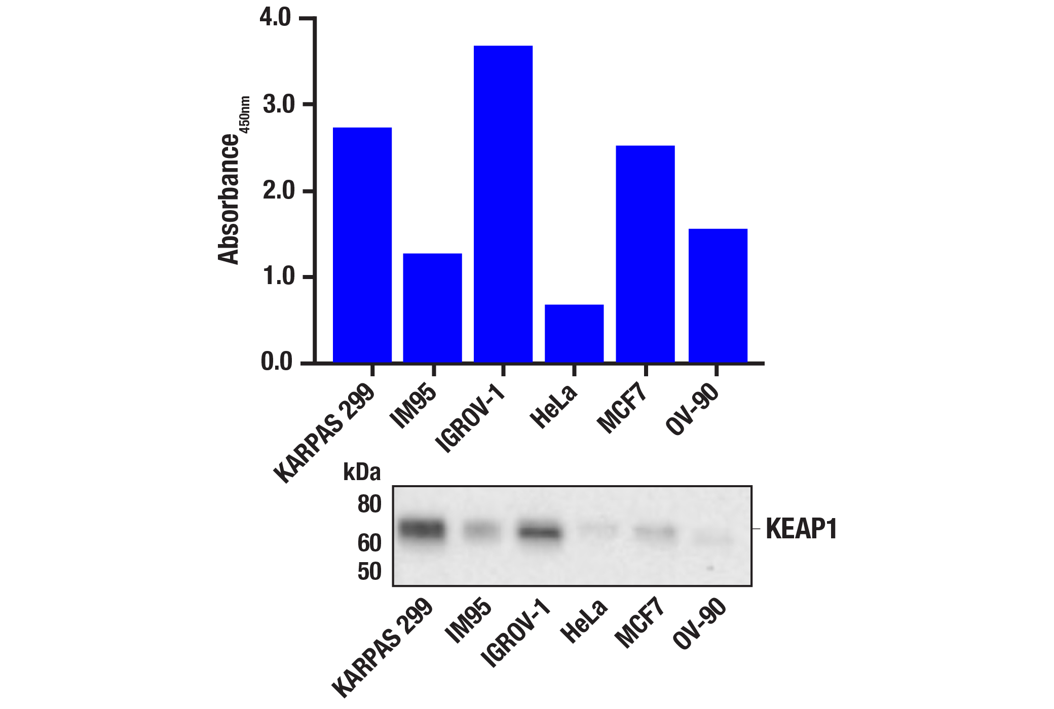
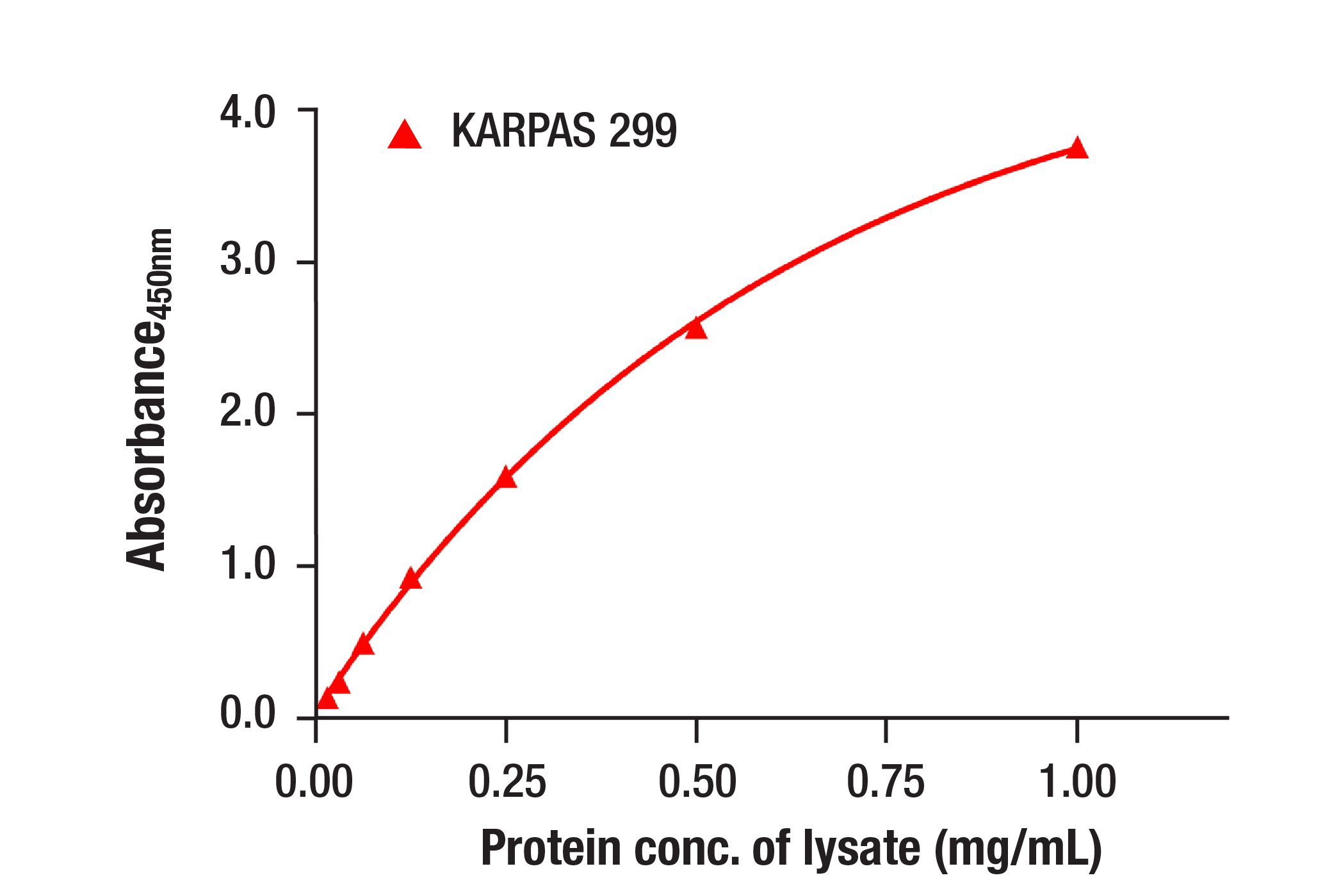
The rapid protocol (RP) PathScan® RP KEAP1 Sandwich ELISA Kit is a solid phase sandwich enzyme-linked immunosorbent assay (ELISA) that detects endogenous levels of KEAP1 protein in a reduced assay time of 1.5 hours. Incubation of cell lysates and detection antibody on the coated microwell plate forms a sandwich with KEAP1 protein in a single step. The plate is then extensively washed and TMB reagent is added for signal development. The magnitude of absorbance for the developed color is proportional to the quantity of KEAP1 protein. Learn more about your ELISA kit options here.

*Antibodies in this kit are custom formulations specific to kit.

Protocol

PRINT

View >Collapse >

PathScan® Sandwich ELISA Protocol (Rapid Protocol)

NOTE: This protocol is for PathScan® kits that use an HRP directly conjugated to the detection antibody (Rapid Protocol), rather than a 2-step method where the detection antibody and a secondary-HRP are added sequentially.

A. Solutions and Reagents

NOTE: Prepare solutions with deionized/purified water or equivalent.

  1. Microwell strips: Bring all to room temperature before opening bag/use. Unused microwell strips should be returned to the original re-sealable bag containing the desiccant pack and stored at 4°C.
  2. Detection Antibody: Reconstitute lyophilized Detection Antibody (red colored cake) with 5.5 mL HRP Diluent. Incubate at room temperature for 5 min with occasional gentle mixing to fully reconstitute. For best results, use immediately following antibody reconstitution. Unused reconstituted Detection Antibody may be stored for up to 4 weeks at 4°C, although there may be some loss of signal compared to freshly reconstituted antibody.
  3. HRP Diluent: Red colored diluent for reconstitution and dilution of the Detection Antibody that is linked to HRP.
  4. 1X ELISA Wash Buffer: Prepare by diluting ELISA Wash Buffer (20X) (included in each kit) to 1X with deionized water.
  5. 1X Cell Lysis Buffer: Prepare by diluting 10X Cell Lysis Buffer #9803 to 1X with deionized water. This buffer can be stored at 4°C for short-term use (1–2 weeks). Recommended: When using to prepare cell lysates, add Protease/Phosphatase Inhibitor Cocktail (#5872, not supplied) and 1 mM phenylmethyl- sulfonyl fluoride (PMSF, #8553, not supplied) immediately before use.
  6. TMB Substrate (#7004): Bring to room temperature before use.
  7. STOP Solution (#7002): Bring to room temperature before use.

B. Preparing Cell Lysates

For adherent cells

  1. Aspirate media when the culture reaches 80–90% confluence. Treat cells by adding fresh media containing regulator for desired time.
  2. Remove media and rinse cells once with ice-cold 1X PBS.
  3. Remove PBS and add 0.5 mL ice-cold 1X Cell Lysis Buffer including 1 mM PMSF and Protease/Phosphatase Inhibitor Cocktail to each plate (10 cm diameter) and incubate the plate on ice for 5 min.
  4. Scrape cells off the plate and transfer to an appropriate tube. Keep on ice.
  5. Sonicate lysates on ice.
  6. Microcentrifuge for 10 min (14,000 rpm) at 4°C and transfer the supernatant to a new tube. The supernatant is the cell lysate. Store at −80°C in single-use aliquots.

For suspension cells

  1. Remove media by low speed centrifugation (~1200 rpm) when the culture reaches 0.5–1.0 x 10⁶ viable cells/mL. Treat cells by adding fresh media containing regulator for desired time.
  2. Collect cells by low speed centrifugation (~1200 rpm) and wash once with 5-10 mL ice-cold 1X PBS.
  3. Cells harvested from 50 mL of growth media can be lysed in 2.0 mL of 1X Cell Lysis Buffer including 1 mM PMSF and Protease/Phosphatase Inhibitor Cocktail.
  4. Sonicate lysates on ice.
  5. Microcentrifuge for 10 min (14,000 rpm) at 4°C and transfer the supernatant to a new tube. The supernatant is the cell lysate. Store at −80°C in single-use aliquots.

C. Test Procedure

NOTE: Equilibrate all materials and prepared reagents to room temperature prior to running the assay.

  1. Prepare all reagents as indicated above (Section A).
  2. Samples should be undiluted or diluted with 1X Cell Lysis Buffer to a 2X protein concentration in order to achieve a final 1X protein concentration upon addition of the Detection Antibody. Individual datasheets for each kit provide a sensitivity curve that serves as a reference for selection of an appropriate starting lysate concentration. The sensitivity curve shows typical results across a range of lysate concentration points.
  3. Add 50 µL of each sample to the appropriate wells.
  4. Add 50 µL of the Detection Antibody to each well.
  5. Seal the plate and incubate for 1 hour at room temperature on a plate shaker set to 400 rpm (moderate agitation).
  6. Gently remove the tape and wash wells:
    1. Discard plate contents into a receptacle.
    2. Wash 4 times with 1X Wash Buffer, 200 µL each time for each well.
    3. For each wash, strike plates on fresh towels hard enough to remove the residual solution in each well, but do not allow wells to completely dry at any time.
    4. Clean the underside of all wells with a lint-free tissue.
  7. Add 100 µL of TMB Substrate to each well. Seal with tape and incubate the plate in the dark for 15 min at room temperature on a plate shaker (400 rpm, moderate agitation) or alternatively for 10 min at 37°C without shaking.
  8. Add 100 µL of STOP Solution to each well. Shake gently for a few seconds.
  9. NOTE: Initial color of positive reaction is blue, which changes to yellow upon addition of STOP Solution.

  10. Read results:
    1. Visual Determination: Read within 30 min after adding STOP Solution.
    2. Spectrophotometric Determination: Wipe underside of wells with a lint-free tissue. Read absorbance at 450 nm within 30 min after adding STOP Solution.

created July 2020

Protocol Id: 2144

Specificity / Sensitivity

The PathScan® RP KEAP1 Sandwich ELISA Kit detects endogenous levels of KEAP1 protein. The kit sensitivity is shown in Figure 1. This kit detects proteins from the indicated species, as determined through in-house testing, but may also detect homologous proteins from other species.

Background

The nuclear factor-like 2 (NRF2) transcriptional activator binds antioxidant response elements (ARE) of target gene promoter regions to regulate expression of oxidative stress response genes. Under basal conditions, the NRF2 inhibitor INrf2 (also called KEAP1) binds and retains NRF2 in the cytoplasm where it can be targeted for ubiquitin-mediated degradation (1). Small amounts of constitutive nuclear NRF2 maintain cellular homeostasis through regulation of basal expression of antioxidant response genes. Following oxidative or electrophilic stress, KEAP1 releases NRF2, thereby allowing the activator to translocate to the nucleus and bind to ARE-containing genes (2). The coordinated action of NRF2 and other transcription factors mediates the response to oxidative stress (3). Altered expression of NRF2 is associated with chronic obstructive pulmonary disease (COPD) (4). NRF2 activity in lung cancer cell lines directly correlates with cell proliferation rates, and inhibition of NRF2 expression by siRNA enhances anti-cancer drug-induced apoptosis (5).
KEAP1 contains an amino terminal BTB/POZ domain and a carboxyl terminal KELCH domain (6,7). The KELCH domain is required for interaction with NRF2, and the BTB/POZ domain functions in binding Cul3 E3 ubiquitin ligase (8-10). Under normal conditions, the complex leads to the cytoplasmic sequestration and ubiquitin-mediated proteasomal degradation of NRF2. Electrophilic modification of KEAP1 leads to disassociation of the NRF2/KEAP1 complex. KEAP1 also targets the down regulation of NF-κB activity by targeting IKKβ degradation (11). Mutation of the corresponding KEAP1 gene is seen in lung cancer cases and can lead to uncontrolled activation of NRF2 (12-14).

Limited Uses

Except as otherwise expressly agreed in a writing signed by a legally authorized representative of CST, the following terms apply to Products provided by CST, its affiliates or its distributors. Any Customer's terms and conditions that are in addition to, or different from, those contained herein, unless separately accepted in writing by a legally authorized representative of CST, are rejected and are of no force or effect.

Products are labeled with For Research Use Only or a similar labeling statement and have not been approved, cleared, or licensed by the FDA or other regulatory foreign or domestic entity, for any purpose. Customer shall not use any Product for any diagnostic or therapeutic purpose, or otherwise in any manner that conflicts with its labeling statement. Products sold or licensed by CST are provided for Customer as the end-user and solely for research and development uses. Any use of Product for diagnostic, prophylactic or therapeutic purposes, or any purchase of Product for resale (alone or as a component) or other commercial purpose, requires a separate license from CST. Customer shall (a) not sell, license, loan, donate or otherwise transfer or make available any Product to any third party, whether alone or in combination with other materials, or use the Products to manufacture any commercial products, (b) not copy, modify, reverse engineer, decompile, disassemble or otherwise attempt to discover the underlying structure or technology of the Products, or use the Products for the purpose of developing any products or services that would compete with CST products or services, (c) not alter or remove from the Products any trademarks, trade names, logos, patent or copyright notices or markings, (d) use the Products solely in accordance with CST Product Terms of Sale and any applicable documentation, and (e) comply with any license, terms of service or similar agreement with respect to any third party products or services used by Customer in connection with the Products.

For Research Use Only. Not for Use in Diagnostic Procedures.
Cell Signaling Technology is a trademark of Cell Signaling Technology, Inc.
PathScan is a registered trademark of Cell Signaling Technology, Inc.
KARPAS cell line source: Dr. Abraham Karpas at the University of Cambridge.
All other trademarks are the property of their respective owners. Visit our Trademark Information page.