| Cat. # | Size | Qty. | Price |
|---|---|---|---|
| 48444S | 1 Kit |
|
| Product Includes | Quantity (with Count) | ||||
|---|---|---|---|---|---|
| Cell Proliferation Tracer Dye, Violet 450 | 1 x 1 ea | ||||
| Anhydrous DMSO | 1 x 150 µl |
Product Information
NOTE: The following protocol is a general labeling procedure. Because of differences in cell types and variations in culture conditions, optimization of the dye concentration, staining time, and/or staining temperature may be necessary. Higher dye concentrations may be required to track more cell generations, while lower concentrations may be sufficient to track fewer divisions. We recommend using the lowest dye concentration that yields sufficient signal for your assay, because cell proliferation dyes can be toxic to cells at high concentrations.
NOTE: Cell Proliferation Tracer dyes are susceptible to hydrolysis. Therefore, the DMSO stock solution should only be prepared on the day of use, and not subjected to freeze/thaw cycles. The dyes should only be added to aqueous buffer immediately before staining. Do not use buffers containing Tris or other free amines.
Supplied Reagents:
Additional Reagents (Not Supplied):
Note: Staining can be performed in cell culture medium containing serum, however, this results in 5-10 fold lower fluorescent signal compared to labeling in buffer without serum or other proteins.
posted January 2020
Protocol Id: 1950
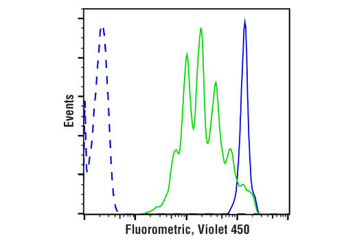
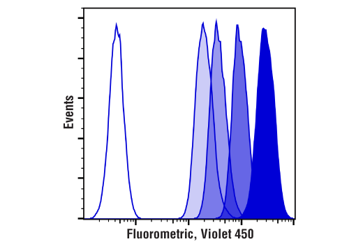
Due to their inherent metabolic stability once inside a cell, fluorescent proliferation dyes partition in an equal manner between daughter cells during the M phase of the cell cycle. This allows the principle of dye dilution to be leveraged as a means to trace multiple rounds of cell proliferation using flow cytometry. Added benefits of proliferation dyes are that they are non-radioactive and do not require cells to be actively synthesizing DNA for efficient uptake (1-3).
Except as otherwise expressly agreed in a writing signed by a legally authorized representative of CST, the following terms apply to Products provided by CST, its affiliates or its distributors. Any Customer's terms and conditions that are in addition to, or different from, those contained herein, unless separately accepted in writing by a legally authorized representative of CST, are rejected and are of no force or effect.
Products are labeled with For Research Use Only or a similar labeling statement and have not been approved, cleared, or licensed by the FDA or other regulatory foreign or domestic entity, for any purpose. Customer shall not use any Product for any diagnostic or therapeutic purpose, or otherwise in any manner that conflicts with its labeling statement. Products sold or licensed by CST are provided for Customer as the end-user and solely for research and development uses. Any use of Product for diagnostic, prophylactic or therapeutic purposes, or any purchase of Product for resale (alone or as a component) or other commercial purpose, requires a separate license from CST. Customer shall (a) not sell, license, loan, donate or otherwise transfer or make available any Product to any third party, whether alone or in combination with other materials, or use the Products to manufacture any commercial products, (b) not copy, modify, reverse engineer, decompile, disassemble or otherwise attempt to discover the underlying structure or technology of the Products, or use the Products for the purpose of developing any products or services that would compete with CST products or services, (c) not alter or remove from the Products any trademarks, trade names, logos, patent or copyright notices or markings, (d) use the Products solely in accordance with CST Product Terms of Sale and any applicable documentation, and (e) comply with any license, terms of service or similar agreement with respect to any third party products or services used by Customer in connection with the Products.