View Featured Offers >>
13296
Mitochondrial Membrane Potential Assay Kit (II)
Cellular Assay Kits
Assay Kit

Mitochondrial Membrane Potential Assay Kit (II) #13296

Citations (0)
Figure 1. Flow cytometric analysis of Jurkat cells, unlabeled (black) or labeled with 200 nM TMRE and treated with 0 μM CCCP (blue), 3.2 μM CCCP (green), or 80 μM CCCP (red).
Figure 2. Confocal immunofluorescent analysis of NIH/3T3 cells (2x10⁵ cell/ml) seeded in a 96-well black plate with a clear bottom and incubated overnight. Cells were untreated or treated with CCCP (400 μM, 20 min) followed by labeling with TMRE (200 nM, 30 min).
Figure 3. HeLa cells (3x10⁵ cell/ml) were treated with various concentrations of CCCP for 15 minutes prior to labeling with 200 nM TMRE.
To Purchase # 13296
Cat. # Size Qty. Price
13296S
1 Kit  (500 assays (96 well format))

Product Includes Quantity (with Count) Solution Color
TMRE 1 x 29 µg Purple
CCCP 1 x 100 µl Yellow
Phosphate Buffered Saline (PBS-20X) 9808 1 x 25 ml

Protocol

PRINT

View >Collapse >

#13296 Mitochondrial Membrane Potential Assay Kit (II) Protocol

A. Solutions and Reagents

  1. 1X PBS: Prepare by diluting 20X PBS (included in each kit, #9808) in reverse osmosis deionized (RODI) or equivalently purified water.

    NOTE: For flow cytometry, adding 0.5% BSA to wash buffer may help to prevent cell loss during the process.

  2. TMRE: Add 55 µl DMSO (#12611) to each vial of TMRE to make a 1000µM stock solution. Each vial includes enough TMRE for five 96-well plates (0.1 µl/well) or 50 flow cytometry assays (10 µl/assay). Aliquot if desired and store at -20°C.
    Dilute TMRE to 1:500 with full cell culture medium to make 10X TMRE Labeling Solution. 2 µM is used in this protocol; 0.1 to 10 µM is recommended based on different cell lines.
  3. CCCP: If CCCP is used as positive control, allow CCCP solution to equilibrate to room temperature before use.

B. Test Procedure

For Suspension Cells

  1. Suspend cells in warm medium or PBS at 1 x 10⁶ cell/ml. Prepare 1 ml aliquots; each 1 ml cell aliquot is one assay point. Make sure there are enough cells for your experiment. For example, if one compound is going to be assayed at three different concentrations, a total of 4 x 1 ml samples will be needed (this includes a positive control).
  2. Add test compound(s) to sample tubes at desired concentration and incubate cells for desired time. For best results, a compound titration and incubation time course can help to determine the best assay conditions. To prepare the positive control (mitochondrial membrane potential loss), add 1 µl of 50 mM CCCP (supplied with this kit) to the control tube for a 50 µM final concentration; incubate cells at 37°C for 15 min.
  3. Add 100 µl of the 2 µM TMRE Labeling Solution to each sample (200 nM final concentration) and incubate cells in the incubator (37°C and 5% CO2) for 15 to 30 min.

    Note: 200 nM TMRE is recommended in this protocol. For best results, a titration of TMRE is recommended.

  4. Centrifuge sample at 300 g for 5 min, then remove the supernatant.
  5. Wash cells once with 1 ml warm 1X PBS wash buffer, repeat step 4.
  6. Resuspend cells into 1000 µl warm 1X PBS.
  7. Analyze sample on a flow cytometer. If samples are analyzed on plate reader, transfer 100 µl/ cell suspension/well to a black 96-well plate with a clear bottom and read plate on the plate reader. The settings are excitation about 550 nm and emission about 580 nm.

For Adherent Cells

  1. Plate cells to a 96-well plate in warm culture medium and culture cells in incubator overnight to allow cells to attach to the plate. A typical cell number is between 1-5 x 10⁴ cells/ well. A cell number titration may be necessary for optimal results.
  2. Aspirate the medium from the plate and add test compounds in growth medium or 1X PBS to plate at 100 µl/well at desired concentration and incubate cells for desired time. Compound titration and incubation time course can help to determine the best assay conditions. For positive control (mitochondrial membrane potential loss), add CCCP (supplied with this kit) to the control wells for a 50 µM final concentration and incubate cells at 37°C for 15 min.

    Example: Add 1 µl of 50 mM stock CCCP to 100 µl medium to make 500 µM CCCP; then add 10 µl of this 500 µM CCCP to each well containing 100 µl medium to get final concentration of 50 µM.

  3. Add 10 µl of 2 µM TMRE Labeling Solution to each well to get a final concentration of 200 nM and place plate in an incubator (37°C and 5% CO2) for 20 min.

    Note: 200 nM TMRE is recommended in this protocol. For best results, a titration of TMRE is recommended.

  4. Aspirate the solution from the plate.
  5. Wash plate 3 times with warm 1X PBS and then add 100 µl/well 1X PBS to the plate.
  6. Analyze samples on the plate reader. The settings are excitation about 550 nm and emission about 580 nm.

posted June 2020

Protocol Id: 2057

Product Description

The Mitochondrial Membrane Potential Assay Kit (II) is a fluorescent assay that detects the mitochondrial membrane potential in living cells. The kit includes the cationic dye TMRE (tetramethylrhodamine ethyl ester perchlorate) and a mitochondrial membrane potential disruptor CCCP (carbonyl cyanide 3-chlorophenylhydrazone). TMRE is a cell membrane permeable, fluorescent dye that accumulates in intact mitochondria. Depolarized or inactive mitochondria exhibit decreased membrane potential, resulting in reduced TMRE accumulation.

Specificity / Sensitivity

The Mitochondrial Membrane Potential Assay Kit (II) is expected to detect the mitochondrial membrane potential in living cells cross all species. For the best result, a cell number titration is recommended when using a plate-reader and a 96-well plate.

Species Reactivity:

All Species Expected

Background

Mitochondria are the main power house in cells and play important roles in processes such as steroid metabolism, calcium homeostasis, apoptosis and cellular proliferation. Mitochondrial membrane potential is a key indicator of its function and cell health (1,2). The dissipation of mitochondrial membrane potential is established as an early indicator for apoptosis (3).

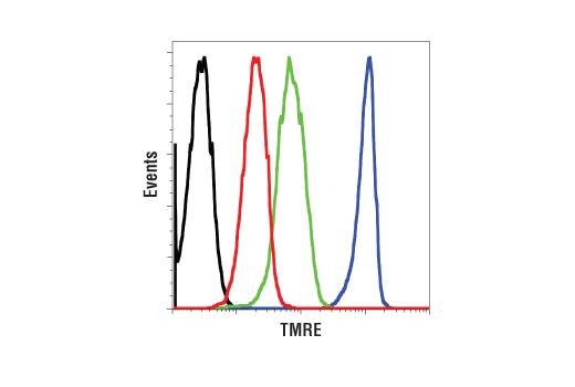
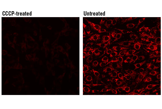
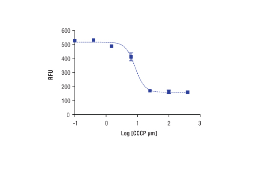
TMRE (tetramethylrhodamine, ethyl ester) is a cell membrane permeable cationic dye. In normal cells, TMRE accumulates in the mitochondria in response to their high membrane potential and negative charge. When excited at 550 nm, TMRE emits an orange-red fluorescence with a maximum at 575 nm (orange-red). Cells that have lost membrane potential or mitochondria activity cannot accumulate TMRE. Therefore, the fluorescence intensity of the orange-red emission can be used to measure mitochondria membrane potential and is an indicator for cell health (4).

Limited Uses

Except as otherwise expressly agreed in a writing signed by a legally authorized representative of CST, the following terms apply to Products provided by CST, its affiliates or its distributors. Any Customer's terms and conditions that are in addition to, or different from, those contained herein, unless separately accepted in writing by a legally authorized representative of CST, are rejected and are of no force or effect.

Products are labeled with For Research Use Only or a similar labeling statement and have not been approved, cleared, or licensed by the FDA or other regulatory foreign or domestic entity, for any purpose. Customer shall not use any Product for any diagnostic or therapeutic purpose, or otherwise in any manner that conflicts with its labeling statement. Products sold or licensed by CST are provided for Customer as the end-user and solely for research and development uses. Any use of Product for diagnostic, prophylactic or therapeutic purposes, or any purchase of Product for resale (alone or as a component) or other commercial purpose, requires a separate license from CST. Customer shall (a) not sell, license, loan, donate or otherwise transfer or make available any Product to any third party, whether alone or in combination with other materials, or use the Products to manufacture any commercial products, (b) not copy, modify, reverse engineer, decompile, disassemble or otherwise attempt to discover the underlying structure or technology of the Products, or use the Products for the purpose of developing any products or services that would compete with CST products or services, (c) not alter or remove from the Products any trademarks, trade names, logos, patent or copyright notices or markings, (d) use the Products solely in accordance with CST Product Terms of Sale and any applicable documentation, and (e) comply with any license, terms of service or similar agreement with respect to any third party products or services used by Customer in connection with the Products.

For Research Use Only. Not for Use in Diagnostic Procedures.
Cell Signaling Technology is a trademark of Cell Signaling Technology, Inc.
All other trademarks are the property of their respective owners. Visit our Trademark Information page.