View Featured Offers >>
8794
SAPK/JNK Kinase Assay Kit (Nonradioactive)
Cellular Assay Kits
Assay Kit

SAPK/JNK Kinase Assay Kit (Nonradioactive) #8794

Citations (1)
SAPK-induced phosphorylation of c-Jun was measured by quantitative immunoblotting using Phospho-c-Jun (Ser63) Antibody (upper) and c-Jun (L70B11) Mouse mAb #2315 (lower). Lysate titrations were performed by immuoprecipitating phospho-SAPK/JNK from the indicated amounts of HeLa cell extracts, untreated or UV-treated. Phosphorylation of c-Jun at Ser63 is observed in UV-treated lysates.
Inquiry Info.# 8794

Protocol

PRINT

View >Collapse >

Western Blotting Protocol

For western blots, incubate membrane with diluted primary antibody in 5% w/v BSA, 1X TBS, 0.1% Tween® 20 at 4°C with gentle shaking, overnight.

NOTE: Please refer to primary antibody product webpage for recommended antibody dilution.

A. Solutions and Reagents

From sample preparation to detection, the reagents you need for your Western Blot are now in one convenient kit: #12957 Western Blotting Application Solutions Kit

NOTE: Prepare solutions with reverse osmosis deionized (RODI) or equivalent grade water.

  1. 20X Phosphate Buffered Saline (PBS): (#9808) To prepare 1 L 1X PBS: add 50 ml 20X PBS to 950 ml dH2O, mix.
  2. 10X Tris Buffered Saline (TBS): (#12498) To prepare 1 L 1X TBS: add 100 ml 10X to 900 ml dH2O, mix.
  3. 1X SDS Sample Buffer: Blue Loading Pack (#7722) or Red Loading Pack (#7723) Prepare fresh 3X reducing loading buffer by adding 1/10 volume 30X DTT to 1 volume of 3X SDS loading buffer. Dilute to 1X with dH2O.
  4. 10X Tris-Glycine SDS Running Buffer: (#4050) To prepare 1 L 1X running buffer: add 100 ml 10X running buffer to 900 ml dH2O, mix.
  5. 10X Tris-Glycine Transfer Buffer: (#12539) To prepare 1 L 1X Transfer Buffer: add 100 ml 10X Transfer Buffer to 200 ml methanol + 700 ml dH2O, mix.
  6. 10X Tris Buffered Saline with Tween® 20 (TBST): (#9997) To prepare 1 L 1X TBST: add 100 ml 10X TBST to 900 ml dH2O, mix.
  7. Nonfat Dry Milk: (#9999).
  8. Blocking Buffer: 1X TBST with 5% w/v nonfat dry milk; for 150 ml, add 7.5 g nonfat dry milk to 150 ml 1X TBST and mix well.
  9. Wash Buffer: (#9997) 1X TBST.
  10. Bovine Serum Albumin (BSA): (#9998).
  11. Primary Antibody Dilution Buffer: 1X TBST with 5% BSA; for 20 ml, add 1.0 g BSA to 20 ml 1X TBST and mix well.
  12. Biotinylated Protein Ladder Detection Pack: (#7727).
  13. Blue Prestained Protein Marker, Broad Range (11-250 kDa): (#59329).
  14. Blotting Membrane and Paper: (#12369) This protocol has been optimized for nitrocellulose membranes. Pore size 0.2 µm is generally recommended.
  15. Secondary Antibody Conjugated to HRP: Anti-rabbit IgG, HRP-linked Antibody (#7074).
  16. Detection Reagent: SignalFire™ ECL Reagent (#6883).

B. Protein Blotting

A general protocol for sample preparation.

  1. Treat cells by adding fresh media containing regulator for desired time.
  2. Aspirate media from cultures; wash cells with 1X PBS; aspirate.
  3. Lyse cells by adding 1X SDS sample buffer (100 µl per well of 6-well plate or 500 µl for a 10 cm diameter plate). Immediately scrape the cells off the plate and transfer the extract to a microcentrifuge tube. Keep on ice.
  4. Sonicate for 10–15 sec to complete cell lysis and shear DNA (to reduce sample viscosity).
  5. Heat a 20 µl sample to 95–100°C for 5 min; cool on ice.
  6. Microcentrifuge for 5 min.
  7. Load 20 µl onto SDS-PAGE gel (10 cm x 10 cm).

    NOTE: Loading of prestained molecular weight markers (#59329, 10 µl/lane) to verify electrotransfer and biotinylated protein ladder (#7727, 10 µl/lane) to determine molecular weights are recommended.

  8. Electrotransfer to nitrocellulose membrane (#12369).

C. Membrane Blocking and Antibody Incubations

NOTE: Volumes are for 10 cm x 10 cm (100 cm2) of membrane; for different sized membranes, adjust volumes accordingly.

I. Membrane Blocking

  1. (Optional) After transfer, wash nitrocellulose membrane with 25 ml TBS for 5 min at room temperature.
  2. Incubate membrane in 25 ml of blocking buffer for 1 hr at room temperature.
  3. Wash three times for 5 min each with 15 ml of TBST.

II. Primary Antibody Incubation

  1. Incubate membrane and primary antibody (at the appropriate dilution and diluent as recommended in the product webpage) in 10 ml primary antibody dilution buffer with gentle agitation overnight at 4°C.
  2. Wash three times for 5 min each with 15 ml of TBST.
  3. Incubate membrane with Anti-rabbit IgG, HRP-linked Antibody (#7074 at 1:2000) and anti-biotin, HRP-linked Antibody (#7075 at 1:1000–1:3000) to detect biotinylated protein markers in 10 ml of blocking buffer with gentle agitation for 1 hr at room temperature.
  4. Wash three times for 5 min each with 15 ml of TBST.
  5. Proceed with detection (Section D).

D. Detection of Proteins

Directions for Use:

  1. Wash membrane-bound HRP (antibody conjugate) three times for 5 minutes in TBST.
  2. Prepare 1X SignalFire™ ECL Reagent (#6883) by diluting one part 2X Reagent A and one part 2X Reagent B (e.g. for 10 ml, add 5 ml Reagent A and 5 ml Reagent B). Mix well.
  3. Incubate substrate with membrane for 1 minute, remove excess solution (membrane remains wet), wrap in plastic and expose to X-ray film.

* Avoid repeated exposure to skin.

posted June 2005

revised June 2020

Protocol Id: 10

Immunoprecipitation for Analysis by Western Blotting

This protocol is intended for immunoprecipitation of native proteins for analysis by western immunoblot or kinase activity.

A. Solutions and Reagents

NOTE: Prepare solutions with reverse osmosis deionized (RODI) or equivalently purified water.

  1. 20X Phosphate Buffered Saline (PBS): (#9808).
  2. 10X Cell Lysis Buffer: (#9803) 20 mM Tris (pH 7.5), 150 mM NaCl, 1 mM EDTA, 1 mM EGTA, 1% Triton X-100, 2.5 mM Sodium pyrophosphate, 1 mM β-glycerophosphate, 1 mM Na3VO4, 1 μg/ml Leupeptin

    NOTE: CST recommends adding 1 mM PMSF (#8553) before use*.

  1. 3X SDS Sample Buffer: (#7722) 187.5 mM Tris-HCl (pH 6.8 at 25°C), 6% w/v SDS, 30% glycerol, 150 mM DTT, 0.03% w/v bromophenol blue
  2. 10X Kinase Buffer (for kinase assays): (#9802) To Prepare 1 ml of 1X kinase buffer, add 100 µl 10X kinase buffer to 900 µl dH2O, mix.
  3. ATP (10 mM) (for kinase assays): (#9804) To prepare 0.5 ml of ATP (200 µM), add 10 µl ATP (10 mM) to 490 µl 1X kinase buffer.

B. Preparing Cell Lysates

  1. Aspirate media. Treat cells by adding fresh media containing regulator for desired time.
  2. To harvest cells under nondenaturing conditions, remove media and rinse cells once with ice-cold PBS.
  3. Remove PBS and add 0.5 ml 1X ice-cold cell lysis buffer to each plate (10 cm) and incubate the plates on ice for 5 minutes.
  4. Scrape cells off the plates and transfer to microcentrifuge tubes. Keep on ice.
  5. Sonicate samples on ice three times for 5 seconds each.
  6. Microcentrifuge for 10 minutes at 4°C, 14,000 x g, and transfer the supernatant to a new tube. If necessary, lysate can be stored at –80°C.

C. Immunoprecipitation

  1. Take 200 μl cell lysate and add 10 μl of the immobilized antibody, incubate with rotation overnight at 4°C.
  2. Microcentrifuge for 30 seconds at 4°C. Wash pellet five times with 500 μl of 1X cell lysis buffer. Keep on ice during washes.
  3. Proceed to sample analysis by western blotting or kinase activity (section D).

D. Sample Analysis

Proceed to one of the following specific set of steps.

For Analysis by Western Immunoblotting

  1. Resuspend the pellet with 20 µl 3X SDS sample buffer. Vortex, then microcentrifuge for 30 sec at 14,000 x g.
  2. Heat the sample to 95–100°C for 2-5 min and microcentrifuge for 1 min at 14,000 x g.
  3. Load the sample (15–30 µl) on a 4–20% gel for SDS-PAGE.
  4. Analyze sample by western blot (see Western Immunoblotting Protocol).

NOTE: When using primary antibodies produced in rabbit to detect proteins with a molecular weight in the range of 50 kDa, we recommend using Mouse Anti-Rabbit IgG (Light-Chain Specific) (D4W3E) mAb (#45262) or Mouse Anti-Rabbit IgG (Conformation Specific) (L27A9) mAb (#3678) (or HRP conjugate #5127) as a secondary antibody to minimize interference produced by denatured rabbit heavy chain. For proteins with a molecular weight in the range of 25 kDa, Mouse Anti-Rabbit IgG (Conformation Specific) (L27A9) mAb (#3678) (or HRP conjugate #5127) is recommended to minimize interference produced by denatured mouse light chain.

When using primary antibodies produced in mouse to detect proteins with a molecular weight in the range of 50 kDa, we recommend using Rabbit Anti-Mouse IgG (Light Chain Specific) (D3V2A) mAb (HRP Conjugate) (#58802) as a secondary antibody to minimize interference produced by denatured mouse heavy chain.

For Analysis by Kinase Assay

  1. Wash pellet twice with 500 µl 1X kinase buffer. Keep on ice.
  2. Suspend pellet in 40 µl 1X kinase buffer supplemented with 200 µM ATP and appropriate substrate.
  3. Incubate for 30 min at 30°C.
  4. Terminate reaction with 20 µl 3X SDS sample buffer. Vortex, then microcentrifuge for 30 sec.
  5. Transfer supernatant containing phosphorylated substrate to another tube.
  6. Heat the sample to 95–100°C for 2–5 min and microcentrifuge for 1 min at 14,000 x g.
  7. Load the sample (15–30 µl) on SDS-PAGE (4–20%).

posted December 2007

revised October 2021

Protocol Id: 27

Product Description

The SAPK/JNK Kinase Assay Kit (Nonradioactive) provides all the reagents necessary to measure SAPK/JNK activity in the cell. A phospho-SAPK/JNK Rabbit mAb linked to agarose beads is used to pull down SAPK enzyme from cell extracts. Upon addition of kinase buffer, c-Jun fusion protein and ATP, SAPK phosphorylates the c-Jun substrate. Phospho-c-Jun (Ser63) Antibody can then be used to measure SAPK activity by immunoblotting.

Specificity / Sensitivity

Species Reactivity:

Human, Mouse, Rat, Monkey, Pig

Background

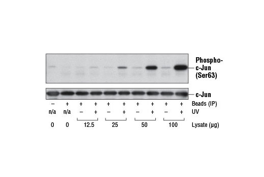
The stress-activated protein kinase/Jun-amino-terminal kinase SAPK/JNK is potently and preferentially activated by a variety of environmental stresses, including UV and gamma radiation, ceramides, inflammatory cytokines, and in some instances, growth factors and GPCR agonists (1-6). As with the other MAPKs, the core signaling unit is composed of a MAPKKK, typically MEKK1-MEKK4, or by one of the mixed lineage kinases (MLKs), which phosphorylate and activate MKK4/7. Upon activation, MKKs phosphorylate and activate the SAPK/JNK kinase (2). Stress signals are delivered to this cascade by small GTPases of the Rho family (Rac, Rho, cdc42) (3). Both Rac1 and cdc42 mediate the stimulation of MEKKs and MLKs (3). Alternatively, MKK4/7 can be activated in a GTPase-independent mechanism via stimulation of a germinal center kinase (GCK) family member (4). There are three SAPK/JNK genes each of which undergoes alternative splicing, resulting in numerous isoforms (3). SAPK/JNK, when active as a dimer, can translocate to the nucleus and regulate transcription through its effects on c-Jun, ATF-2, and other transcription factors (3,5).

Pathways

Explore pathways related to this product.

Limited Uses

Except as otherwise expressly agreed in a writing signed by a legally authorized representative of CST, the following terms apply to Products provided by CST, its affiliates or its distributors. Any Customer's terms and conditions that are in addition to, or different from, those contained herein, unless separately accepted in writing by a legally authorized representative of CST, are rejected and are of no force or effect.

Products are labeled with For Research Use Only or a similar labeling statement and have not been approved, cleared, or licensed by the FDA or other regulatory foreign or domestic entity, for any purpose. Customer shall not use any Product for any diagnostic or therapeutic purpose, or otherwise in any manner that conflicts with its labeling statement. Products sold or licensed by CST are provided for Customer as the end-user and solely for research and development uses. Any use of Product for diagnostic, prophylactic or therapeutic purposes, or any purchase of Product for resale (alone or as a component) or other commercial purpose, requires a separate license from CST. Customer shall (a) not sell, license, loan, donate or otherwise transfer or make available any Product to any third party, whether alone or in combination with other materials, or use the Products to manufacture any commercial products, (b) not copy, modify, reverse engineer, decompile, disassemble or otherwise attempt to discover the underlying structure or technology of the Products, or use the Products for the purpose of developing any products or services that would compete with CST products or services, (c) not alter or remove from the Products any trademarks, trade names, logos, patent or copyright notices or markings, (d) use the Products solely in accordance with CST Product Terms of Sale and any applicable documentation, and (e) comply with any license, terms of service or similar agreement with respect to any third party products or services used by Customer in connection with the Products.

For Research Use Only. Not for Use in Diagnostic Procedures.
Cell Signaling Technology is a trademark of Cell Signaling Technology, Inc.
All other trademarks are the property of their respective owners. Visit our Trademark Information page.