Product Information
Human
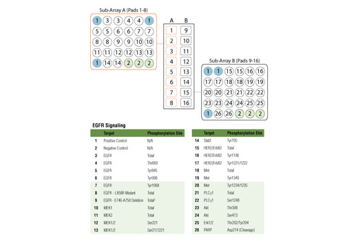
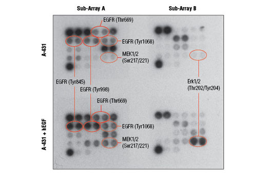
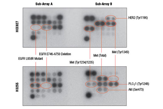
The Epidermal Growth Factor Receptor (EGFR) is a receptor tyrosine kinase (RTK) that constitutes an important disease driver, as well as a validated drug target. The potency of EGFR in driving tumorigenesis can be attributed to its pleiotropic intracellular signaling. Activated EGFR initiates a wide range of signaling modules and switches such as the Ras-Erk/MAP kinase, Akt, Src, Stat, and PKC. Two of the most common EGFR mutations occurring in lung cancer are the E746-A750 deletion and L858R point mutation. This array utilizes unique antibodies made by Cell Signaling Technology that are sensitive to each of these EGFR mutants, allowing specific target detection in cell extracts.
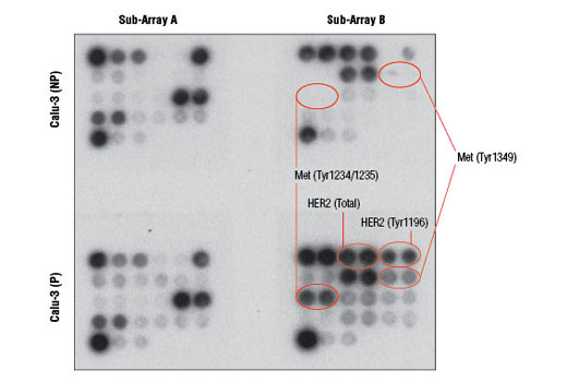
EGFR can interact and heterodimerize with other RTKs. HER2 (also known as ErbB2) is an oncogenic RTK belonging to the EGFR/HER family of RTKs and is an important heterodimerization partner of all HER family members. Another prominent heterodimerization partner of EGFR is c-Met. c-Met is an RTK serving as a receptor for the hepatocyte growth factor (HGF). c-Met can induce cell scattering, migration, and invasion. It has been shown that c-Met is responsible for some cases of tumor resistance to EGFR-targeted therapies and is a contributing factor to tumor metastasis.
PLCγ is a phosphoinositide-specific phospholipase. EGFR can activate PLCγ that, in turn, hydrolyzes phosphoinositide phospholipids residing within the inner leaflet of the plasma membrane. This hydrolysis generates two important second messengers: inositol 1,4,5-triphosphate (IP3) and diacylglycerol (DAG). IP3 causes calcium mobilization from intracellular storage pools, while DAG (together with calcium) activates PKC. MEK1 is a dual-specificity protein kinase and serves as the MAP kinase kinase for Erk1 and Erk2. Upon EGFR activation, MEK1 is phosphorylated by Raf and, in turn, phosphorylates the Erk kinases at Thr202 and Tyr204, leading to their activation. Activated Erk MAP kinase is a major signaling node with a multitude of substrates and primarily transmits growth and proliferation signals. Akt is another important protein kinase downstream of EGFR. Akt is activated by many RTKs and has a large number of intracellular substrates. Akt generates anabolic growth and survival signals. Stat3 is activated in response to EGFR stimulation, as well as in response to activation of a variety of cytokine receptors. Stat3 is a well-established oncogene that is also a transcription factor.
The oncogenic signals generated by activated EGFR are a focus of intense drug discovery efforts. It has become clear that in many cases a single agent inhibiting only one target is unable to cause tumor cell death in vivo. To monitor the blockade of EGFR signals alongside markers of cell death, cleaved PARP is included in this array. PARP is an enzyme involved in DNA repair. As a part of the apoptotic process, PARP is irreversibly inactivated by endoproteolytic cleavage executed by activated cell death proteases, such as caspase-3 and caspasae-7.
Explore pathways related to this product.
STRING - Known and Predicted Protein-Protein Interactions.
Except as otherwise expressly agreed in a writing signed by a legally authorized representative of CST, the following terms apply to Products provided by CST, its affiliates or its distributors. Any Customer's terms and conditions that are in addition to, or different from, those contained herein, unless separately accepted in writing by a legally authorized representative of CST, are rejected and are of no force or effect.
Products are labeled with For Research Use Only or a similar labeling statement and have not been approved, cleared, or licensed by the FDA or other regulatory foreign or domestic entity, for any purpose. Customer shall not use any Product for any diagnostic or therapeutic purpose, or otherwise in any manner that conflicts with its labeling statement. Products sold or licensed by CST are provided for Customer as the end-user and solely for research and development uses. Any use of Product for diagnostic, prophylactic or therapeutic purposes, or any purchase of Product for resale (alone or as a component) or other commercial purpose, requires a separate license from CST. Customer shall (a) not sell, license, loan, donate or otherwise transfer or make available any Product to any third party, whether alone or in combination with other materials, or use the Products to manufacture any commercial products, (b) not copy, modify, reverse engineer, decompile, disassemble or otherwise attempt to discover the underlying structure or technology of the Products, or use the Products for the purpose of developing any products or services that would compete with CST products or services, (c) not alter or remove from the Products any trademarks, trade names, logos, patent or copyright notices or markings, (d) use the Products solely in accordance with CST Product Terms of Sale and any applicable documentation, and (e) comply with any license, terms of service or similar agreement with respect to any third party products or services used by Customer in connection with the Products.