View Featured Offers >>
12116
PathScan® Phospho-Acetyl-CoA Carboxylase (Ser79) Chemiluminescent Sandwich ELISA Kit
ELISA Kits
ELISA Kit

PathScan® Phospho-Acetyl-CoA Carboxylase (Ser79) Chemiluminescent Sandwich ELISA Kit #12116

Citations (0)
Figure 1. Relationship between protein concentration of lysates from untreated and H2O2-treated Hep G2 cells and immediate light generation with chemiluminescent substrate is shown. Hep G2 cells (80-90% confluent) were treated with H2O2 (10 mM, 10 min) and lysed with PathScan® Sandwich ELISA Lysis Buffer (1X) #7018. Graph inset corresponding to the shaded area shows high sensitivity and a linear response at the low protein concentration range.
To Purchase # 12116
Cat. # Size Qty. Price
12116C
1 Kit  (96 assays)

Important Ordering Details

Custom Ordering Details:

When ordering five or more kits, please contact us for processing time and pricing.

Looking for this ELISA kit in a 384-well format? Inquire for availability, processing time, and pricing.

Supporting Data

REACTIVITY H

Application Key:

  • WB-Western Blot
  • IP-Immunoprecipitation
  • IHC-Immunohistochemistry
  • ChIP-Chromatin Immunoprecipitation
  • C&R-CUT&RUN
  • C&T-CUT&Tag
  • DB-Dot Blot
  • eCLIP-eCLIP
  • IF-Immunofluorescence
  • F-Flow Cytometry

Species Cross-Reactivity Key:

  • H-Human
  • M-Mouse
  • R-Rat
  • Hm-Hamster
  • Mk-Monkey
  • Vir-Virus
  • Mi-Mink
  • C-Chicken
  • Dm-D. melanogaster
  • X-Xenopus
  • Z-Zebrafish
  • B-Bovine
  • Dg-Dog
  • Pg-Pig
  • Sc-S. cerevisiae
  • Ce-C. elegans
  • Hr-Horse
  • GP-Guinea Pig
  • Rab-Rabbit
  • All-All Species Expected
Product Includes Volume Solution Color
Phospho-ACC (Ser79) Rabbit mAb Coated Microwells 96 tests
Acetyl-CoA Carboxylase Mouse Detection mAb 1 ea Green (Lyophilized)
Anti-mouse IgG, HRP-linked Antibody (ELISA Formulated) 1 ea Red (Lyophilized)
Detection Antibody Diluent 5.5 ml Green
HRP Diluent 5.5 ml Red
Luminol/Enhancer Solution 3 ml
Stable Peroxide Buffer 3 ml
Sealing Tape 2 ea
ELISA Wash Buffer (20X) 9801 25 ml
ELISA Sample Diluent 25 ml Blue
PathScan® Sandwich ELISA Lysis Buffer (1X) 7018 30 ml

Product Description

The PathScan® Phospho-Acetyl-CoA Carboxylase (Ser79) Chemiluminescent Sandwich ELISA Kit is a solid phase sandwich enzyme-linked immunosorbent assay (ELISA) that detects endogenous levels of acetyl-CoA carboxylase (ACC) protein phosphorylated at Ser79 with a chemiluminescent readout. Chemiluminescent ELISAs often have a wider dynamic range and higher sensitivity than conventional chromogenic detection. This chemiluminescent ELISA, which is offered in low volume microplates, shows increased signal and sensitivity while using smaller samples. A Phospho-ACC (Ser79) Rabbit Antibody has been coated onto the microwells. After incubation with cell lysates, phospho-ACC protein is captured by the coated antibody. Following extensive washing, an ACC Mouse Detection mAb is added to detect the captured ACC protein. Anti-mouse IgG, HRP-linked Antibody is then used to recognize the bound detection antibody. Chemiluminescent reagent is added for signal development. The magnitude of light emission, measured in relative light units (RLU), is proportional to the quantity of phospho-ACC (Ser79) protein.

*Antibodies in this kit are custom formulations specific to kit.

Protocol

PRINT

View >Collapse >

ELISA Chemiluminescent (Lyophilized)

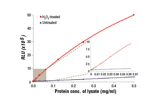
NOTE: Refer to product-specific datasheets for assay incubation temperature. This chemiluminescent ELISA is offered in low volume microplates. Only 50 μl of samples or reagents are required in each microwell.

A. Solutions and Reagents

NOTE: Prepare solutions with purified water.

  1. Microwell strips: Bring all to room temperature before use.
  2. Detection Antibody: Supplied lyophilized as a green colored cake or powder. Add 0.5 ml of Detection Antibody Diluent (green solution) to yield a concentrated stock solution. Incubate at room temperature for 5 min with occasional gentle mixing to fully reconstitute. To make the final working solution, add the full 0.5 ml volume of reconstituted Detection Antibody to 5.0 ml of Detection Antibody Diluent in a clean tube and gently mix. Unused working solution may be stored for 4 weeks at 4°C.
  3. HRP-Linked Antibody*: Supplied lyophilized as a red colored cake or powder. Add 0.5 ml of HRP Diluent (red solution) to yield a concentrated stock solution. Incubate at room temperature for 5 min with occasional gentle mixing to fully reconstitute. To make the final working solution, add the full 0.5 ml volume of reconstituted HRP-Linked Antibody to 5.0 ml of HRP Diluent in a clean tube and gently mix. Unused working solution may be stored for 4 weeks at 4°C.
  4. Detection Antibody Diluent: Green colored diluent for reconstitution and dilution of the detection antibody (5.5 ml provided).
  5. HRP Diluent: Red colored diluent for reconstitution and dilution of the HRP‑Linked Antibody (5.5 ml provided).
  6. Sample Diluent: Blue colored diluent for dilution of cell lysates.
  7. 1X Wash Buffer: Prepare by diluting 20X Wash Buffer (included in each PathScan® Sandwich ELISA Kit) in purified water.
  8. Cell Lysis Buffer: PathScan® Sandwich ELISA Lysis Buffer (1X) #7018: This buffer can be stored at 4°C for short-term use (1–2 weeks). Recommended: Add 1 mM phenylmethylsulfonyl fluoride (PMSF) immediately before use.
  9. Luminol/Enhancer Solution and Stable Peroxide Buffer.

*NOTE: Some PathScan® ELISA Kits may include HRP-Linked Streptavidin in place of HRP-Linked Antibody.

B. Preparing Cell Lysates

For adherent cells.

  1. Aspirate media when the culture reaches 80–90% confluence. Treat cells by adding fresh media containing regulator for desired time.
  2. Remove media and rinse cells once with ice-cold 1X PBS.
  3. Remove PBS and add 0.5 ml to 1 ml ice-cold PathScan® Sandwich ELISA Lysis Buffer (1X) #7018 plus 1 mM PMSF to each plate (10 cm diameter) and incubate the plate on ice for 2 min.
  4. Collect cell lysate in a clean tube.
  5. Centrifuge for 10 min (14,000 x g) at 4°C and transfer the supernatant to a new tube. Store supernatant at -80°C in single-use aliquots.

For suspension cells

  1. Remove media by low speed centrifugation (~1200 rpm) when the culture reaches 0.5–1.0 x 10⁶ viable cells/ml. Treat cells by adding fresh media containing regulator for desired time.
  2. Collect cells by low speed centrifugation (~1200 rpm) and wash once with 5–10 ml ice-cold 1X PBS.
  3. Cells harvested from 50 ml of growth media can be lysed in 2.0 ml of 1X Cell Lysis Buffer plus 1 mM PMSF.
  4. Resuspend the cell pellet and incubate the tube for 2 min.
  5. Microcentrifuge for 10 min (x14,000 rpm) at 4°C and transfer the supernatant to a new tube. The supernatant is the cell lysate. Store at −80°C in single-use aliquots.

C. Test Procedure

  1. After the microwell strips have reached room temperature, break off the required number of microwells. Place the microwells in the strip holder. Unused microwells must be resealed and stored at 4°C immediately.
  2. Cell lysates can be undiluted or diluted with Sample Diluent (supplied in each PathScan® Sandwich ELISA Kit, blue color). Individual datasheets for each kit provide a sensitivity curve that serves as a reference for selection of an appropriate starting lysate concentration. The sensitivity curve shows typical kit assay results across a range of lysate concentration points.
  3. Add 50 μl of each undiluted or diluted cell lysate to the appropriate well. Seal with tape and press firmly onto top of microwells. Incubate the plate for 2 hr at room temperature. Alternatively, the plate can be incubated overnight at 4°C.
  4. Gently remove the tape and wash wells:
    1. Discard plate contents into a receptacle.
    2. Wash 4 times with 1X Wash Buffer, 150 μl each time for each well.
    3. For each wash, strike plates on fresh towels hard enough to remove the residual solution in each well, but do not allow wells to completely dry at any time.
    4. Clean the underside of all wells with a lint-free tissue.
  5. Add 50 μl of reconstituted Detection Antibody (green color) to each well (refer to Section A, Step 2). Seal with tape and incubate the plate at room temperature for 1 hr.
  6. Repeat wash procedure (Section C, Step 4).
  7. Add 50 μl of reconstituted HRP-linked secondary antibody (red color) to each well (refer to Section A, Step 3). Seal with tape and incubate the plate at room temperature for 30 min.
  8. Repeat wash procedure (Section C, Step 4).
  9. Prepare Detection Reagent Working Solution by mixing equal parts Luminol/Enhancer Solution and Stable Peroxide Buffer.
  10. Add 50 μl of the Detection Reagent Working Solution to each well.
  11. Use a plate-based luminometer to measure Relative Light Units (RLU) at 425 nm within 1–10 min following addition of the substrate. Optimal signal intensity is achieved when read within 10 min.

posted November 2013

revised January 2016

Protocol Id: 203

Specificity / Sensitivity

PathScan® Phospho-Acetyl-CoA Carboxylase (Ser79) Chemiluminescent Sandwich ELISA Kit recognizes endogenous levels of phospho-ACC (Ser79) in human cells. This kit detects proteins from the indicated species, as determined through in-house testing, but may also detect homologous proteins from other species.

Background

Acetyl-CoA carboxylase (ACC) catalyzes the carboxylation of acetyl-CoA to malonyl-CoA (1). It is the key enzyme in the biosynthesis and oxidation of fatty acids (1). In rodents, the 265 kDa ACC1 (ACCα) form is primarily expressed in lipogenic tissues, while 280 kDa ACC2 (ACCβ) is the main isoform in oxidative tissues (1,2). However, in humans, ACC2 is the predominant isoform in both lipogenic and oxidative tissues (1,2). Phosphorylation by AMPK at Ser79 or by PKA at Ser1200 inhibits the enzymatic activity of ACC (3). ACC is a potential target of anti-obesity drugs (4,5).

Pathways

Explore pathways related to this product.

Limited Uses

Except as otherwise expressly agreed in a writing signed by a legally authorized representative of CST, the following terms apply to Products provided by CST, its affiliates or its distributors. Any Customer's terms and conditions that are in addition to, or different from, those contained herein, unless separately accepted in writing by a legally authorized representative of CST, are rejected and are of no force or effect.

Products are labeled with For Research Use Only or a similar labeling statement and have not been approved, cleared, or licensed by the FDA or other regulatory foreign or domestic entity, for any purpose. Customer shall not use any Product for any diagnostic or therapeutic purpose, or otherwise in any manner that conflicts with its labeling statement. Products sold or licensed by CST are provided for Customer as the end-user and solely for research and development uses. Any use of Product for diagnostic, prophylactic or therapeutic purposes, or any purchase of Product for resale (alone or as a component) or other commercial purpose, requires a separate license from CST. Customer shall (a) not sell, license, loan, donate or otherwise transfer or make available any Product to any third party, whether alone or in combination with other materials, or use the Products to manufacture any commercial products, (b) not copy, modify, reverse engineer, decompile, disassemble or otherwise attempt to discover the underlying structure or technology of the Products, or use the Products for the purpose of developing any products or services that would compete with CST products or services, (c) not alter or remove from the Products any trademarks, trade names, logos, patent or copyright notices or markings, (d) use the Products solely in accordance with CST Product Terms of Sale and any applicable documentation, and (e) comply with any license, terms of service or similar agreement with respect to any third party products or services used by Customer in connection with the Products.

For Research Use Only. Not for Use in Diagnostic Procedures.
Cell Signaling Technology is a trademark of Cell Signaling Technology, Inc.
PathScan is a registered trademark of Cell Signaling Technology, Inc.
All other trademarks are the property of their respective owners. Visit our Trademark Information page.