View Featured Offers >>
7866
PathScan® Tri-Methyl-Histone H3 (Lys27) Sandwich ELISA Kit
ELISA Kits
ELISA Kit

PathScan® Tri-Methyl-Histone H3 (Lys27) Sandwich ELISA Kit #7866

Citations (0)
Figure 1. Treatment of NIH/3T3 cells with trichostatin A (TSA) increases the acetylation of Histone H3 at Lys 9, detected by PathScan® Acetyl-Histone H3 (Lys9) Sandwich ELISA Kit #7121, and the di-methylation of Histone H3 at Lys4, detected by PathScan® Di-Methyl-Histone H3 (Lys4) Sandwich ELISA Kit #7124. However, TSA treatment does not affect the level of tri-methylation at Lys27, detected by PathScan® Tri-Methyl-Histone H3 (Lys27) Sandwich ELISA Kit #7866, or the level of total Histone H3, detected by Pathscan® Total Histone H3 Sandwich ELISA Kit #7253. NIH/3T3 cells (70-80% confluent) were treated for 16-18 hours with 0.4 μM TSA at 37ºC. Absorbance readings at 450 nm are shown in the top figure while the corresponding Western blots using Histone H3 Antibody #9715 (panel A), Acetyl-Histone H3 (Lys9) Antibody #9671 (panel B), Di-Methyl-Histone H3 (Lys4) (C64G9) Rabbit mAb #9725 (panel C) or Tri-Methyl-Histone H3 (Lys27) Antibody #9756 (panel D) are shown in the bottom figure.
Figure 2. The relationship between the protein concentration of the lysate from HeLa cells and the absorbance at 450 nm is shown.
Figure 3. Kit specificity as demonstrated by Western analysis of the ELISA microwell captured protein. Lysates were prepared from NIH/3T3 cells and incubated in microwells coated with the Tri-Methyl-Histone H3 (Lys27) capture antibody. Wells were washed, and the captured protein was solubilized in SDS gel loading buffer. Western blot analysis of NIH/3T3 cell starting lysate (lanes 1 & 2) and the captured protein (lane 3) was performed using Histone H3 Antibody #9715. The major band detected in the captured material corresponds to Histone H3 tri-methylated at Lys27 (lane 3).
Inquiry Info.# 7866

Supporting Data

REACTIVITY H M Mk

Application Key:

  • WB-Western Blot
  • IP-Immunoprecipitation
  • IHC-Immunohistochemistry
  • ChIP-Chromatin Immunoprecipitation
  • C&R-CUT&RUN
  • C&T-CUT&Tag
  • DB-Dot Blot
  • eCLIP-eCLIP
  • IF-Immunofluorescence
  • F-Flow Cytometry

Species Cross-Reactivity Key:

  • H-Human
  • M-Mouse
  • R-Rat
  • Hm-Hamster
  • Mk-Monkey
  • Vir-Virus
  • Mi-Mink
  • C-Chicken
  • Dm-D. melanogaster
  • X-Xenopus
  • Z-Zebrafish
  • B-Bovine
  • Dg-Dog
  • Pg-Pig
  • Sc-S. cerevisiae
  • Ce-C. elegans
  • Hr-Horse
  • GP-Guinea Pig
  • Rab-Rabbit
  • All-All Species Expected

Product Description

The PathScan® Tri-Methyl-Histone H3 (Lys27) Sandwich ELISA Kit is a solid phase sandwich enzyme-linked immunosorbent assay (ELISA) that detects endogenous levels of histone H3 when tri-methylated at Lys27. A Histone H3 Rabbit mAb has been coated onto the microwells. After incubation with cell lysates, histone H3 is captured by the coated antibody. Following extensive washing, biotinylated Tri-Methyl Histone H3 (Lys27) Rabbit Antibody is added to detect the captured histone H3 protein. HRP-linked streptavidin is then used to recognize the bound detection antibody. HRP substrate, TMB, is added to develop color. The magnitude of the absorbance for this developed color is proportional to the quantity of histone H3 tri-methylated at Lys27.
Antibodies in kit are custom formulations specific to kit.

Protocol

PRINT

View >Collapse >

ELISA Colorimetric

NOTE: Refer to product-specific datasheets or product webpage for assay incubation temperature.

A. Solutions and Reagents

NOTE: Prepare solutions with reverse osmosis deionized (RODI) or equivalent grade water.

  1. 20X Phosphate Buffered Saline (PBS): (#9808) To prepare 1 L PBS: add 50 ml 10X PBS to 950 ml dH2O, mix.
  2. Bring all microwell strips to room temperature before use.
  3. Prepare 1X Wash Buffer by diluting 20X Wash Buffer (included in each PathScan® Sandwich ELISA Kit) in dH2O.
  4. 1X Cell Lysis Buffer: 10X Cell Lysis Buffer (#9803): To prepare 10 ml of 1X Cell Lysis Buffer, add 1 ml of 10X Cell Lysis Buffer to 9 ml of dH2O, mix. Buffer can be stored at 4°C for short-term use (1–2 weeks).

    Recommended: Add 1 mM phenylmethylsulfonyl fluoride (PMSF) (#8553) immediately before use.

    NOTE: Refer to product-specific datasheet or webpage for lysis buffer recommendation.

  5. TMB Substrate: (#7004).
  6. STOP Solution: (#7002).

B. Preparing Cell Lysates

For adherent cells

  1. Aspirate media when the culture reaches 80–90% confluence. Treat cells by adding fresh media containing regulator for desired time.
  2. Remove media and rinse cells once with ice-cold 1X PBS.
  3. Remove PBS and add 0.5 ml ice-cold 1X cell lysis buffer plus 1 mM PMSF to each plate (10 cm diameter) and incubate the plate on ice for 5 min.
  4. Scrape cells off the plate and transfer to an appropriate tube. Keep on ice.
  5. Sonicate lysates on ice.
  6. Microcentrifuge for 10 min (x14,000 rpm) at 4°C and transfer the supernatant to a new tube. The supernatant is the cell lysate. Store at -80°C in single-use aliquots.

For suspension cells

  1. Remove media by low speed centrifugation (~1,200 rpm) when the culture reaches 0.5–1.0 x 10⁶ viable cells/ml. Treat cells by adding fresh media containing regulator for desired time.
  2. Collect cells by low speed centrifugation (~1,200 rpm) and wash once with 5–10 ml ice-cold 1X PBS.
  3. Cells harvested from 50 ml of growth media can be lysed in 2.0 ml of 1X cell lysis buffer plus 1 mM PMSF.
  4. Sonicate lysates on ice.
  5. Microcentrifuge for 10 min (x14,000 rpm) at 4°C and transfer the supernatant to a new tube. The supernatant is the cell lysate. Store at -80°C in single-use aliquots.

C. Test Procedure

  1. After the microwell strips have reached room temperature, break off the required number of microwells. Place the microwells in the strip holder. Unused microwells must be resealed in the storage bag and stored at 4°C immediately.
  2. Cell lysates can be undiluted or diluted with sample diluent (supplied in each PathScan® Sandwich ELISA Kit, blue color). Individual datasheets or product webpage for each kit provide information regarding an appropriate dilution factor for lysates and kit assay results.
  3. Add 100 µl of each undiluted or diluted cell lysate to the appropriate well. Seal with tape and press firmly onto top of microwells. Incubate the plate for 2 hr at 37°C. Alternatively, the plate can be incubated overnight at 4°C.
  4. Gently remove the tape and wash wells:
    1. Discard plate contents into a receptacle.
    2. Wash 4 times with 1X wash buffer, 200 µl each time per well.
    3. For each wash, strike plates on fresh paper towels hard enough to remove the residual solution in each well, but do not allow wells to completely dry at any time.
    4. Clean the underside of all wells with a lint-free tissue.
  5. Add 100 µl of detection antibody (green color) to each well. Seal with tape and incubate the plate at 37°C for 1 hr.
  6. Repeat wash procedure (Section C, Step 4).
  7. Add 100 µl of HRP-linked secondary antibody (red color) to each well. Seal with tape and incubate the plate for 30 min at 37°C.
  8. Repeat wash procedure (Section C, Step 4).
  9. Add 100 µl of TMB substrate to each well. Seal with tape and incubate the plate for 10 min at 37°C or 30 min at 25°C.
  10. Add 100 µl of STOP solution to each well. Shake gently for a few seconds.

    NOTE: Initial color of positive reaction is blue, which changes to yellow upon addition of STOP solution.

  11. Read results
    1. Visual Determination: Read within 30 min after adding STOP solution.
    2. Spectrophotometric Determination: Wipe underside of wells with a lint-free tissue. Read absorbance at 450 nm within 30 min after adding STOP solution.

posted June 2005

revised November 2013

Protocol Id: 21

Specificity / Sensitivity

CST's PathScan® Tri-Methyl-Histone H3 (Lys27) Sandwich ELISA Kit #7866 detects endogenous levels of histone H3 when tri-methylated at Lys27. As shown in Figure 1 using the Tri-Methyl-Histone H3 (Lys27) Sandwich ELISA Kit #7866, a high level of tri-methylation at Lys27 is detected on Histone H3 in NIH/3T3 cells. These levels are unchanged in response to TSA-treatment. The level of total histone H3 (modified and unmodified) remains unchanged as shown by Western analysis (Figure 1). Similar results are obtained when COS and Jurkat cells are treated with TSA (data not shown). Note: For this assay, it is recommended that lysates be thoroughly sonicated to ensure complete extraction of Histone H3 and an accurate absorbance reading. This kit detects proteins from the indicated species, as determined through in-house testing, but may also detect homologous proteins from other species.

Background

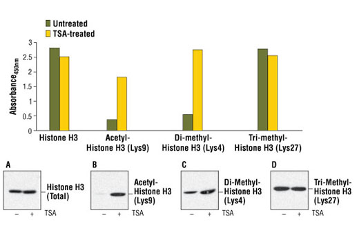
Modulation of chromatin structure plays an important role in the regulation of transcription in eukaryotes. The nucleosome, made up of DNA wound around eight core histone proteins (two each of H2A, H2B, H3, and H4), is the primary building block of chromatin (1). The amino-terminal tails of core histones undergo various posttranslational modifications, including acetylation, phosphorylation, methylation, and ubiquitination (2-5). These modifications occur in response to various stimuli and have a direct effect on the accessibility of chromatin to transcription factors and, therefore, gene expression (6). In most species, histone H2B is primarily acetylated at Lys5, 12, 15, and 20 (4,7). Histone H3 is primarily acetylated at Lys9, 14, 18, 23, 27, and 56. Acetylation of H3 at Lys9 appears to have a dominant role in histone deposition and chromatin assembly in some organisms (2,3). Phosphorylation at Ser10, Ser28, and Thr11 of histone H3 is tightly correlated with chromosome condensation during both mitosis and meiosis (8-10). Phosphorylation at Thr3 of histone H3 is highly conserved among many species and is catalyzed by the kinase haspin. Immunostaining with phospho-specific antibodies in mammalian cells reveals mitotic phosphorylation at Thr3 of H3 in prophase and its dephosphorylation during anaphase (11).

Pathways

Explore pathways related to this product.

Limited Uses

Except as otherwise expressly agreed in a writing signed by a legally authorized representative of CST, the following terms apply to Products provided by CST, its affiliates or its distributors. Any Customer's terms and conditions that are in addition to, or different from, those contained herein, unless separately accepted in writing by a legally authorized representative of CST, are rejected and are of no force or effect.

Products are labeled with For Research Use Only or a similar labeling statement and have not been approved, cleared, or licensed by the FDA or other regulatory foreign or domestic entity, for any purpose. Customer shall not use any Product for any diagnostic or therapeutic purpose, or otherwise in any manner that conflicts with its labeling statement. Products sold or licensed by CST are provided for Customer as the end-user and solely for research and development uses. Any use of Product for diagnostic, prophylactic or therapeutic purposes, or any purchase of Product for resale (alone or as a component) or other commercial purpose, requires a separate license from CST. Customer shall (a) not sell, license, loan, donate or otherwise transfer or make available any Product to any third party, whether alone or in combination with other materials, or use the Products to manufacture any commercial products, (b) not copy, modify, reverse engineer, decompile, disassemble or otherwise attempt to discover the underlying structure or technology of the Products, or use the Products for the purpose of developing any products or services that would compete with CST products or services, (c) not alter or remove from the Products any trademarks, trade names, logos, patent or copyright notices or markings, (d) use the Products solely in accordance with CST Product Terms of Sale and any applicable documentation, and (e) comply with any license, terms of service or similar agreement with respect to any third party products or services used by Customer in connection with the Products.

For Research Use Only. Not for Use in Diagnostic Procedures.
Cell Signaling Technology is a trademark of Cell Signaling Technology, Inc.
PathScan is a registered trademark of Cell Signaling Technology, Inc.
U.S. Patent No. 7,429,487, foreign equivalents, and child patents deriving therefrom.
All other trademarks are the property of their respective owners. Visit our Trademark Information page.