| Cat. # | Size | Qty. | Price |
|---|---|---|---|
| 3640T | 1 Kit (4 x 20 microliters) |
|
| Product Includes | Quantity | Applications | Reactivity | MW(kDa) | Isotype |
|---|---|---|---|---|---|
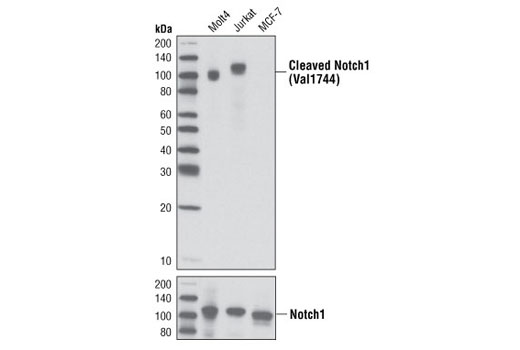
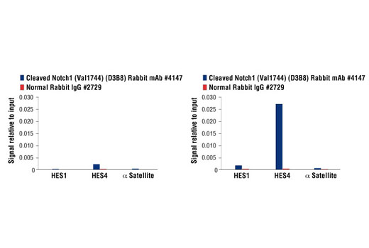
| Cleaved Notch1 (Val1744) (D3B8) Rabbit mAb 4147 | 20 µl |
|
H M R | 110 | Rabbit IgG |
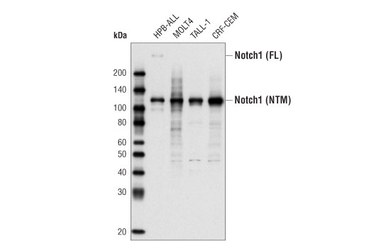
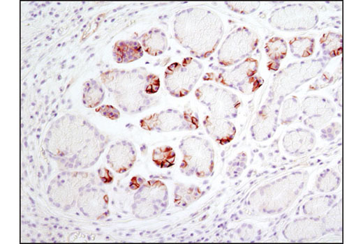
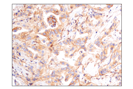
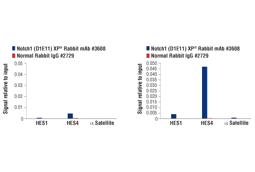
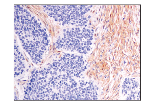
| Notch1 (D1E11) XP® Rabbit mAb 3608 | 20 µl |
|
H M R | 120, 300 | Rabbit IgG |
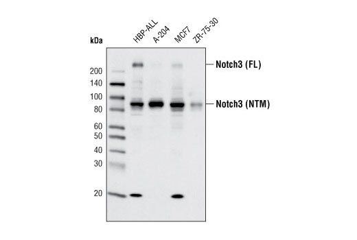
| Notch3 (D11B8) Rabbit mAb 5276 | 20 µl |
|
H | 90, 270 | Rabbit IgG |
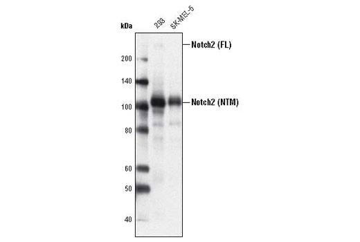
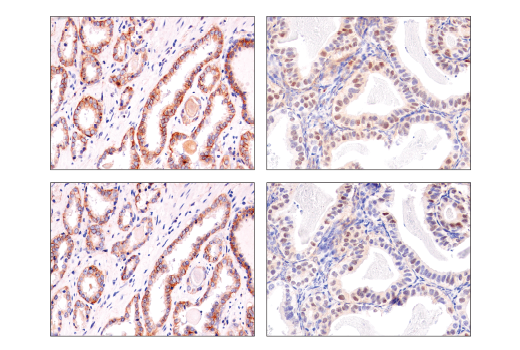
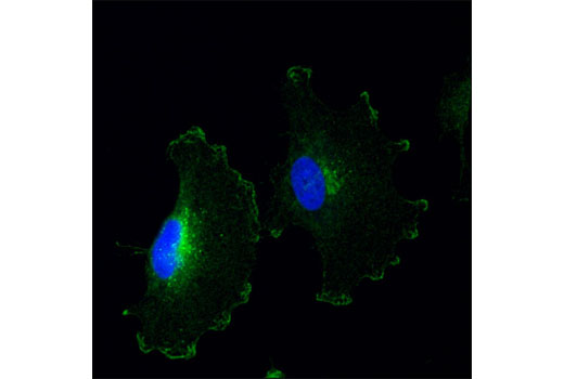
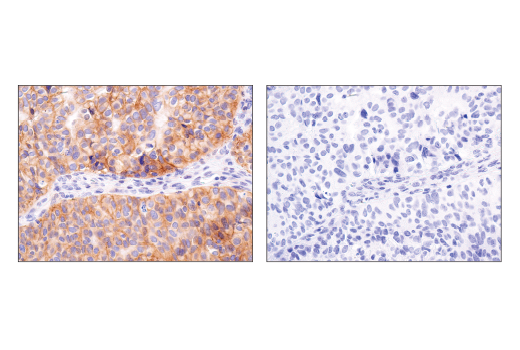
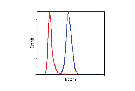
| Notch2 (D76A6) XP® Rabbit mAb 5732 | 20 µl |
|
H M R | 110, 300 | Rabbit |
| Anti-rabbit IgG, HRP-linked Antibody 7074 | 100 µl |
|
Rab | Goat |
Product Information
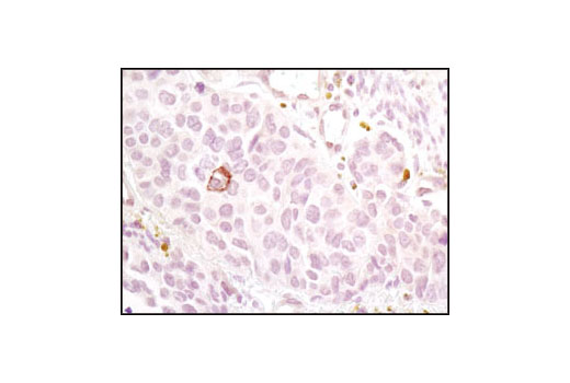
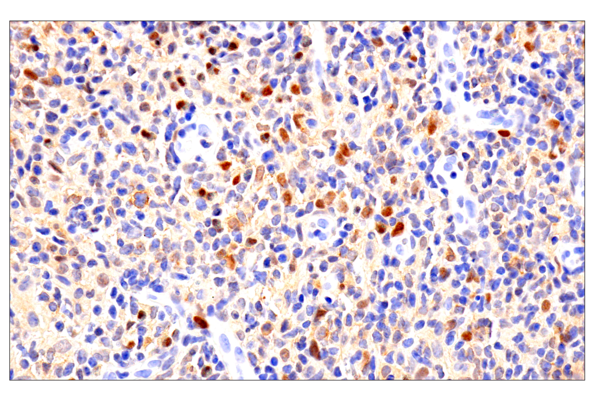
Monoclonal antibodies are produced by immunizing animals with a synthetic peptide corresponding to the sequence at the Val1754 cleavage site in human Notch1 (equivalent to Val1744 in mouse Notch1), Pro2438 of human Notch1, Ala2738 of human Notch2, or Glu2312 of human Notch3 protein.
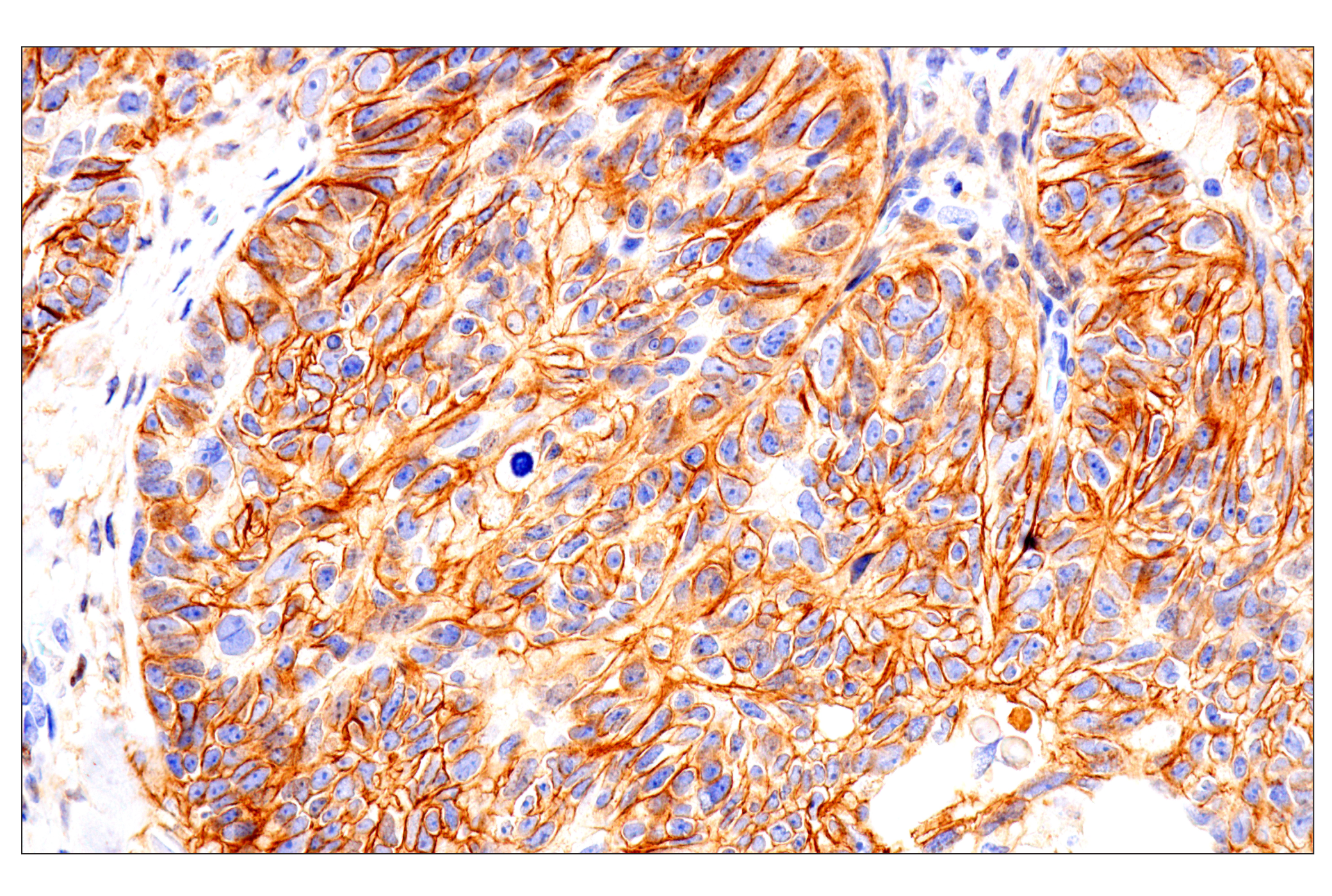
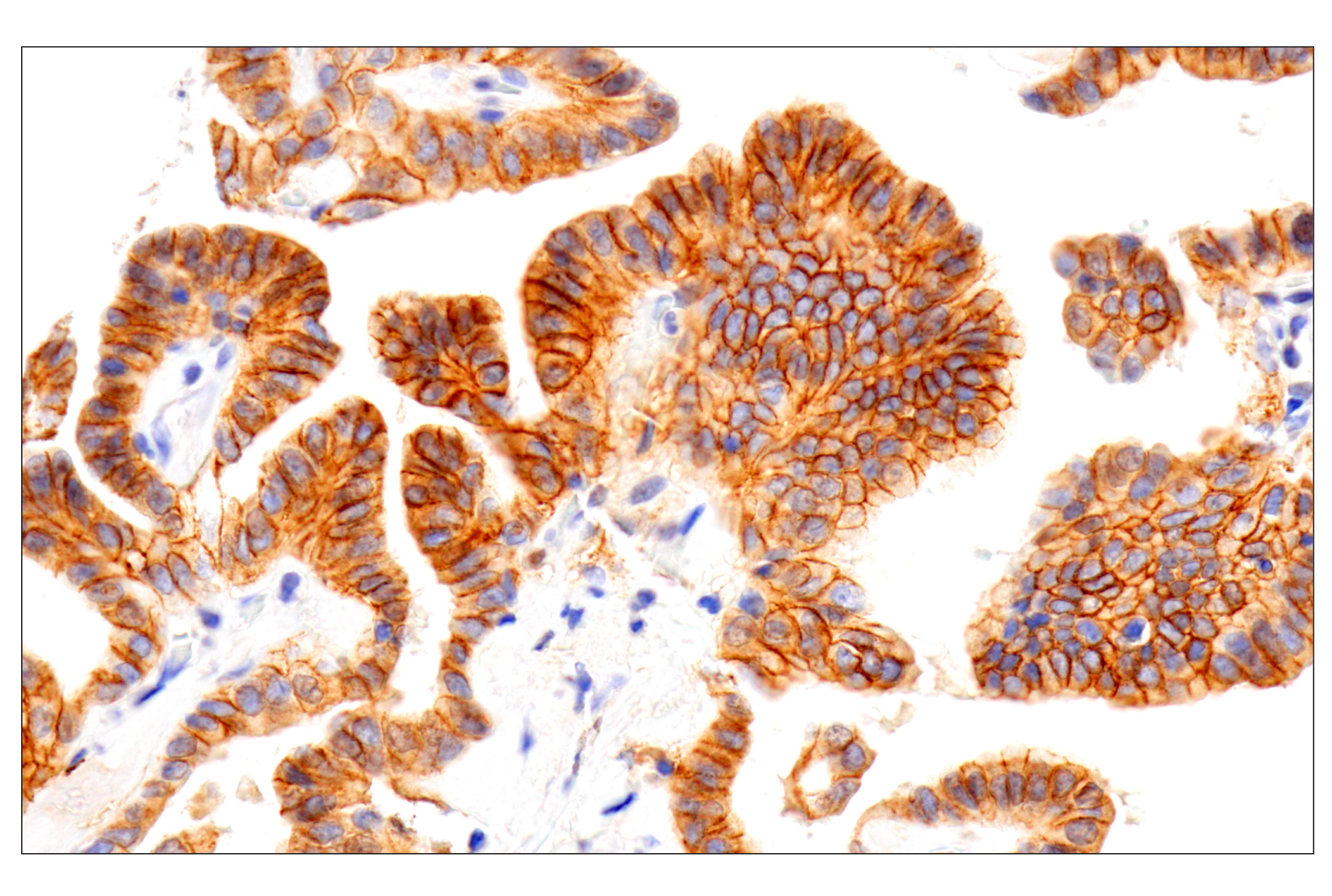
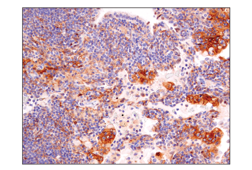
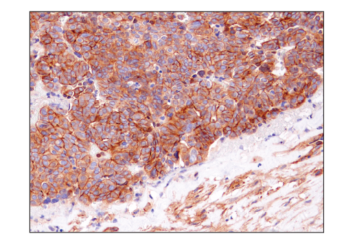
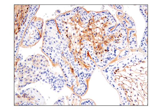
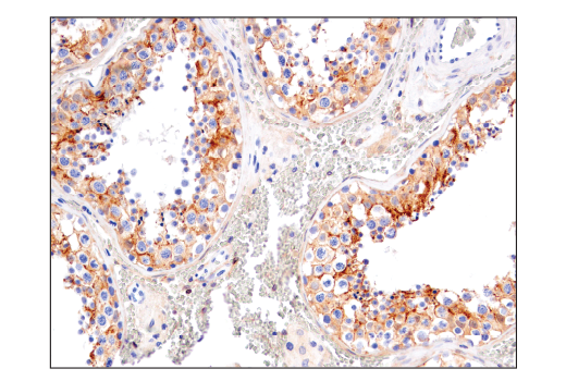
Notch proteins (Notch1-4) are a family of transmembrane receptors that play important roles in development and the determination of cell fate (1). Mature Notch receptors are processed and assembled as heterodimeric proteins, with each dimer comprised of a large extracellular ligand-binding domain, a single-pass transmembrane domain, and a smaller cytoplasmic subunit (Notch intracellular domain, NICD) (2). Binding of Notch receptors to ligands of the Delta-Serrate-Lag2 (DSL) family triggers heterodimer dissociation, exposing the receptors to proteolytic cleavages; these result in release of the NICD, which translocates to the nucleus and activates transcription of downstream target genes (3,4).
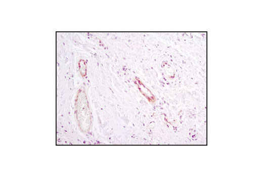
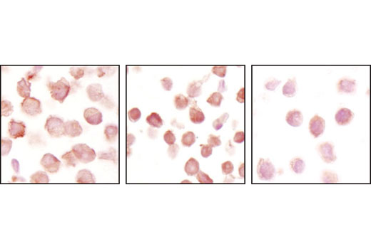
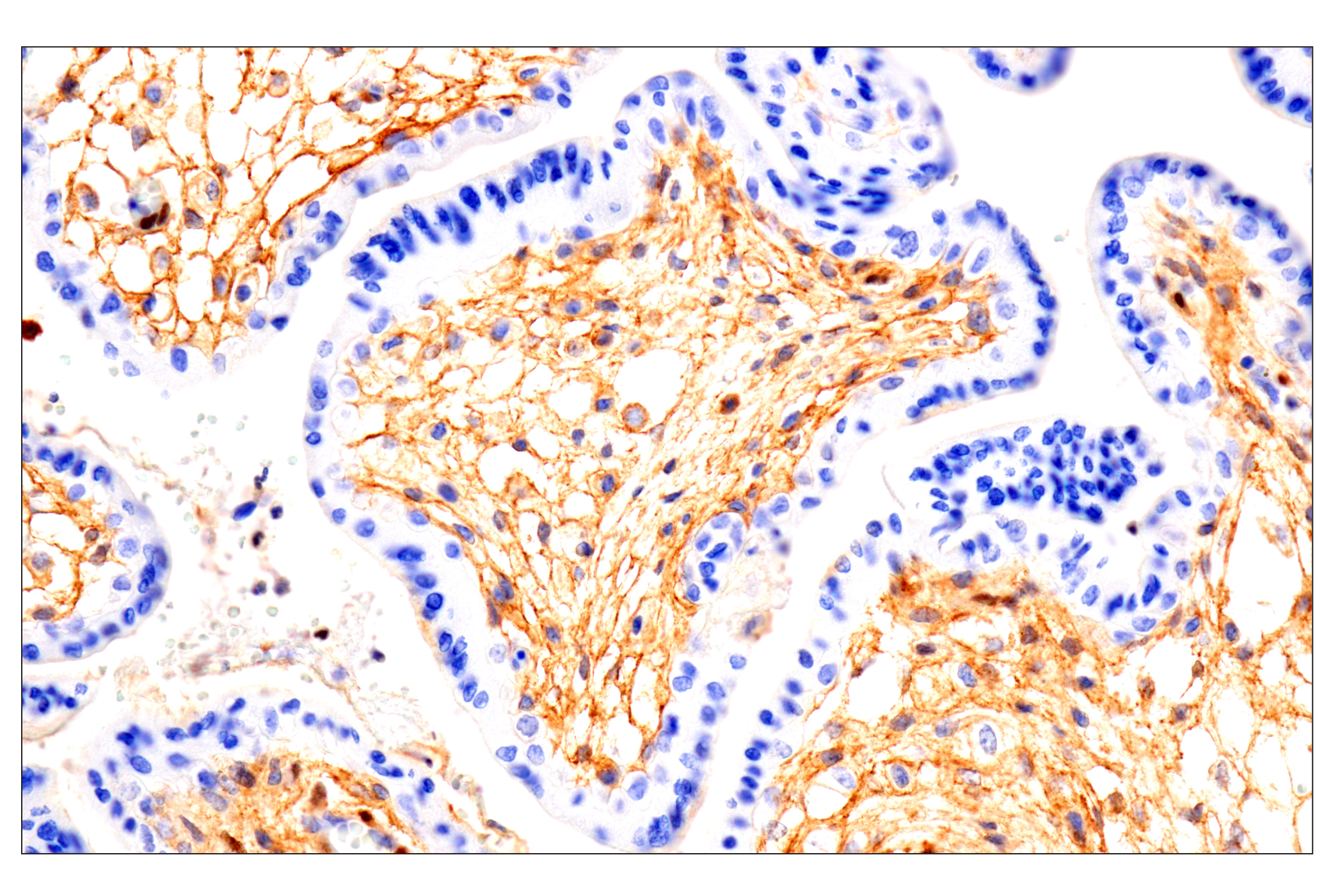
Constitutively activated Notch1 signaling is associated with the majority of cases of T cell acute lymphoblastic leukemia (T-ALL). The activation is either due to mutations in Notch1 itself or in the components of ubiquitin ligase complex, namely FBW7 (5-6). Notch2 is a member of the Notch family and mutation in Notch2 is associated with Alagille syndrome (7). Notch3 is a member of the Notch family and is processed similar to Notch1 (8). It is expressed primarily in arterial smooth muscle cells (SMC). Mutations altering the number of cysteine residues in the notch3 extracellular region are associated with cerebral autosomal dominant arteriopathy with subcortical infarcts and leukoencephalopathy (CADASIL), a hereditary angiopathy leading to strokes and dementia in adults (9-11). Recent studies indicate that Notch3 is overexpressed in many types of cancer (12-14).
Except as otherwise expressly agreed in a writing signed by a legally authorized representative of CST, the following terms apply to Products provided by CST, its affiliates or its distributors. Any Customer's terms and conditions that are in addition to, or different from, those contained herein, unless separately accepted in writing by a legally authorized representative of CST, are rejected and are of no force or effect.
Products are labeled with For Research Use Only or a similar labeling statement and have not been approved, cleared, or licensed by the FDA or other regulatory foreign or domestic entity, for any purpose. Customer shall not use any Product for any diagnostic or therapeutic purpose, or otherwise in any manner that conflicts with its labeling statement. Products sold or licensed by CST are provided for Customer as the end-user and solely for research and development uses. Any use of Product for diagnostic, prophylactic or therapeutic purposes, or any purchase of Product for resale (alone or as a component) or other commercial purpose, requires a separate license from CST. Customer shall (a) not sell, license, loan, donate or otherwise transfer or make available any Product to any third party, whether alone or in combination with other materials, or use the Products to manufacture any commercial products, (b) not copy, modify, reverse engineer, decompile, disassemble or otherwise attempt to discover the underlying structure or technology of the Products, or use the Products for the purpose of developing any products or services that would compete with CST products or services, (c) not alter or remove from the Products any trademarks, trade names, logos, patent or copyright notices or markings, (d) use the Products solely in accordance with CST Product Terms of Sale and any applicable documentation, and (e) comply with any license, terms of service or similar agreement with respect to any third party products or services used by Customer in connection with the Products.