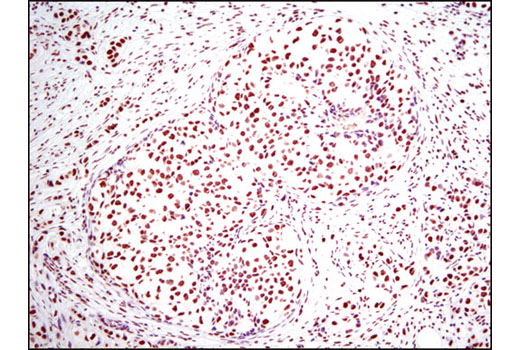
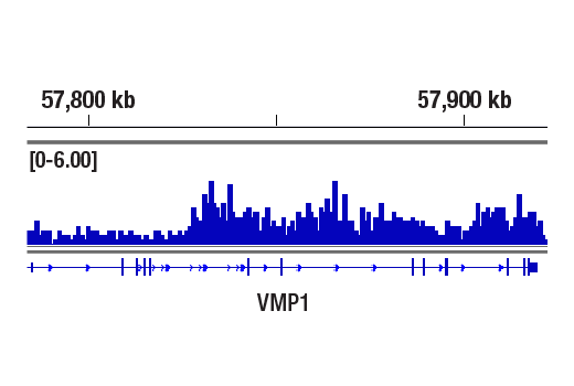
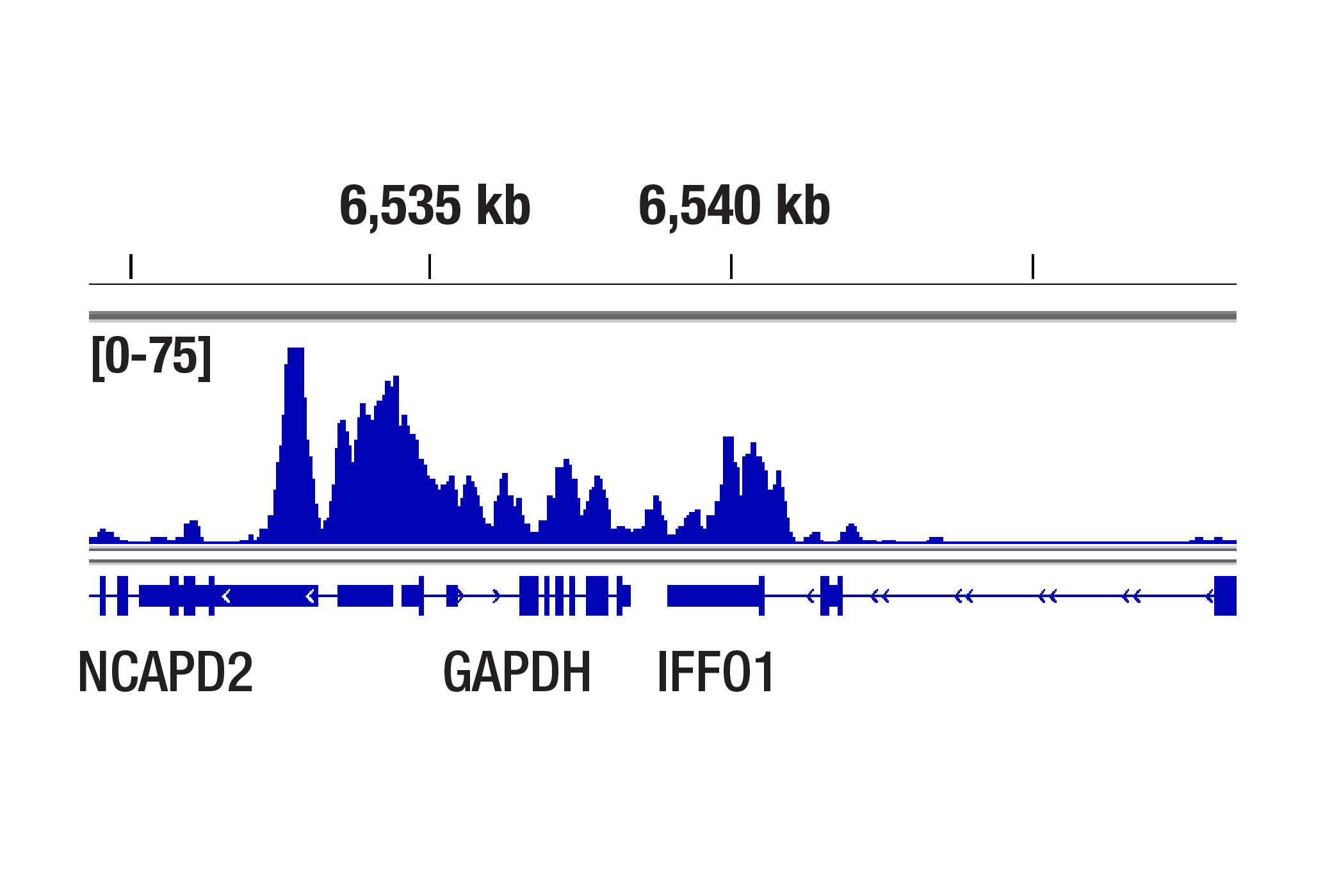
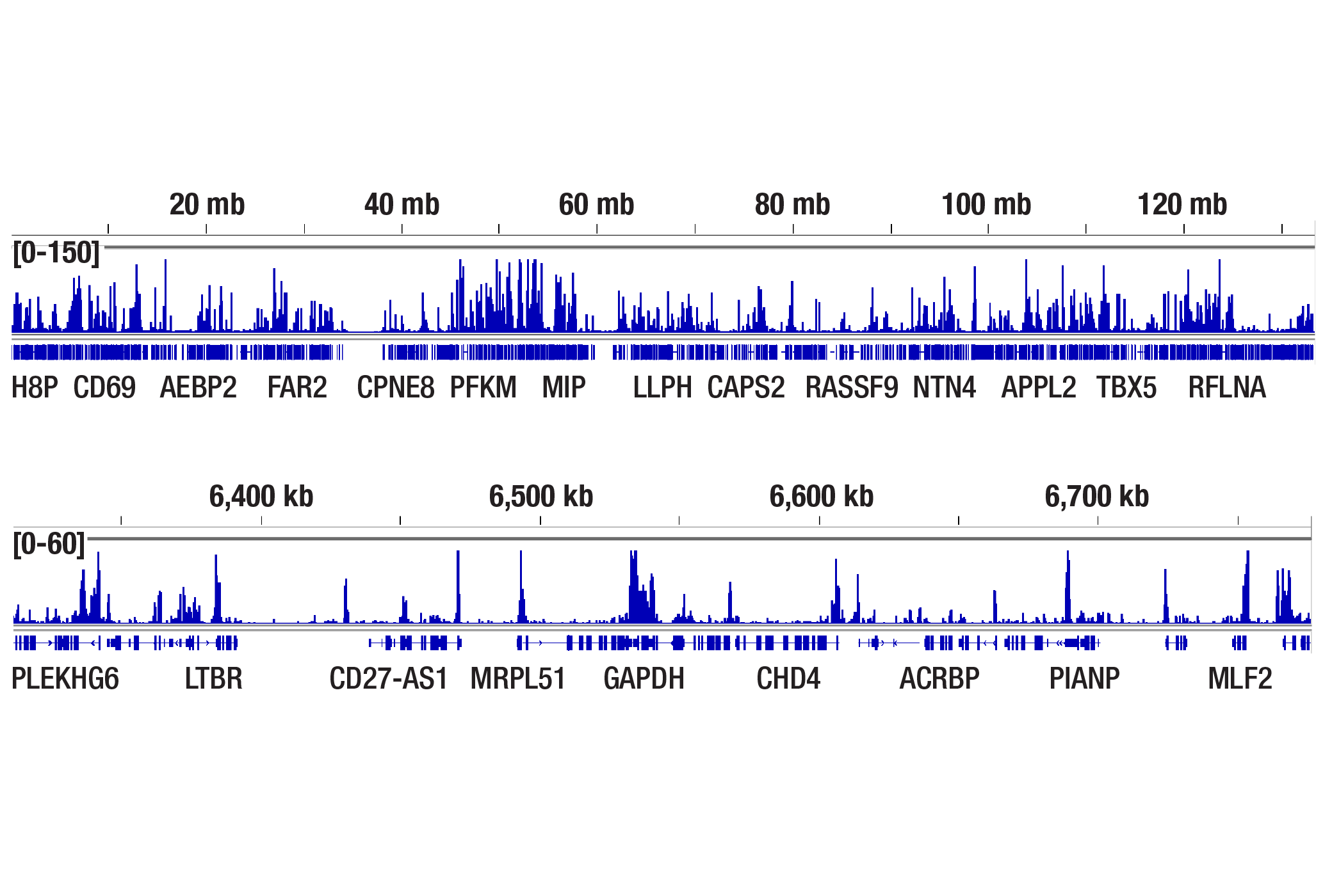
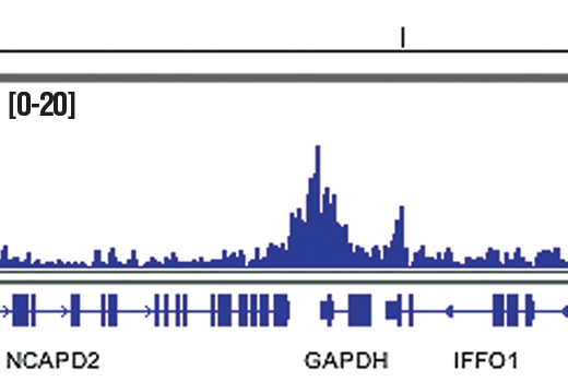
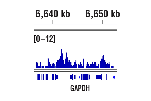
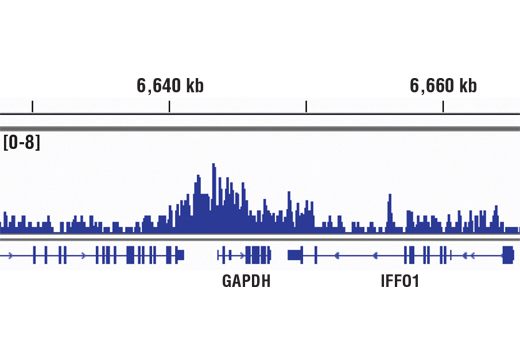
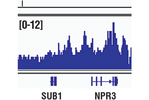
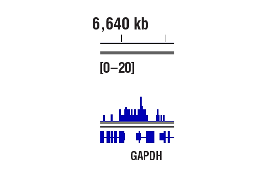
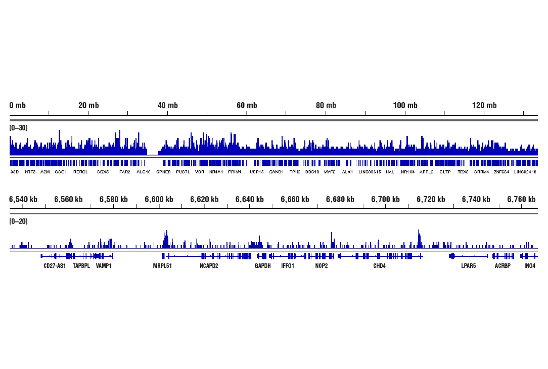
Chromatin immunoprecipitations were performed with cross-linked chromatin from HeLa cells and Acetyl-Histone H3 (Lys14) (D4B9) Rabbit mAb, using SimpleChIP® Plus Enzymatic Chromatin IP Kit (Magnetic Beads) #9005. DNA Libraries were prepared using SimpleChIP® ChIP-seq DNA Library Prep Kit for Illumina Systems #56795. The figure shows binding across VMP1 gene.
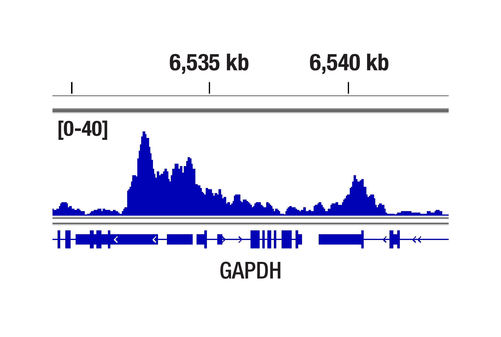
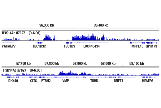
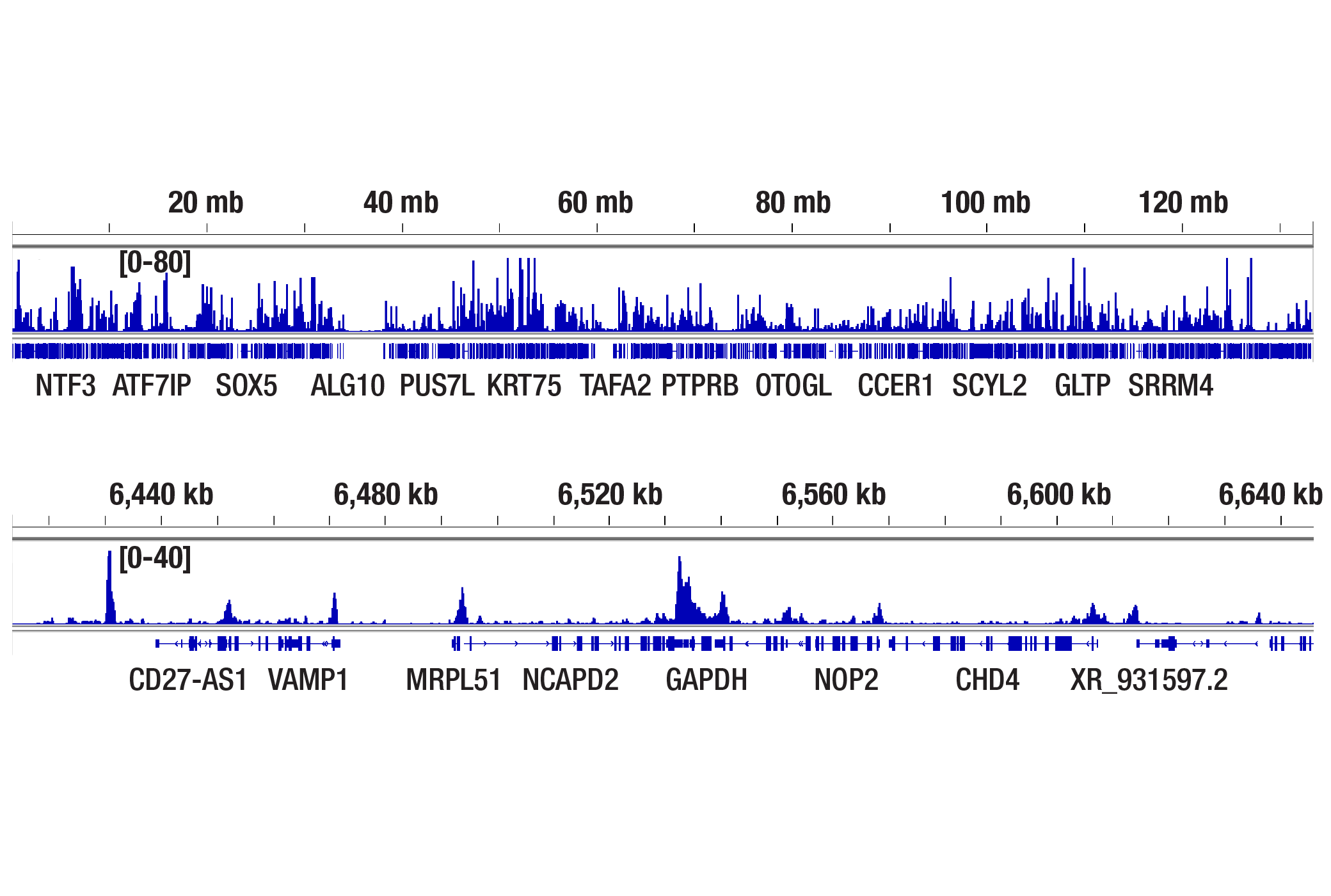
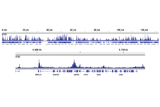
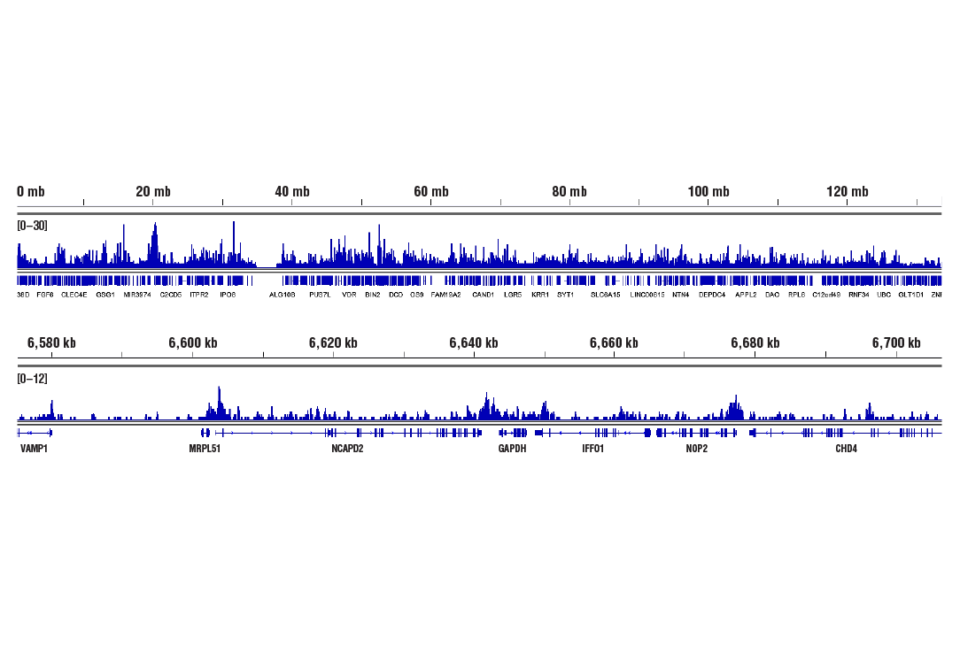
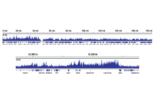
Chromatin immunoprecipitations were performed with cross-linked chromatin from HeLa cells and Acetyl-Histone H3 (Lys14) (D4B9) Rabbit mAb, using SimpleChIP® Plus Enzymatic Chromatin IP Kit (Magnetic Beads) #9005. DNA Libraries were prepared using DNA Library Prep Kit for Illumina Systems (ChIP-seq, CUT&RUN) #56795. The figure shows binding across chromosome 17 (upper), including VMP1 gene (lower).
| Cat. # | Size | Qty. | Price |
|---|---|---|---|
| 9927T | 1 Kit (6 x 20 microliters) |
|
| Product Includes | Quantity | Applications | Reactivity | MW(kDa) | Isotype |
|---|---|---|---|---|---|
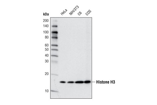
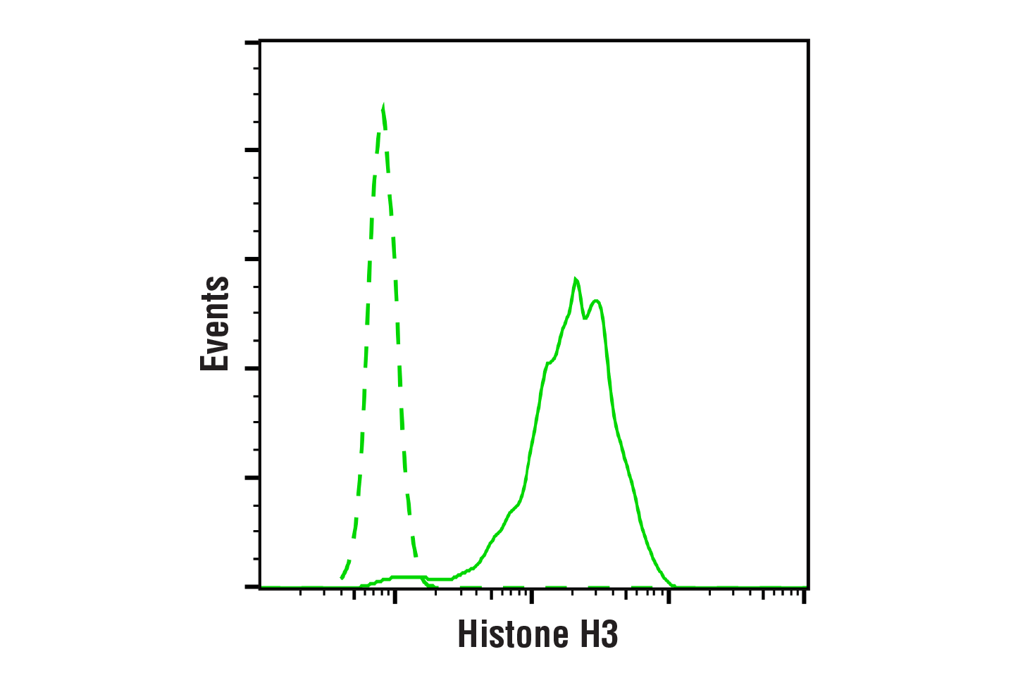
| Histone H3 (D1H2) XP® Rabbit mAb 4499 | 20 µl |
|
H M R Mk | 17 | Rabbit IgG |
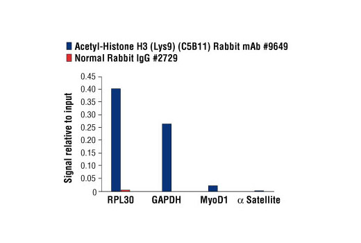
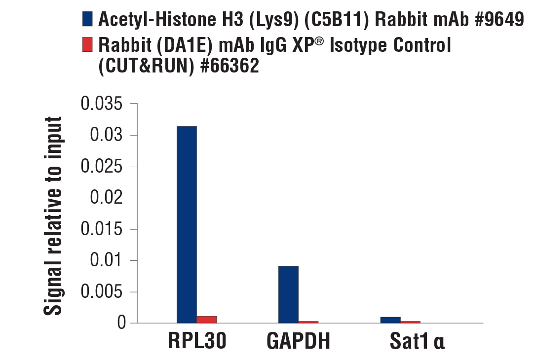
| Acetyl-Histone H3 (Lys9) (C5B11) Rabbit mAb 9649 | 20 µl |
|
H M R Mk Z | 17 | Rabbit IgG |
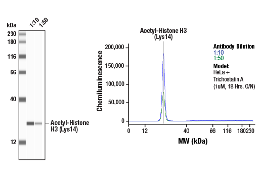
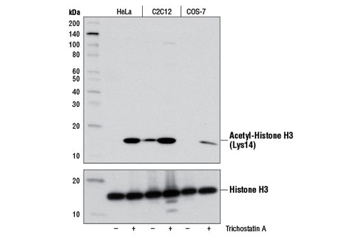
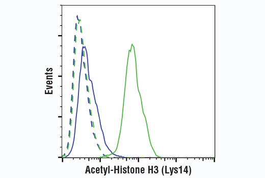
| Acetyl-Histone H3 (Lys14) (D4B9) Rabbit mAb 7627 | 20 µl |
|
H M R Mk | 17 | Rabbit IgG |
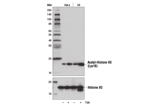
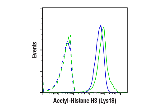
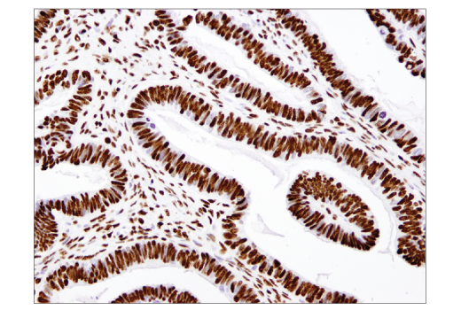
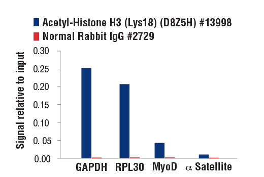
| Acetyl-Histone H3 (Lys18) (D8Z5H) Rabbit mAb 13998 | 20 µl |
|
H M R Mk Sc | 17 | Rabbit IgG |
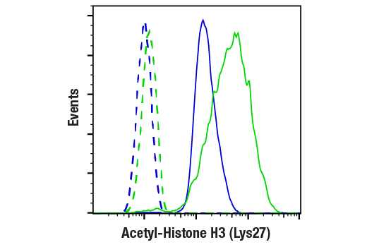
| Acetyl-Histone H3 (Lys27) (D5E4) XP® Rabbit mAb 8173 | 20 µl |
|
H M R Mk | 17 | Rabbit IgG |
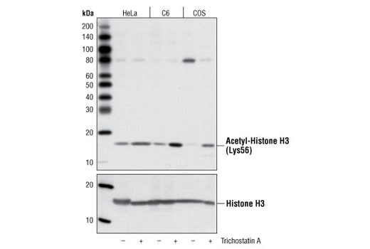
| Acetyl-Histone H3 (Lys56) Antibody 4243 | 20 µl |
|
H M R Mk | 17 | Rabbit |
| Anti-rabbit IgG, HRP-linked Antibody 7074 | 100 µl |
|
Goat |
Product Information
Polyclonal antibodies are produced by immunizing rabbits with synthetic acetylated peptides corresponding to residues surrounding Lys56 of human Histone H3. Antibodies are purified by protein A and peptide affinity chromatography. Monoclonal antibody is produced by immunizing animals with a synthetic peptide corresponding to residues surrounding acetylated Lys18 of human histone H3 protein, Lys14 of human histone H3 protein, acetylated Lys27 of human Histone H3 protein, or the amino terminus of histone H3 in which Lys9 is acetylated.
Modulation of chromatin structure plays an important role in the regulation of transcription in eukaryotes. The nucleosome, made up of DNA wound around eight core histone proteins (two each of H2A, H2B, H3, and H4), is the primary building block of chromatin (1). The amino-terminal tails of core histones undergo various posttranslational modifications, including acetylation, phosphorylation, methylation, and ubiquitination (2-5). These modifications occur in response to various stimuli and have a direct effect on the accessibility of chromatin to transcription factors and, therefore, gene expression (6). In most species, histone H2B is primarily acetylated at Lys5, 12, 15, and 20 (4,7). Histone H3 is primarily acetylated at Lys9, 14, 18, 23, 27, and 56. Acetylation of H3 at Lys9 appears to have a dominant role in histone deposition and chromatin assembly in some organisms (2,3). Phosphorylation at Ser10, Ser28, and Thr11 of histone H3 is tightly correlated with chromosome condensation during both mitosis and meiosis (8-10). Phosphorylation at Thr3 of histone H3 is highly conserved among many species and is catalyzed by the kinase haspin. Immunostaining with phospho-specific antibodies in mammalian cells reveals mitotic phosphorylation at Thr3 of H3 in prophase and its dephosphorylation during anaphase (11).
Explore pathways related to this product.
STRING - Known and Predicted Protein-Protein Interactions.
Except as otherwise expressly agreed in a writing signed by a legally authorized representative of CST, the following terms apply to Products provided by CST, its affiliates or its distributors. Any Customer's terms and conditions that are in addition to, or different from, those contained herein, unless separately accepted in writing by a legally authorized representative of CST, are rejected and are of no force or effect.
Products are labeled with For Research Use Only or a similar labeling statement and have not been approved, cleared, or licensed by the FDA or other regulatory foreign or domestic entity, for any purpose. Customer shall not use any Product for any diagnostic or therapeutic purpose, or otherwise in any manner that conflicts with its labeling statement. Products sold or licensed by CST are provided for Customer as the end-user and solely for research and development uses. Any use of Product for diagnostic, prophylactic or therapeutic purposes, or any purchase of Product for resale (alone or as a component) or other commercial purpose, requires a separate license from CST. Customer shall (a) not sell, license, loan, donate or otherwise transfer or make available any Product to any third party, whether alone or in combination with other materials, or use the Products to manufacture any commercial products, (b) not copy, modify, reverse engineer, decompile, disassemble or otherwise attempt to discover the underlying structure or technology of the Products, or use the Products for the purpose of developing any products or services that would compete with CST products or services, (c) not alter or remove from the Products any trademarks, trade names, logos, patent or copyright notices or markings, (d) use the Products solely in accordance with CST Product Terms of Sale and any applicable documentation, and (e) comply with any license, terms of service or similar agreement with respect to any third party products or services used by Customer in connection with the Products.