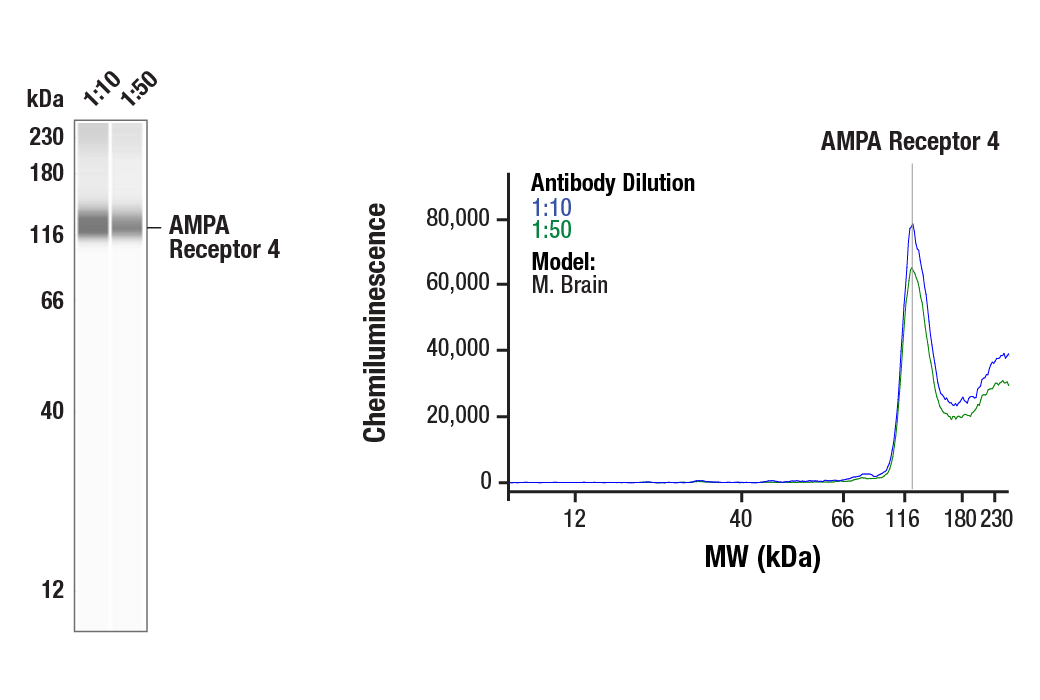
Simple Western™ analysis of lysates (0.75 mg/mL) from Mouse Brain cells using AMPA Receptor 4 (GluA 4) (D41A11) XP® Rabbit mAb #8070. The virtual lane view (left) shows the target band (as indicated) at 1:10 and 1:50 dilutions of primary antibody. The corresponding electropherogram view (right) plots chemiluminescence by molecular weight along the capillary at 1:10 (blue line) and 1:50 (green line) dilutions of primary antibody. This experiment was performed under reducing conditions on the Jess™ Simple Western instrument from ProteinSimple, a BioTechne brand, using the 12-230 kDa separation module.
| Cat. # | Size | Qty. | Price |
|---|---|---|---|
| 8652T | 1 Kit (6 x 20 microliters) |
|
| Product Includes | Quantity | Applications | Reactivity | MW(kDa) | Isotype |
|---|---|---|---|---|---|
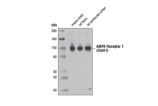
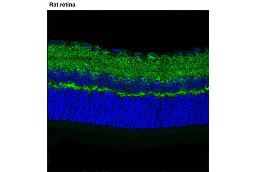
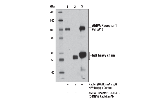
| AMPA Receptor 1 (GluA1) (D4N9V) Rabbit mAb 13185 | 20 µl |
|
M R | 100 | Rabbit IgG |
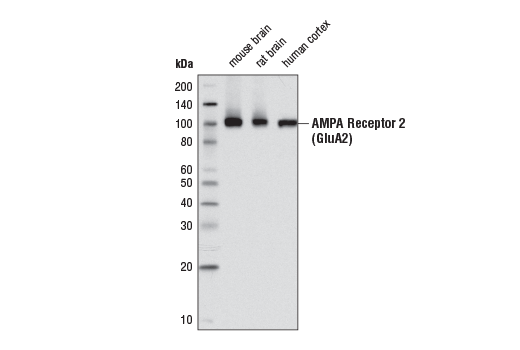
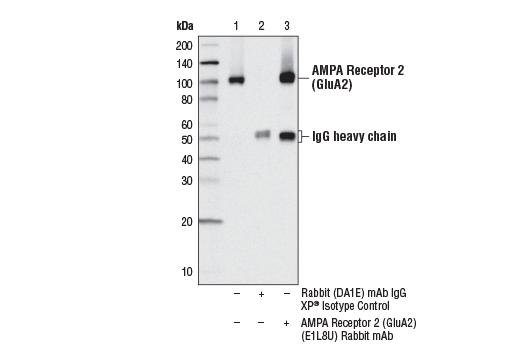
| AMPA Receptor 2 (GluA2) (E1L8U) Rabbit mAb 13607 | 20 µl |
|
H M R | 100 | Rabbit IgG |
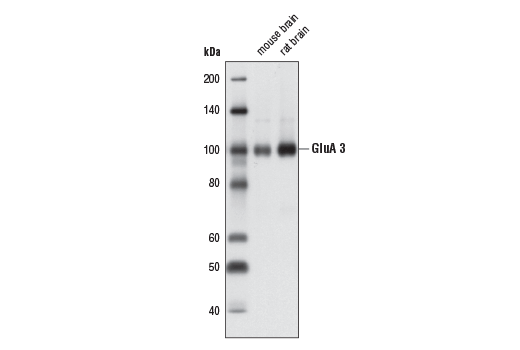
| AMPA Receptor 3 (GluA 3) (D47E3) Rabbit mAb 4676 | 20 µl |
|
H M R | 100 | Rabbit IgG |
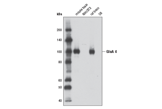
| AMPA Receptor 4 (GluA 4) (D41A11) XP® Rabbit mAb 8070 | 20 µl |
|
H M R | 100 | Rabbit IgG |
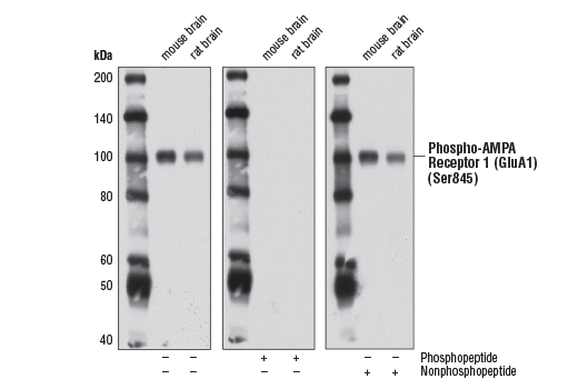
| Phospho-AMPA Receptor 1 (GluA1) (Ser845) (D10G5) Rabbit mAb 8084 | 20 µl |
|
H M R | 100 | Rabbit IgG |
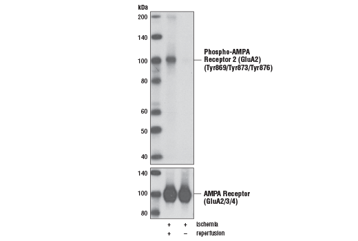
| Phospho-AMPA Receptor 2 (GluA2) (Tyr869/Tyr873/Tyr876) Antibody 3921 | 20 µl |
|
R | 100 | Rabbit |
| Anti-rabbit IgG, HRP-linked Antibody 7074 | 100 µl |
|
Goat |
Product Information
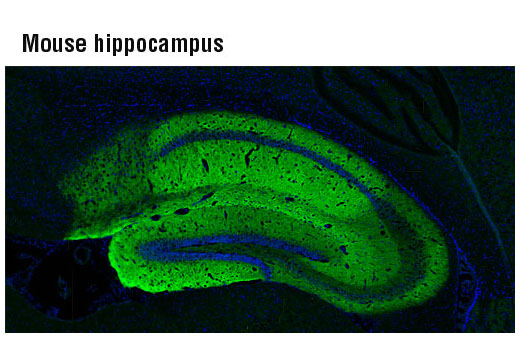
AMPA- (α-amino-3-hydroxy-5-methyl-4-isoxazolepropionic acid), kainate-, and NMDA- (N-methyl-D-aspartate) receptors are the three main families of ionotropic glutamate-gated ion channels. AMPA receptors (AMPARs) are comprised of four subunits (GluR 1-4), which assemble as homo- or hetero-tetramers to mediate the majority of fast excitatory transmissions in the central nervous system. AMPARs are implicated in synapse formation, stabilization, and plasticity (1). In contrast to GluR 2-containing AMPARs, AMPARs that lack GluR 2 are permeable to calcium (2). Post-transcriptional modifications (alternative splicing, nuclear RNA editing) and post-translational modifications (glycosylation, phosphorylation) result in a very large number of permutations, fine-tuning the kinetic properties of AMPARs. Research studies have implicated activity changes in AMPARs in a variety of diseases including Alzheimer’s, amyotrophic lateral sclerosis (ALS), stroke, and epilepsy (1).
Explore pathways related to this product.
STRING - Known and Predicted Protein-Protein Interactions.
Except as otherwise expressly agreed in a writing signed by a legally authorized representative of CST, the following terms apply to Products provided by CST, its affiliates or its distributors. Any Customer's terms and conditions that are in addition to, or different from, those contained herein, unless separately accepted in writing by a legally authorized representative of CST, are rejected and are of no force or effect.
Products are labeled with For Research Use Only or a similar labeling statement and have not been approved, cleared, or licensed by the FDA or other regulatory foreign or domestic entity, for any purpose. Customer shall not use any Product for any diagnostic or therapeutic purpose, or otherwise in any manner that conflicts with its labeling statement. Products sold or licensed by CST are provided for Customer as the end-user and solely for research and development uses. Any use of Product for diagnostic, prophylactic or therapeutic purposes, or any purchase of Product for resale (alone or as a component) or other commercial purpose, requires a separate license from CST. Customer shall (a) not sell, license, loan, donate or otherwise transfer or make available any Product to any third party, whether alone or in combination with other materials, or use the Products to manufacture any commercial products, (b) not copy, modify, reverse engineer, decompile, disassemble or otherwise attempt to discover the underlying structure or technology of the Products, or use the Products for the purpose of developing any products or services that would compete with CST products or services, (c) not alter or remove from the Products any trademarks, trade names, logos, patent or copyright notices or markings, (d) use the Products solely in accordance with CST Product Terms of Sale and any applicable documentation, and (e) comply with any license, terms of service or similar agreement with respect to any third party products or services used by Customer in connection with the Products.