| Cat. # | Size | Qty. | Price |
|---|---|---|---|
| 58214T | 1 Kit (7 x 20 microliters) |
|
| Product Includes | Quantity | Applications | Reactivity | MW(kDa) | Isotype |
|---|---|---|---|---|---|
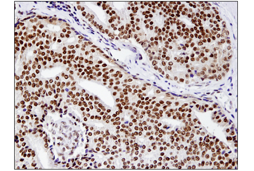
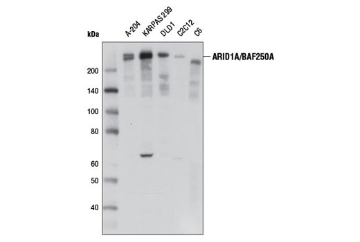
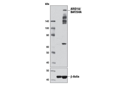
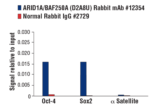
| ARID1A/BAF250A (D2A8U) Rabbit mAb 12354 | 20 µl |
|
H M R Mk | 270 | Rabbit IgG |
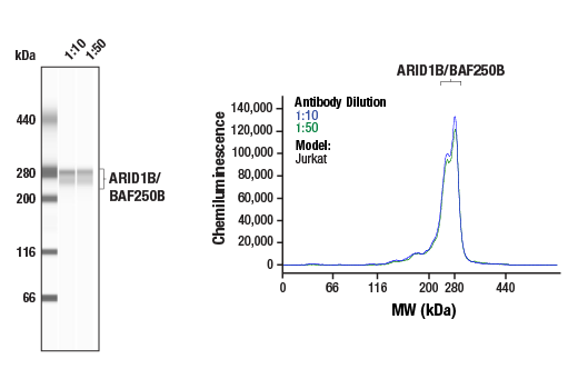
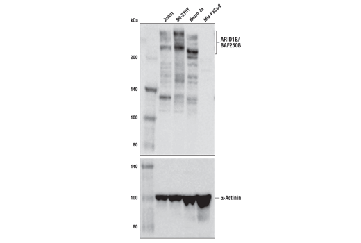
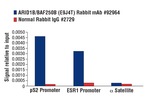
| ARID1B/BAF250B (E9J4T) Rabbit mAb 92964 | 20 µl |
|
H M | 250, 280 | Rabbit IgG |
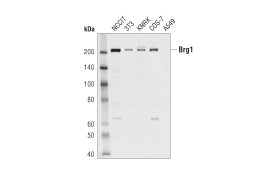
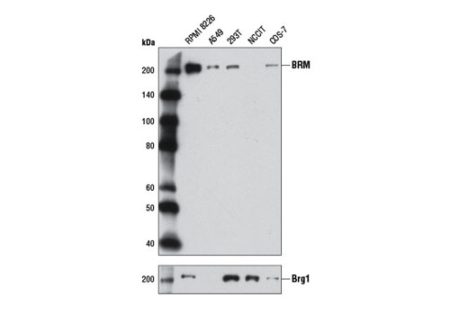
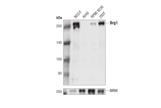
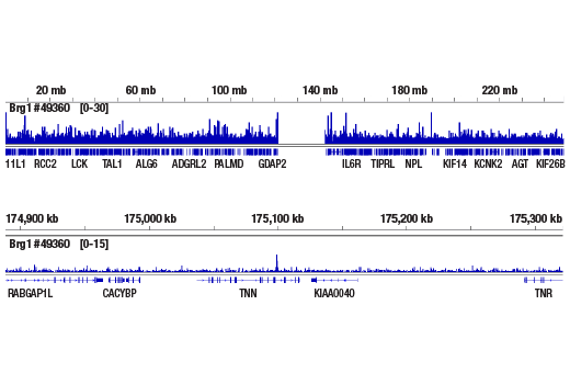
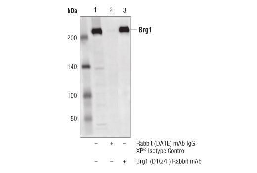
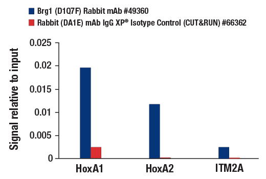
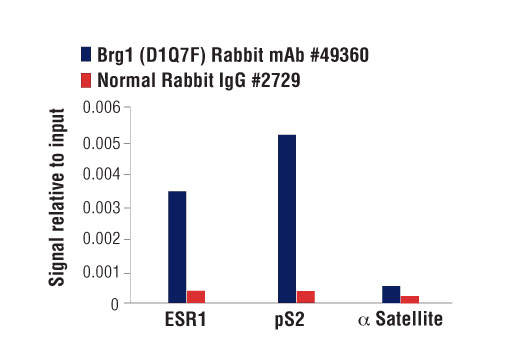
| Brg1 (D1Q7F) Rabbit mAb 49360 | 20 µl |
|
H M R Mk | 220 | Rabbit IgG |
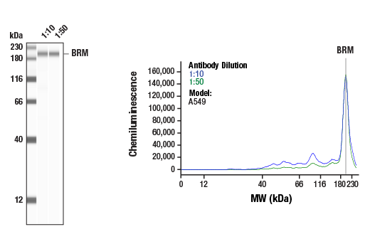
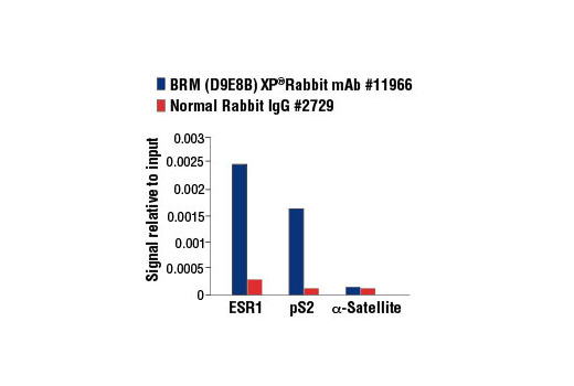
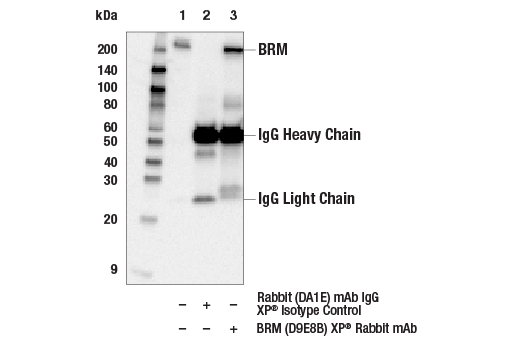
| BRM (D9E8B) XP® Rabbit mAb 11966 | 20 µl |
|
H Mk | 200 | Rabbit IgG |
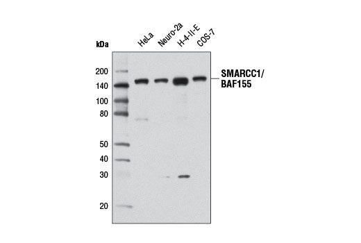
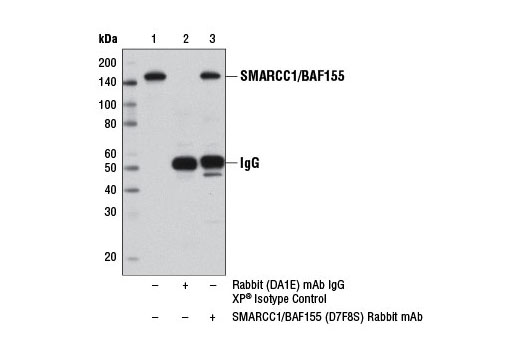
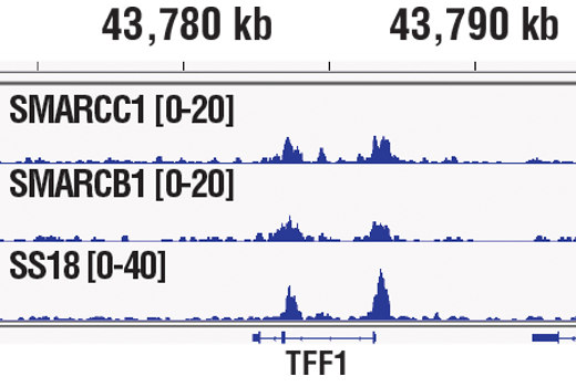
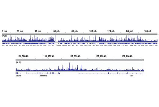
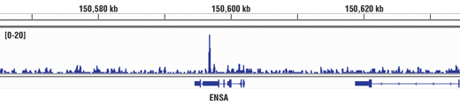
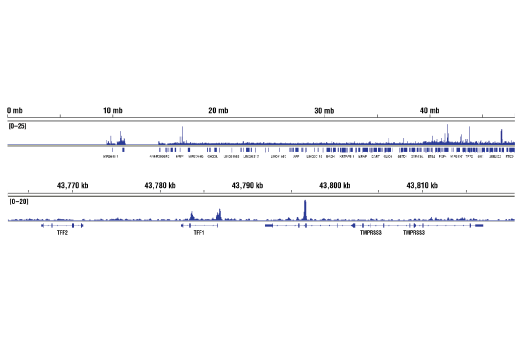
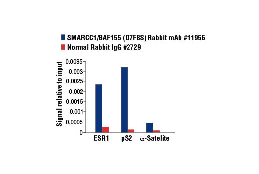
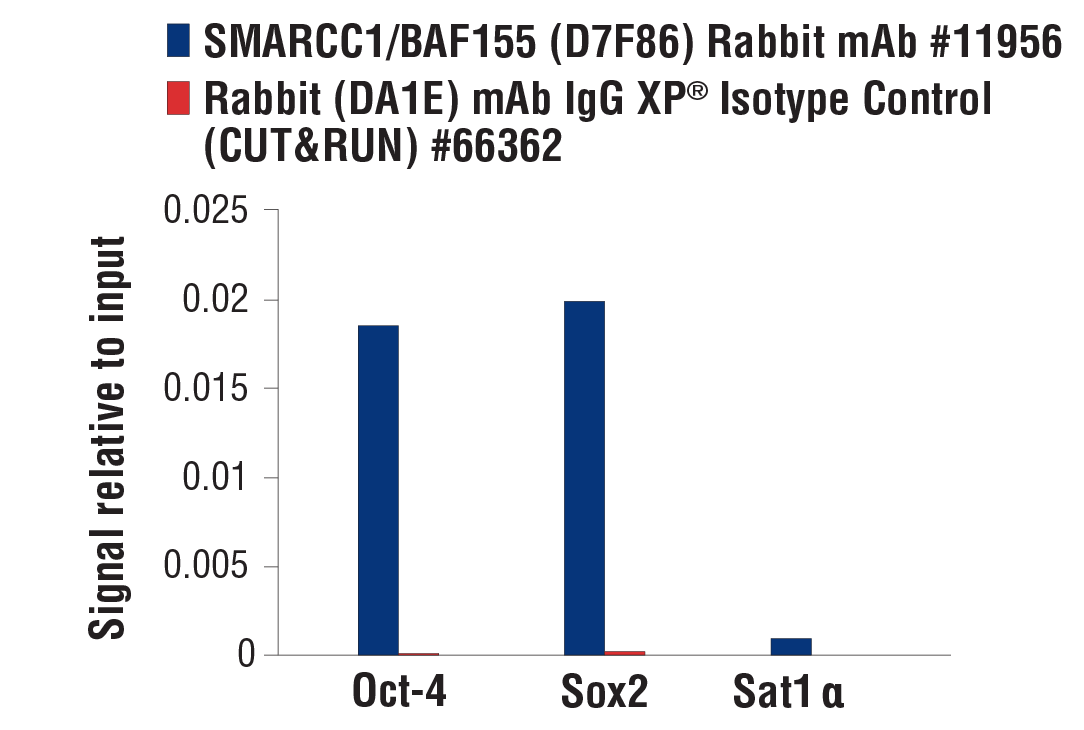
| SMARCC1/BAF155 (D7F8S) Rabbit mAb 11956 | 20 µl |
|
H M R Mk | 155 | Rabbit IgG |
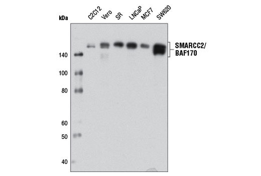
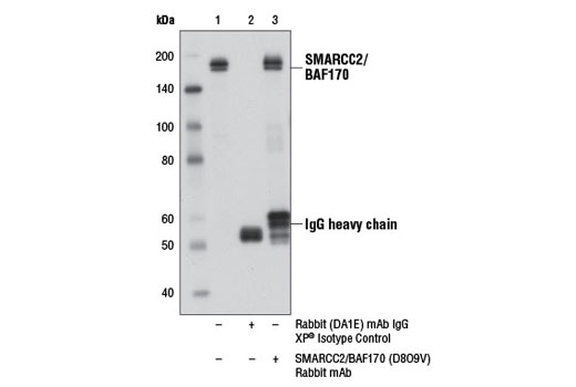
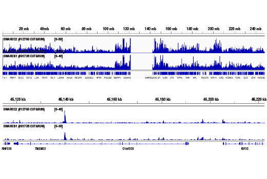
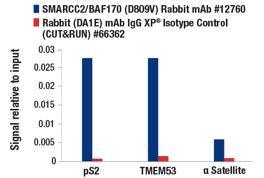
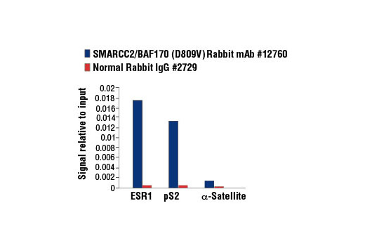
| SMARCC2/BAF170 (D8O9V) Rabbit mAb 12760 | 20 µl |
|
H M R Mk | 162, 170 | Rabbit IgG |
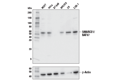
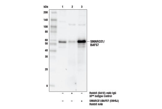
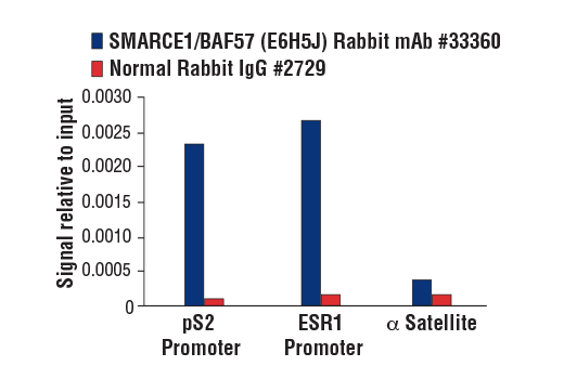
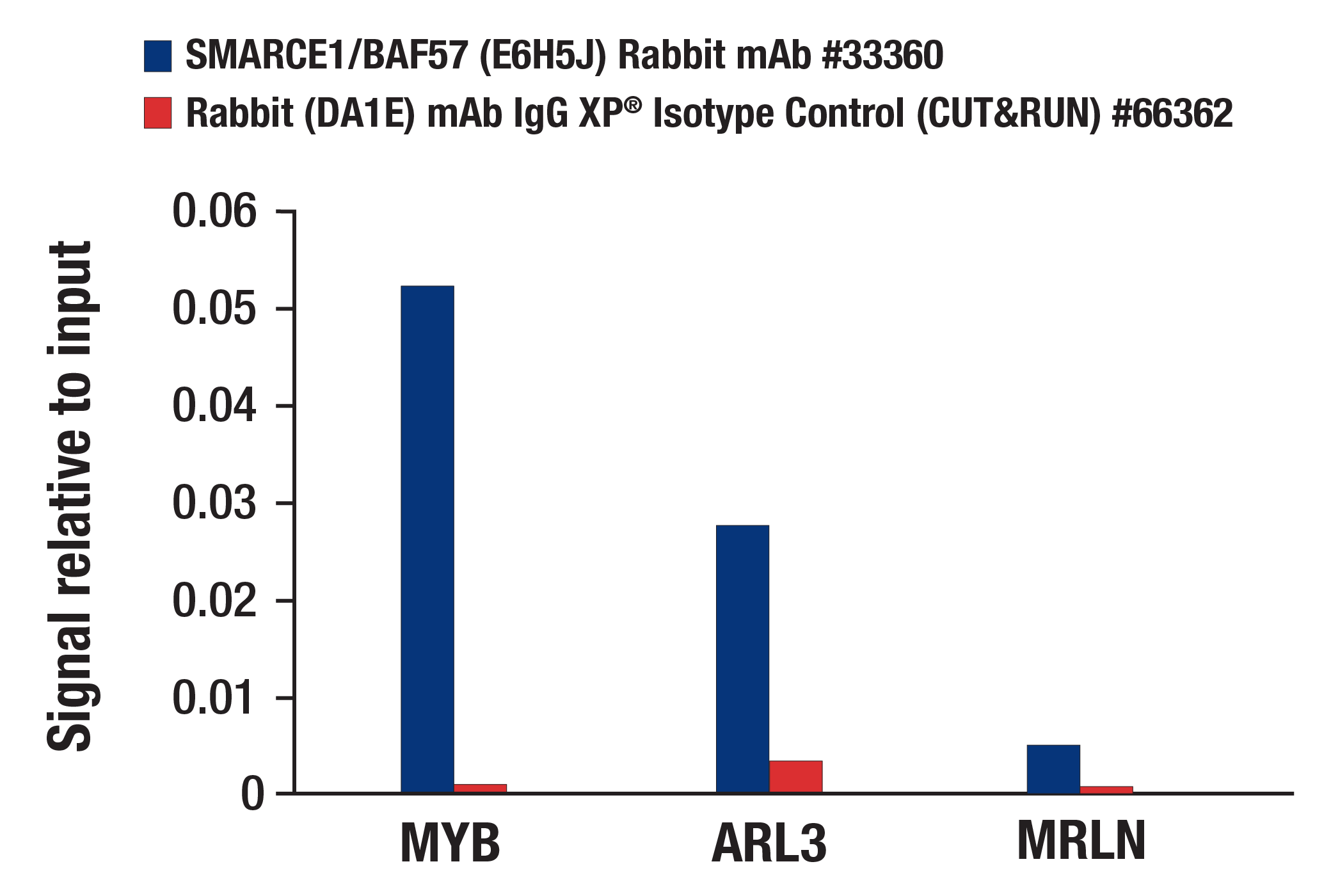
| SMARCE1/BAF57 (E6H5J) Rabbit mAb 33360 | 20 µl |
|
H M R Mk | 57 | Rabbit IgG |
| Anti-rabbit IgG, HRP-linked Antibody 7074 | 100 µl |
|
Goat |
Product Information
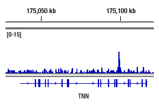
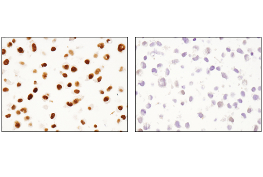
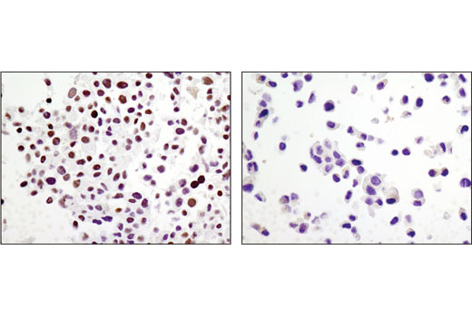
Monoclonal antibody is produced by immunizing animals with a synthetic peptide corresponding to residues surrounding Gly1293 of human ARID1A/BAF250A protein, Ala1320 of human ARID1B/BAF250B protein, residues near the amino terminus of human Brg1 protein, resides surrounding Gly264 of human BRM protein, Gly975 of human SMARCC1/BAF155 protein, Ile818 of human SMARCC2/BAF170, and Leu34 of human SMARCE1/BAF57 protein.
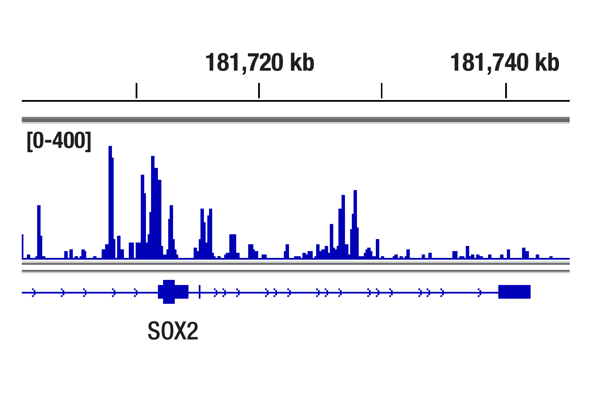
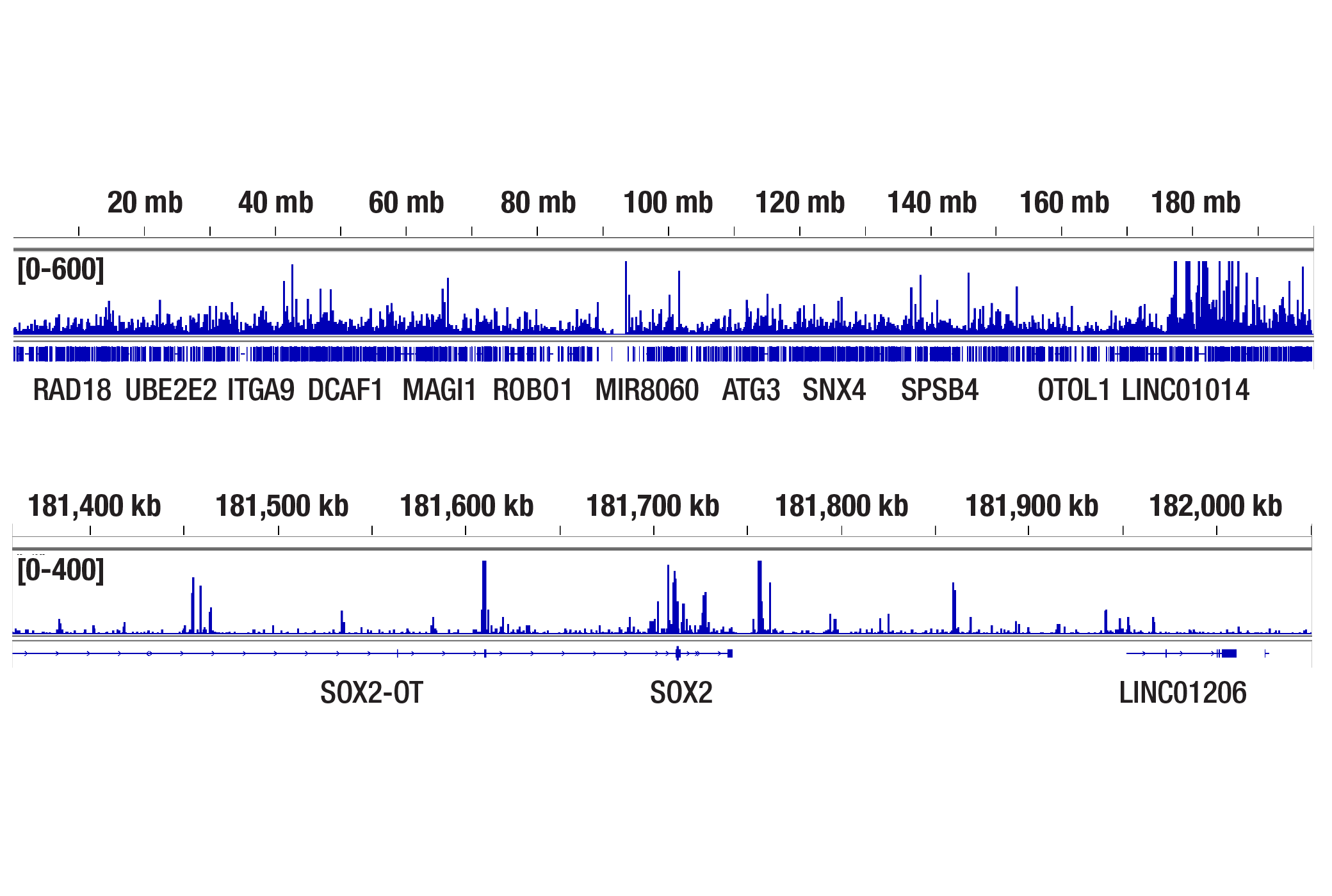
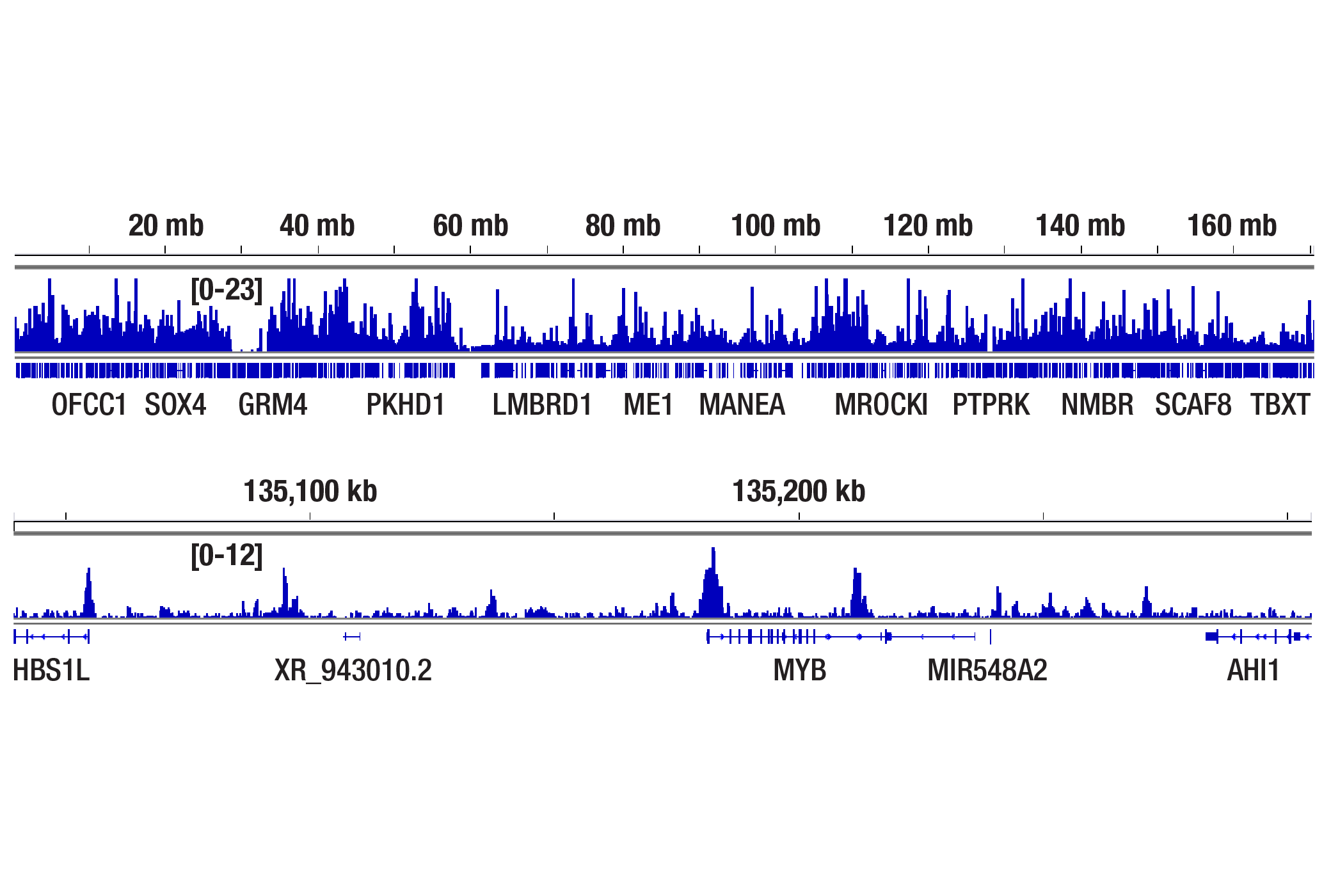
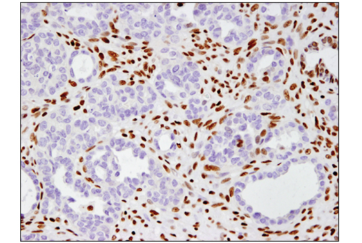
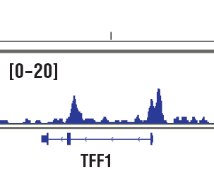
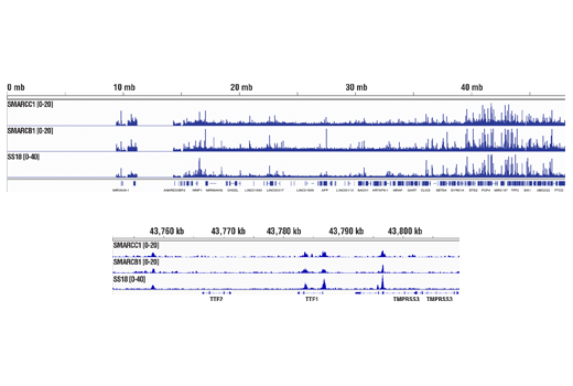
ATP-dependent chromatin remodeling complexes play an essential role in the regulation of various nuclear processes, such as gene expression, DNA replication, and repair (1,2). The SWI/SNF chromatin remodeling complex consists of more than 10 subunits with a single molecule of the ATPase catalytic subunit BRM or BRG1, but not both. The activities of these two subunits drive the disruption of histone-DNA contacts that lead to changes in accessibility of crucial regulatory elements within chromatin (2-5). The BRM/BRG1 containing SWI/SNF complexes are recruited to target promoters by transcription factors, such as nuclear receptors, p53, RB, and BRCA1 to regulate gene activation, cell growth, the cell cycle, and differentiation processes (1,6-9). BRM and BRG1 are also considered to be tumor suppressors and their expression levels are severely reduced in several cancer cell lines (10-13). SMARCC1/BAF155, SMARCC2/BAF170, and SMARCB1/BAF47 are members of the core subunits of the SWI/SNF complex, which is necessary for efficient nucleosome remodeling by BRG1 in vitro (14). ARID1A/BAF250A and ARID1B/BAF250B are DNA-binding members of the complex. They are highly homologous and mutually exclusive, with ARID1B/BAF250B being a critical vulnerability in ARID1A/BAF250A mutant cancers (15-17). SMARCC1, SMARCB1, and ARID1A are an essential part of the mouse embryonic stem cell specific SWI/SNF complex (esBAF). SMARCC1 is necessary for early embryogenesis, especially proper brain and visceral endoderm development (18-20). SMARCB1 is necessary for early embryogenesis and hepatocyte differentiation (21,22). ARID1A is critical for embryonic stem (ES) cell pluripotency and differentiation into mesoderm-derived cardiomyocytes and adipocytes (15). While SMARCC2 has been shown to be part of the SWI/SNF complex in non-pluripotent cells, it is absent in pluripotent ES cells. Expression of SMARCC2 has been shown to be up-regulated in neurons/neuronal progenitors upon differentiation of mouse ES cells with retinoic acid, and exogenous expression of SMARCC2 leads to loss of stem cell pluripotency and self renewal (23).
Except as otherwise expressly agreed in a writing signed by a legally authorized representative of CST, the following terms apply to Products provided by CST, its affiliates or its distributors. Any Customer's terms and conditions that are in addition to, or different from, those contained herein, unless separately accepted in writing by a legally authorized representative of CST, are rejected and are of no force or effect.
Products are labeled with For Research Use Only or a similar labeling statement and have not been approved, cleared, or licensed by the FDA or other regulatory foreign or domestic entity, for any purpose. Customer shall not use any Product for any diagnostic or therapeutic purpose, or otherwise in any manner that conflicts with its labeling statement. Products sold or licensed by CST are provided for Customer as the end-user and solely for research and development uses. Any use of Product for diagnostic, prophylactic or therapeutic purposes, or any purchase of Product for resale (alone or as a component) or other commercial purpose, requires a separate license from CST. Customer shall (a) not sell, license, loan, donate or otherwise transfer or make available any Product to any third party, whether alone or in combination with other materials, or use the Products to manufacture any commercial products, (b) not copy, modify, reverse engineer, decompile, disassemble or otherwise attempt to discover the underlying structure or technology of the Products, or use the Products for the purpose of developing any products or services that would compete with CST products or services, (c) not alter or remove from the Products any trademarks, trade names, logos, patent or copyright notices or markings, (d) use the Products solely in accordance with CST Product Terms of Sale and any applicable documentation, and (e) comply with any license, terms of service or similar agreement with respect to any third party products or services used by Customer in connection with the Products.