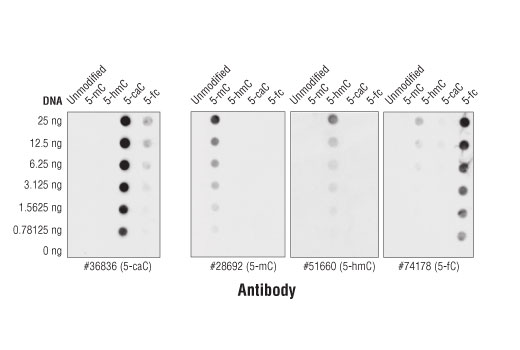
View Featured Offers >>
97763
DNA Cytosine Modification Antibody Sampler Kit
Primary Antibodies
Antibody Sampler Kit

DNA Cytosine Modification Antibody Sampler Kit #97763

Citations (0)
Confocal immunofluorescent analysis of 293T cells transfected with a construct expressing DYKDDDDK-tagged TET1 catalytic domain (TET1-CD) using 5-Methylcytosine (5-mC) (D3S2Z) Rabbit mAb (green) and DYKDDDDK Tag (9A3) Mouse mAb #8146 (red). Blue pseudocolor = DRAQ5® #4084 (fluorescent DNA dye). As expected, 293T cells expressing TET1-CD (red) exhibit decreased levels of 5-methylcytosine (green).
Specificity of various modified cytosine antibodies were determined by dot blot. The same sequence of a 387-base pair DNA fragment was generated by PCR using exclusively unmodified cytosine, 5-methylcytosine (5-mC), 5-hydroxymethylcytosine (5-hmC), 5-carboxylcytosine (5-caC), or 5-formylcytosine (5-fC). The respective DNA fragments were blotted onto a nylon membrane, UV cross-linked, and probed with 5-Carboxylcytosine (5-caC) (D7S8U) Rabbit mAb #36836, 5-Methylcytosine (5-mC) (D3S2Z) Rabbit mAb #28692, 5-Hydroxymethylcytosine (5-hmC) (HMC31) Mouse mAb #51660, and 5-Formylcytosine (5-fC) (D5D4K) Rabbit mAb #74178. As shown, the 5-Carboxylcytosine (5-caC) (D7S8U) Rabbit mAb shows specificity for 5-caC.
After the primary antibody is bound to the target protein, a complex with HRP-linked secondary antibody is formed. The LumiGLO® is added and emits light during enzyme catalyzed decomposition.
After the primary antibody is bound to the target protein, a complex with HRP-linked secondary antibody is formed. The LumiGLO* is added and emits light during enzyme catalyzed decomposition.
Confocal immunofluorescent analysis of 293T cells transfected with a construct expressing DDK-tagged TET1 catalytic domain (TET1-CD) using 5-Formylcytosine (5-fC) (D5D4K) Rabbit mAb (green) and DYKDDDDK Tag (9A3) Mouse mAb #8146 (red). Blue pseudocolor = DRAQ5® #4084 (fluorescent DNA dye). As expected, 293T cells expressing TET1-CD (red) exhibit increased levels of 5-formylcytosine (green).
DNA fragments from HCT 116 wild type (WT) and DNMT1/DNMT3B knock-out (KO) cells were blotted onto a nylon membrane, UV cross-linked, and probed with 5-Methylcytosine (5-mC) (D3S2Z) Rabbit mAb. The top panel shows the antibody detecting more methylated cytosine in the wild type cells, while the bottom panel shows the membrane stained with methylene blue.
Confocal immunofluorescent analysis of 293T cells transfected with a construct expressing DYKDDDDK-tagged TET1 catalytic domain (TET1-CD) using 5-Carboxylcytosine (5-caC) (D7S8U) Rabbit mAb (green) and DYKDDDDK Tag (9A3) Mouse mAb #8146 (red). Blue pseudocolor = DRAQ5® #4084 (fluorescent DNA dye). As expected, 293T cells expressing TET1-CD (red) exhibit increased levels of 5-carboxylcytosine (green).
Confocal immunofluorescent analysis of 293T cells transfected with a construct expressing DDK-tagged TET1 catalytic domain (TET1-CD) using 5-Hydroxymethylcytosine (5-hmC) (HMC31) Mouse mAb (green) and DYKDDDDK Tag Antibody #2368 (red). Blue pseudocolor = DRAQ5® #4084 (fluorescent DNA dye). As expected, 293T cells expressing TET1-CD (red) exhibit increased levels of 5-hydroxymethylcytosine (green).
The specificity of 5-Formylcytosine (5-fC) (D5D4K) Rabbit mAb was determined by dot blot. The same sequence of a 387-base pair DNA fragment was generated by PCR using exclusively unmodified cytosine, 5-methylcytosine (5-mC), 5-hydroxymethylcytosine (5-hmC), 5-carboxylcytosine (5-caC), or 5-formylcytosine (5-fC). The respective DNA fragments were blotted onto a nylon membrane, UV cross-linked, and probed with 5-Formylcytosine (5-fC) (D5D4K) Rabbit mAb. The upper panel shows the antibody only binding to the DNA fragment containing 5-fC, while the lower panel shows the membrane stained with methylene blue.
The specificity of 5-Methylcytosine (5-mC) (D3S2Z) Rabbit mAb was determined by dot blot. The same sequence of a 387 base pair DNA fragment was generated by PCR using exclusively unmodified cytosine, 5-methylcytosine (5-mC), 5-hydroxymethylcytosine (5-hmC), 5-carboxylcytosine (5-caC), or 5-formylcytosine (5-fC). The respective DNA fragments were blotted onto a nylon membrane, UV cross-linked, and probed with 5-Methylcytosine (5-mC) (D3S2Z) Rabbit mAb. The top panel shows the antibody only binding to the DNA fragment containing 5-mC, while the bottom panel shows the membrane stained with methylene blue.
5-Hydroxymethylcytosine (5-hmC) (HMC31) Mouse mAb specificity was determined by dot blot. The same sequence of a 387-base pair DNA fragment was generated by PCR using exclusively unmodified cytosine, 5-methylcytosine (5-mC), 5-hydroxymethylcytosine (5-hmC), 5-carboxylcytosine (5-caC), or 5-formylcytosine (5-fC). The respective DNA fragments were blotted onto a nylon membrane, UV cross-linked, and probed with 5-Hydroxymethylcytosine (5-hmC) (HMC31) Mouse mAb. The upper panel shows the antibody only binding to the DNA fragment containing 5-hmC, while the lower panel shows the membrane stained with methylene blue.
To Purchase # 97763
Cat. # Size Qty. Price
97763T
1 Kit  (4 x 20 microliters)

Product Includes Quantity Applications Reactivity MW(kDa) Isotype
5-Methylcytosine (5-mC) (D3S2Z) Rabbit mAb 28692 20 µl
  • IF
  • Dot Blot
All Rabbit IgG
5-Hydroxymethylcytosine (5-hmC) (HMC31) Mouse mAb 51660 20 µl
  • IF
  • Dot Blot
All Mouse IgG1
5-Carboxylcytosine (5-caC) (D7S8U) Rabbit mAb 36836 20 µl
  • IF
  • Dot Blot
All Rabbit IgG
5-Formylcytosine (5-fC) (D5D4K) Rabbit mAb 74178 20 µl
  • IF
  • Dot Blot
All Rabbit IgG
Anti-rabbit IgG, HRP-linked Antibody 7074 100 µl
  • WB
Goat 
Anti-mouse IgG, HRP-linked Antibody 7076 100 µl
  • WB
Horse 

Product Description

The DNA Cytosine Modification Antibody Sampler Kit provides an economical means of detecting the levels of cytosine modifications in DNA by dot blot using antibodies against 5-methylcytosine, 5-hydroxymethylcytosine, 5-formylcytosine, and 5-carboxylcytosine.

Specificity / Sensitivity

5-Methylcytosine (5-mC) (D3S2Z) Rabbit mAb recognizes endogenous levels of 5-methylcytosine. 5-Hydroxymethylcytosine (5-hmC) (HMC31) Mouse mAb recognizes endogenous levels of 5-hydroxymethylcytosine. 5-Formylcytosine (5-fC) (D5D4K) Rabbit mAb recognizes transfected levels of 5-formylcytosine. 5-Caroxylcytosine (5-caC) (D7S8U) Rabbit mAb recognizes transfected levels of 5-methylcytosine. These antibodies have been validated using ELISA and dot blot, and cross-reactivity was not observed with other marks. Many cells and tissues contain very low endogenous levels of 5-hmC, 5-fC, and 5-caC that may fall below the detection limits of these antibodies.

Source / Purification

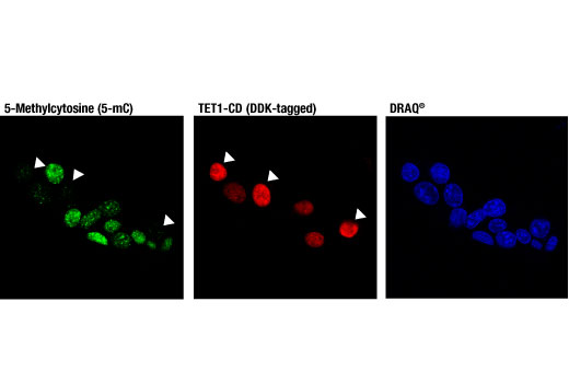
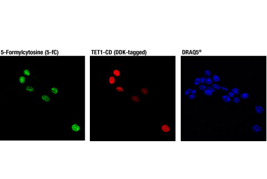
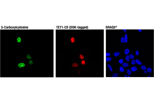
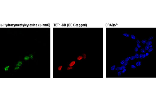
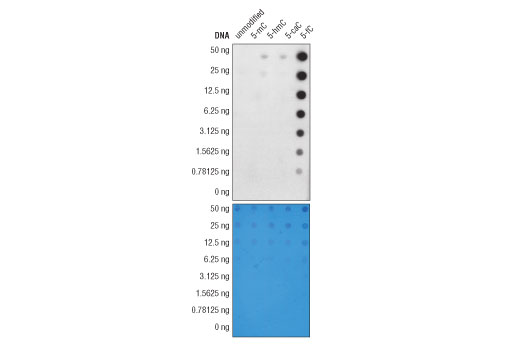
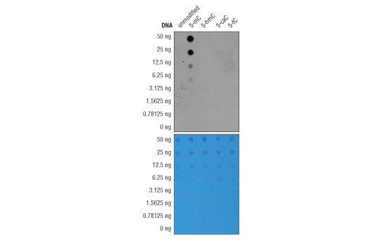
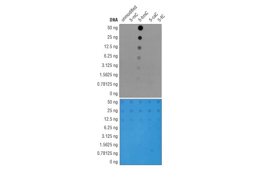
Monoclonal antibodies are produced by immunizing animals with 5-methylcytidine, 5-hydroxymethylcytidine, 5-formyl-2'-deoxycytosine, or 5-carboxylcytidine.

Background

Methylation of DNA at cytosine residues is a heritable, epigenetic modification that is critical for proper regulation of gene expression, genomic imprinting, and mammalian development (1,2). 5-methylcytosine is a repressive epigenetic mark established de novo by two enzymes, DNMT3a and DNMT3b, and is maintained by DNMT1 (3, 4). 5-methylcytosine was originally thought to be passively depleted during DNA replication. However, subsequent studies have shown that Ten-Eleven Translocation (TET) proteins TET1, TET2, and TET3 can catalyze the oxidation of methylated cytosine to 5-hydroxymethylcytosine (5-hmC) (5). Additionally, TET proteins can further oxidize 5-hmC to form 5-formylcytosine (5-fC) and 5-carboxylcytosine (5-caC), both of which are excised by thymine-DNA glycosylase (TDG), effectively linking cytosine oxidation to the base excision repair pathway and supporting active cytosine demethylation (6,7).
TET protein-mediated cytosine hydroxymethylation was initially demonstrated in mouse brain and embryonic stem cells (5, 8). Since then this modification has been discovered in many tissues, with the highest levels found in the brain (9). While 5-fC and 5-caC appear to be short-lived intermediate species, there is mounting evidence showing that 5-hmC is a distinct epigenetic mark with various unique functions (10,11). The modified base itself is stable in vivo and interacts with various readers, including MeCP2 (11,12). The global level of 5-hmC increases during brain development and 5-hmC is enriched at promoter regions and poised enhancers. Furthermore, there is an inverse correlation between levels of 5-hmC and histone H3K9 and H3K27 trimethylation, suggesting a role for 5-hmC in gene activation (12). Lower amounts of 5-hmC have been reported in various cancers, including myeloid leukemia and melanoma (13,14).

Limited Uses

Except as otherwise expressly agreed in a writing signed by a legally authorized representative of CST, the following terms apply to Products provided by CST, its affiliates or its distributors. Any Customer's terms and conditions that are in addition to, or different from, those contained herein, unless separately accepted in writing by a legally authorized representative of CST, are rejected and are of no force or effect.

Products are labeled with For Research Use Only or a similar labeling statement and have not been approved, cleared, or licensed by the FDA or other regulatory foreign or domestic entity, for any purpose. Customer shall not use any Product for any diagnostic or therapeutic purpose, or otherwise in any manner that conflicts with its labeling statement. Products sold or licensed by CST are provided for Customer as the end-user and solely for research and development uses. Any use of Product for diagnostic, prophylactic or therapeutic purposes, or any purchase of Product for resale (alone or as a component) or other commercial purpose, requires a separate license from CST. Customer shall (a) not sell, license, loan, donate or otherwise transfer or make available any Product to any third party, whether alone or in combination with other materials, or use the Products to manufacture any commercial products, (b) not copy, modify, reverse engineer, decompile, disassemble or otherwise attempt to discover the underlying structure or technology of the Products, or use the Products for the purpose of developing any products or services that would compete with CST products or services, (c) not alter or remove from the Products any trademarks, trade names, logos, patent or copyright notices or markings, (d) use the Products solely in accordance with CST Product Terms of Sale and any applicable documentation, and (e) comply with any license, terms of service or similar agreement with respect to any third party products or services used by Customer in connection with the Products.

For Research Use Only. Not for Use in Diagnostic Procedures.
Cell Signaling Technology is a trademark of Cell Signaling Technology, Inc.
All other trademarks are the property of their respective owners. Visit our Trademark Information page.