| Cat. # | Size | Qty. | Price |
|---|---|---|---|
| 38866T | 1 Kit (7 x 20 microliters) |
|
| Product Includes | Quantity | Applications | Reactivity | MW(kDa) | Isotype |
|---|---|---|---|---|---|
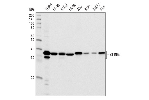
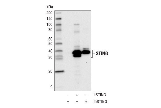
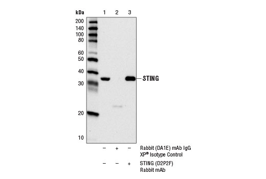
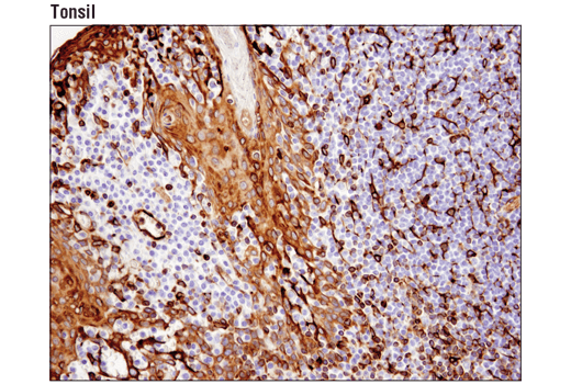
| STING (D2P2F) Rabbit mAb 13647 | 20 µl |
|
H M Hm | 33, 35 | Rabbit IgG |
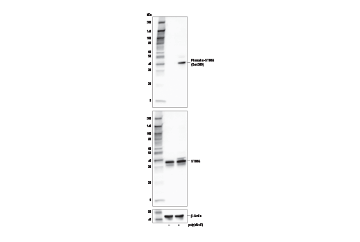
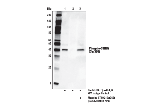
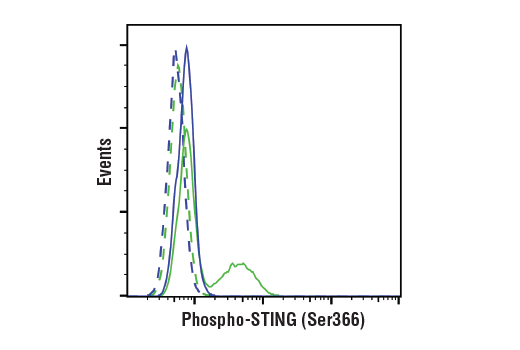
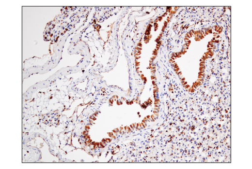
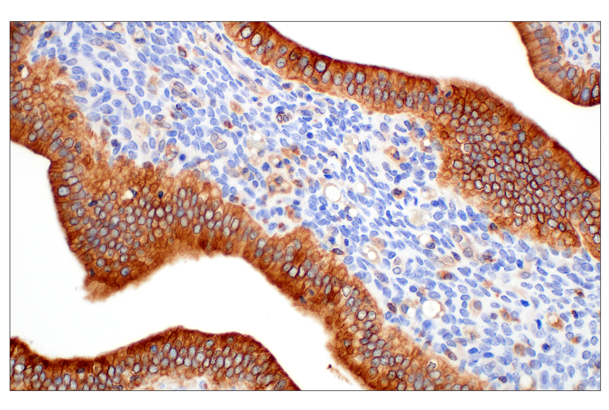
| Phospho-STING (Ser366) (E9A9K) Rabbit mAb 50907 | 20 µl |
|
H | 40 | Rabbit IgG |
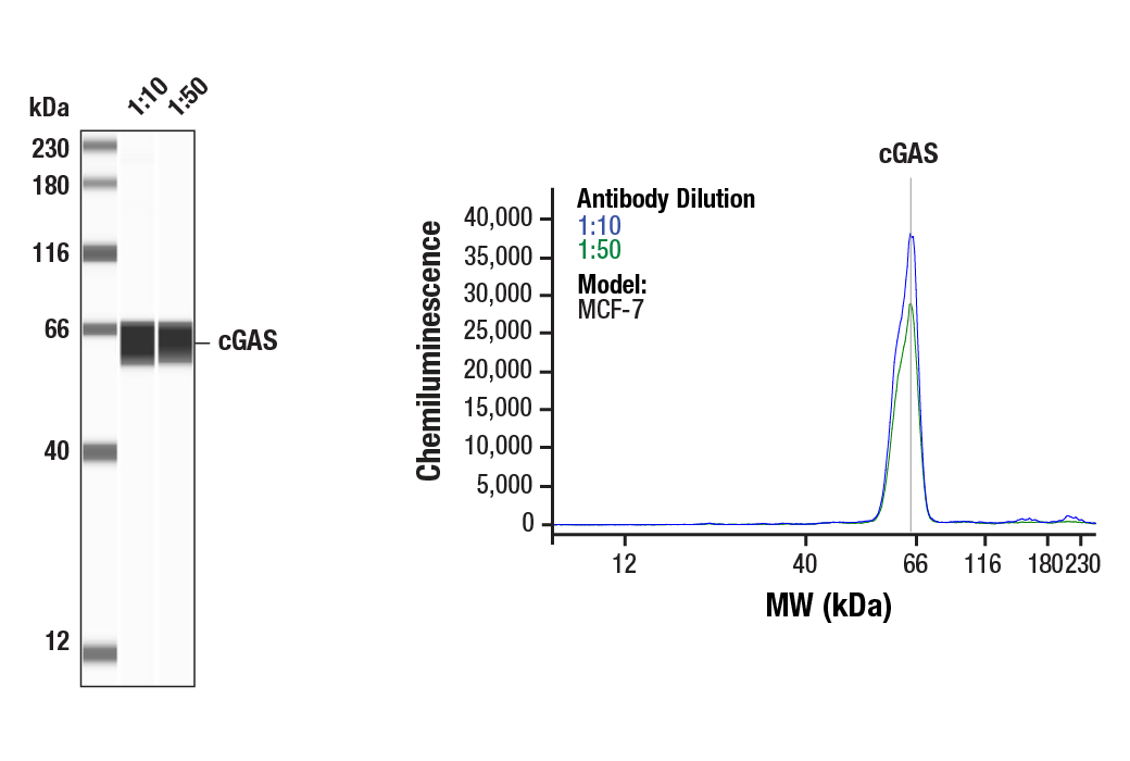
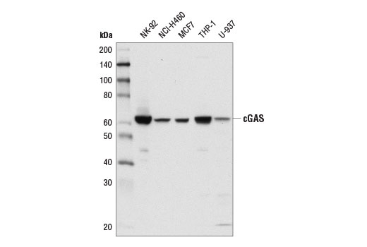
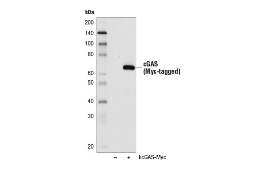
| cGAS (D1D3G) Rabbit mAb 15102 | 20 µl |
|
H | 62 | Rabbit IgG |
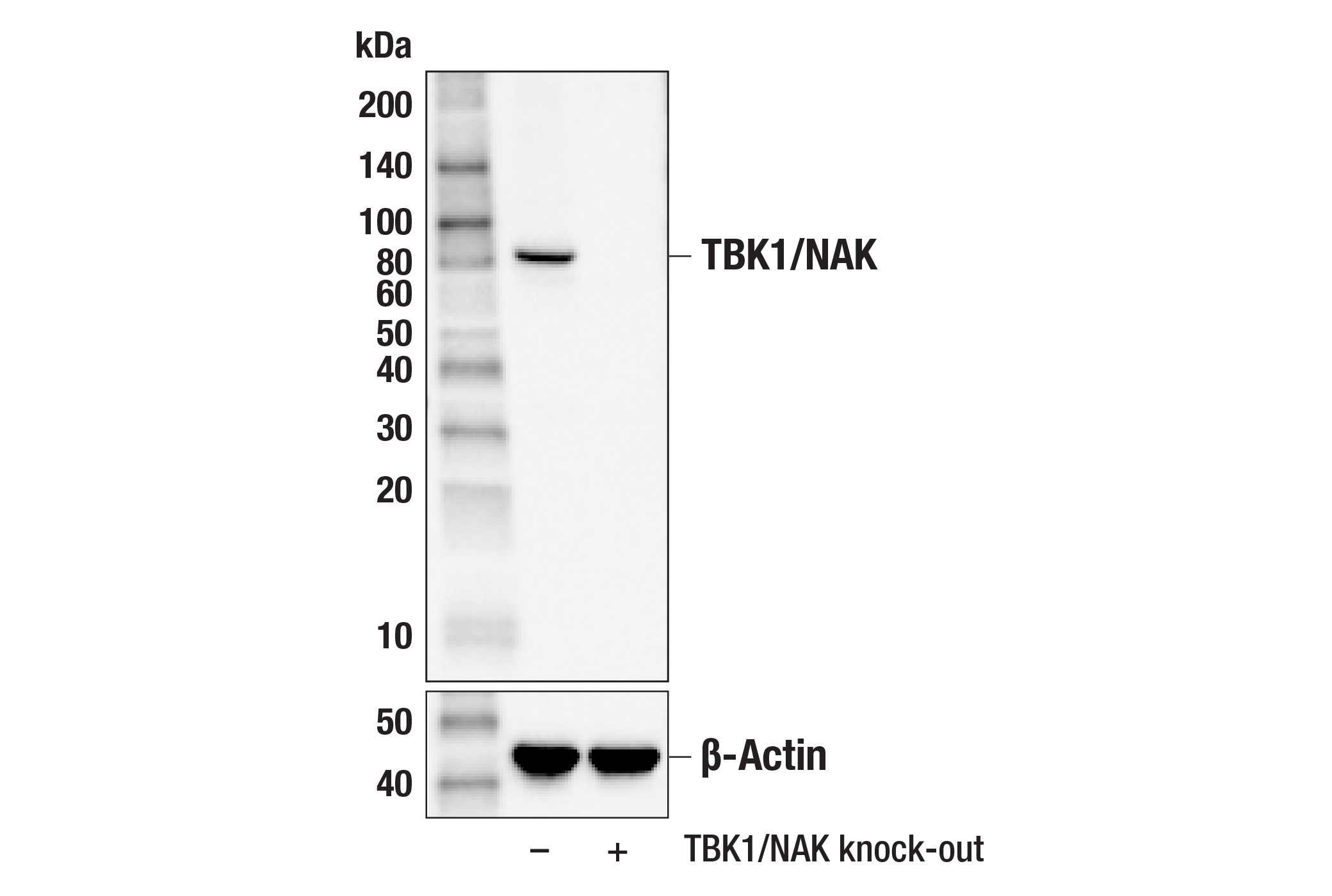
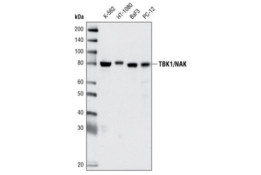
| TBK1/NAK (D1B4) Rabbit mAb 3504 | 20 µl |
|
H M R Mk | 84 | Rabbit IgG |
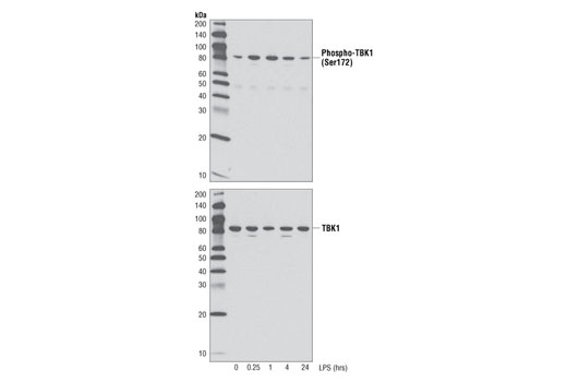
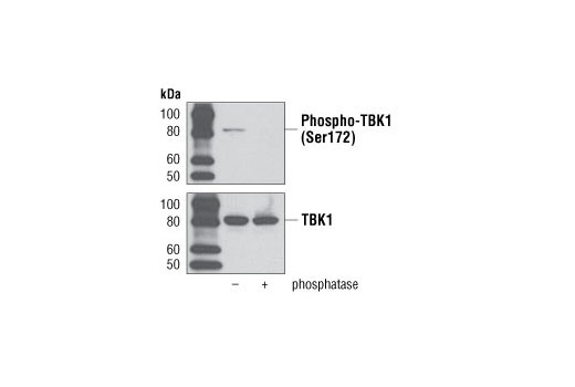
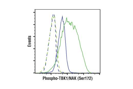
| Phospho-TBK1/NAK (Ser172) (D52C2) XP® Rabbit mAb 5483 | 20 µl |
|
H M | 84 | Rabbit IgG |
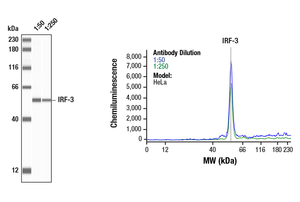
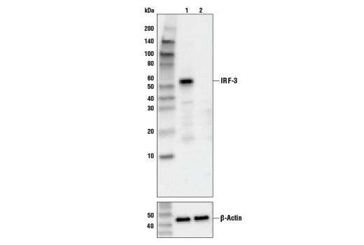
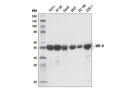
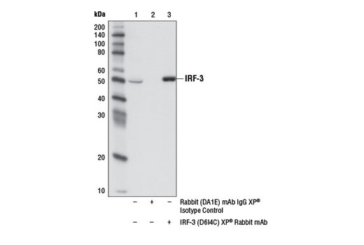
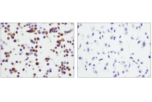
| IRF-3 (D6I4C) XP® Rabbit mAb 11904 | 20 µl |
|
H Mk | 50-55 | Rabbit IgG |
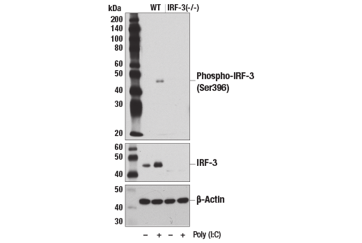
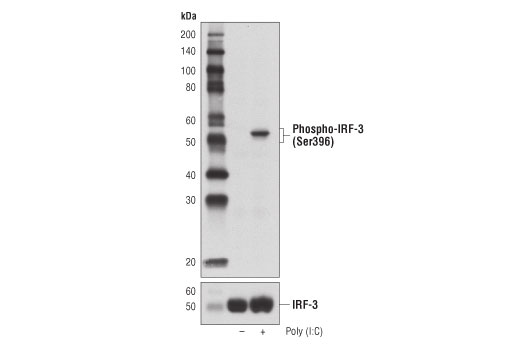
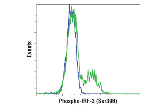
| Phospho-IRF-3 (Ser396) (D6O1M) Rabbit mAb 29047 | 20 µl |
|
H M R | 45-55 | Rabbit IgG |
| Anti-rabbit IgG, HRP-linked Antibody 7074 | 100 µl |
|
Goat |
Product Information
Monoclonal antibodies are produced by immunizing rabbits with synthetic peptides corresponding to residues surrounding Ala19 of human cGAS protein, Ser645 of human TBK1/NAK, Pro226 of human STING, and recombinant human IRF-3 protein. Phosphorylation-specific monoclonal antibodies are produced by immunizing rabbits with synthetic peptides corresponding to residues surrounding Ser366 of human STING protein, Ser172 of human TBK1, and Ser396 of human IRF-3.
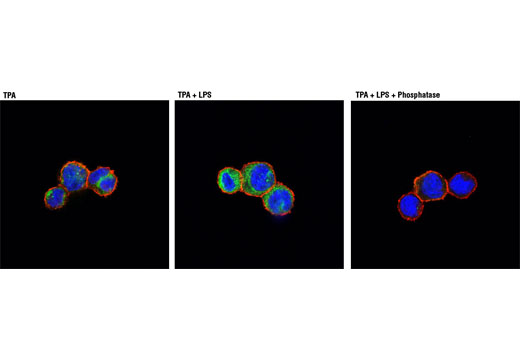
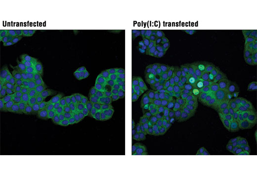
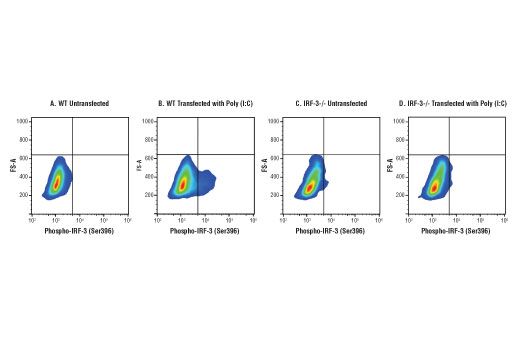
Stimulator of interferon genes (STING, TMEM173, MITA) is a transmembrane adaptor protein that is a critical component of the cellular innate immune response to pathogenic cytoplasmic DNA (1,2). STING is a ubiquitously expressed protein found predominantly in the ER (1). The enzyme cGAMP synthase (cGAS) produces the second messenger cyclic-GMP-AMP (cGAMP) in response to cytoplasmic DNA (3,4). cGAMP binds and activates STING (3,4). In addition, detection of cytoplasmic DNA by nucleic acid sensors, including DDX41 or IFI16, results in STING activation (5,6). Following activation, STING translocates with TBK1 to perinuclear endosomes and gets phosphorylated by ULK1 at Ser366 (Ser365 in mouse) (7,8). The TBK1 kinase phosphorylates and activates IRF-3 and NF-κB, which leads to the induction of type I interferon and other immune response genes (1,2,7).
Explore pathways related to this product.
STRING - Known and Predicted Protein-Protein Interactions.
Except as otherwise expressly agreed in a writing signed by a legally authorized representative of CST, the following terms apply to Products provided by CST, its affiliates or its distributors. Any Customer's terms and conditions that are in addition to, or different from, those contained herein, unless separately accepted in writing by a legally authorized representative of CST, are rejected and are of no force or effect.
Products are labeled with For Research Use Only or a similar labeling statement and have not been approved, cleared, or licensed by the FDA or other regulatory foreign or domestic entity, for any purpose. Customer shall not use any Product for any diagnostic or therapeutic purpose, or otherwise in any manner that conflicts with its labeling statement. Products sold or licensed by CST are provided for Customer as the end-user and solely for research and development uses. Any use of Product for diagnostic, prophylactic or therapeutic purposes, or any purchase of Product for resale (alone or as a component) or other commercial purpose, requires a separate license from CST. Customer shall (a) not sell, license, loan, donate or otherwise transfer or make available any Product to any third party, whether alone or in combination with other materials, or use the Products to manufacture any commercial products, (b) not copy, modify, reverse engineer, decompile, disassemble or otherwise attempt to discover the underlying structure or technology of the Products, or use the Products for the purpose of developing any products or services that would compete with CST products or services, (c) not alter or remove from the Products any trademarks, trade names, logos, patent or copyright notices or markings, (d) use the Products solely in accordance with CST Product Terms of Sale and any applicable documentation, and (e) comply with any license, terms of service or similar agreement with respect to any third party products or services used by Customer in connection with the Products.