| Cat. # | Size | Qty. | Price |
|---|---|---|---|
| 9862T | 1 Kit (5 x 20 microliters) |
|
| Product Includes | Quantity | Applications | Reactivity | MW(kDa) | Isotype |
|---|---|---|---|---|---|
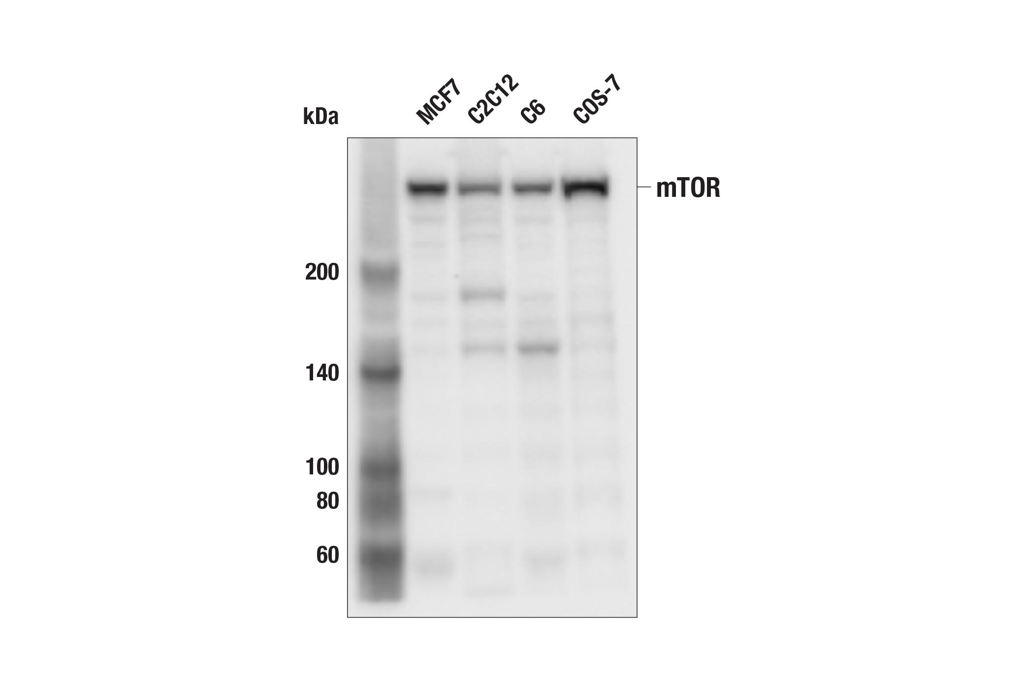
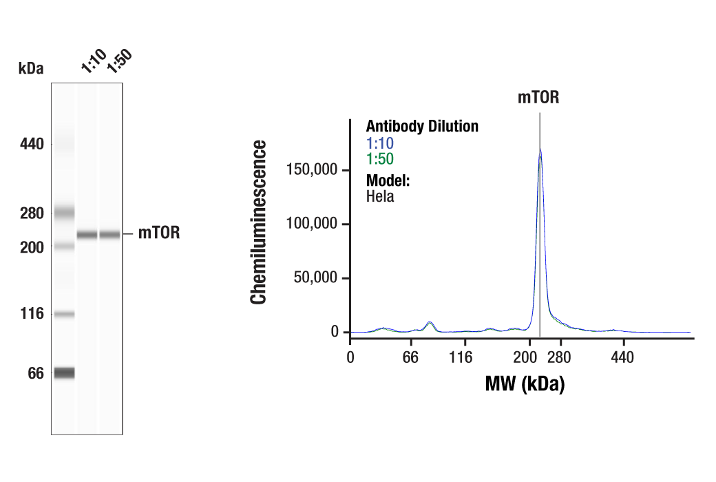
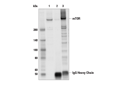
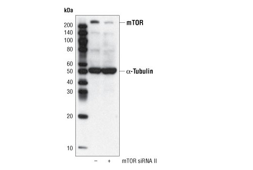
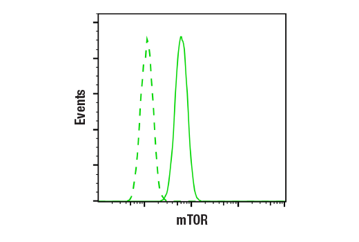
| mTOR (7C10) Rabbit mAb 2983 | 20 µl |
|
H M R Mk | 289 | Rabbit IgG |
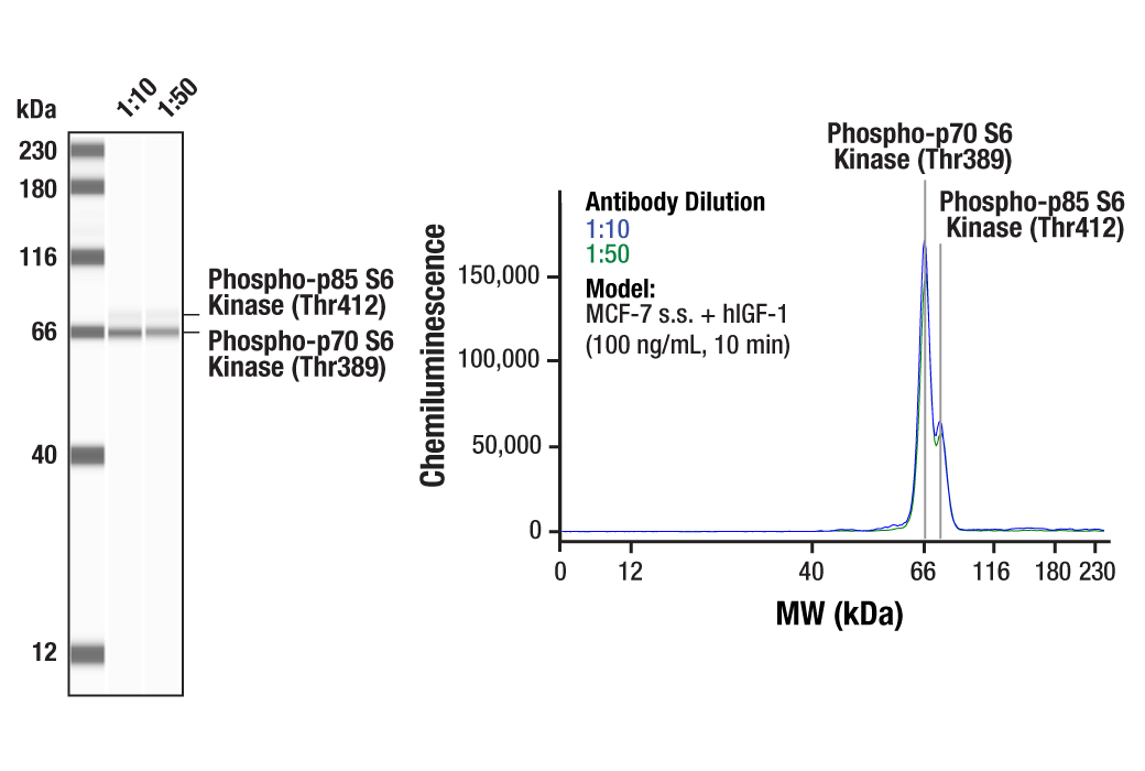
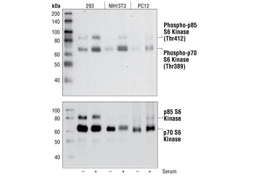
| Phospho-p70 S6 Kinase (Thr389) (108D2) Rabbit mAb 9234 | 20 µl |
|
H M R Mk | 70, 85 | Rabbit IgG |
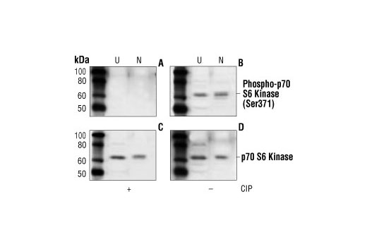
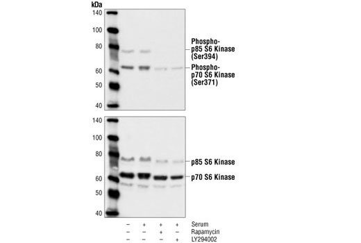
| Phospho-p70 S6 Kinase (Ser371) Antibody 9208 | 20 µl |
|
H M R Mk | 70, 85 | Rabbit |
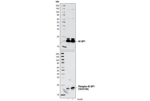
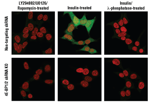
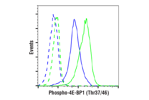
| Phospho-4E-BP1 (Thr37/46) (236B4) Rabbit mAb 2855 | 20 µl |
|
H M R Mk Dm | 15 to 20 | Rabbit IgG |
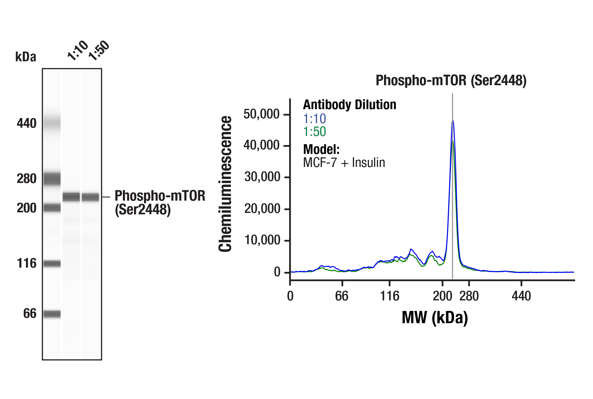
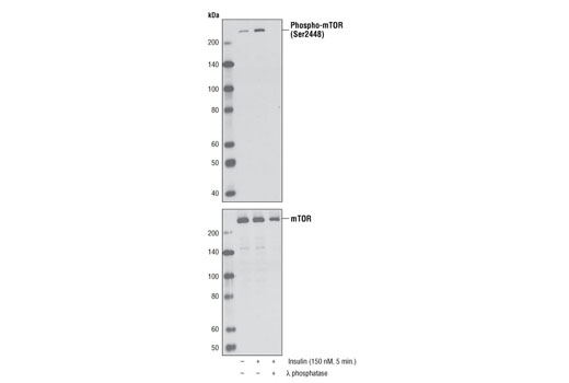
| Phospho-mTOR (Ser2448) (D9C2) XP® Rabbit mAb 5536 | 20 µl |
|
H M R Mk | 289 | Rabbit IgG |
| Anti-rabbit IgG, HRP-linked Antibody 7074 | 100 µl |
|
Goat |
Product Information
Polyclonal antibodies are produced by immunizing animals with a synthetic phosphopeptide corresponding to residues surrounding Ser371 of human p70 S6 kinase. Polyclonal antibodies are purified by protein A and peptide affinity chromatography. Phospho-specific rabbit monoclonal antibodies are produced by immunizing animals with synthetic phosphopeptides corresponding to residues surrounding Thr389 of human p70 S6 kinase, Thr37 and Thr46 of mouse 4E-BP1 and the Ser2448 site of human mTOR. The mTOR (7C10) Monoclonal antibody is produced by immunizing animals with a synthetic peptide corresponding to residues surrounding Ser2481 of human mTOR.
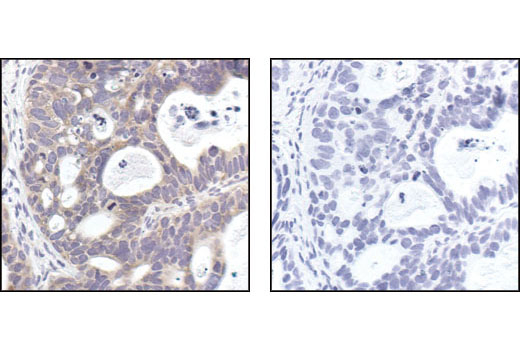
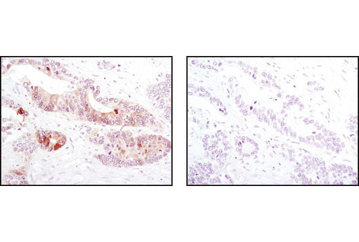
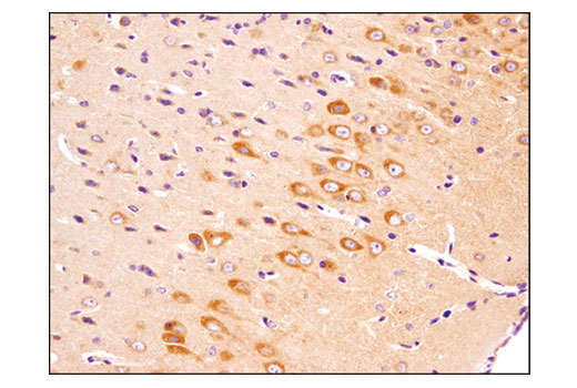
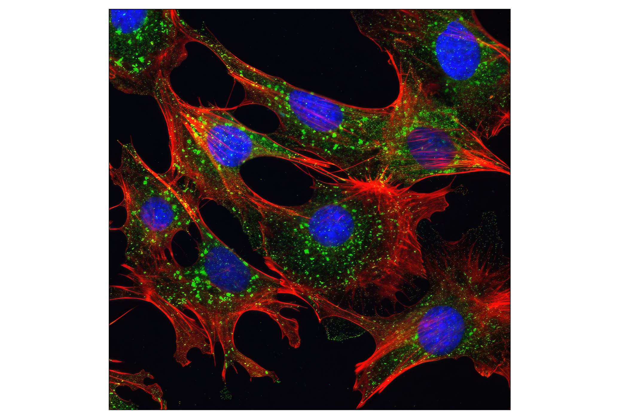
The mammalian target of rapamycin (mTOR, FRAP, RAFT) is a Ser/Thr protein kinase (1-3) that functions as an ATP and amino acid sensor to balance nutrient availability and cell growth (4,5). When sufficient nutrients are available, mTOR responds to a phosphatidic acid-mediated signal to transmit a positive signal to p70 S6 kinase and participate in the inactivation of the eIF4E inhibitor, 4E-BP1 (6). These events result in the translation of specific mRNA subpopulations. mTOR is phosphorylated at Ser2448 via the PI3 kinase/Akt signaling pathway and autophosphorylated at Ser2481 (7,8). mTOR plays a key role in cell growth and homeostasis and may be abnormally regulated in tumors. For these reasons, mTOR is currently under investigation as a potential target for anti-cancer therapy (9).
The regulatory associated protein of mTOR (Raptor) interacts with mTOR to mediate mTOR signaling to downstream targets (10,11). Raptor binds to mTOR substrates, such as 4E-BP1 and p70 S6 kinase, through their TOR signaling (TOS) motifs and is required for mTOR-mediated substrate phosphorylation (12,13). Binding of the FKBP12-rapamycin complex to mTOR inhibits mTOR-raptor interaction, which suggests a mechanism for the inhibition of mTOR signaling by rapamycin (14). This mTOR-raptor interaction and its regulation by nutrients and/or rapamycin are dependent on a protein called GβL (15). GβL is part of the rapamycin-insensitive complex between mTOR and rictor (rapamycin-insensitive companion of mTOR) and may mediate rictor-mTOR signaling to PKCα and other downstream targets (16). The rictor-mTOR complex has been identified as the previously elusive PDK2 responsible for the phosphorylation of Akt/PKB at Ser473, which is required for PDK1 phosphorylation of Akt/PKB at Thr308 and full activation of Akt/PKB (17).
Except as otherwise expressly agreed in a writing signed by a legally authorized representative of CST, the following terms apply to Products provided by CST, its affiliates or its distributors. Any Customer's terms and conditions that are in addition to, or different from, those contained herein, unless separately accepted in writing by a legally authorized representative of CST, are rejected and are of no force or effect.
Products are labeled with For Research Use Only or a similar labeling statement and have not been approved, cleared, or licensed by the FDA or other regulatory foreign or domestic entity, for any purpose. Customer shall not use any Product for any diagnostic or therapeutic purpose, or otherwise in any manner that conflicts with its labeling statement. Products sold or licensed by CST are provided for Customer as the end-user and solely for research and development uses. Any use of Product for diagnostic, prophylactic or therapeutic purposes, or any purchase of Product for resale (alone or as a component) or other commercial purpose, requires a separate license from CST. Customer shall (a) not sell, license, loan, donate or otherwise transfer or make available any Product to any third party, whether alone or in combination with other materials, or use the Products to manufacture any commercial products, (b) not copy, modify, reverse engineer, decompile, disassemble or otherwise attempt to discover the underlying structure or technology of the Products, or use the Products for the purpose of developing any products or services that would compete with CST products or services, (c) not alter or remove from the Products any trademarks, trade names, logos, patent or copyright notices or markings, (d) use the Products solely in accordance with CST Product Terms of Sale and any applicable documentation, and (e) comply with any license, terms of service or similar agreement with respect to any third party products or services used by Customer in connection with the Products.