| Cat. # | Size | Qty. | Price |
|---|---|---|---|
| 9916T | 1 Kit (7 x 20 microliters) |
|
| Product Includes | Quantity | Applications | Reactivity | MW(kDa) | Isotype |
|---|---|---|---|---|---|
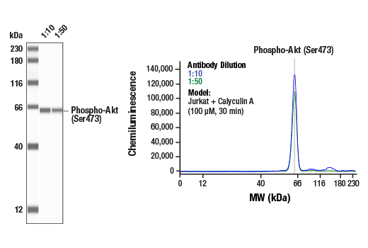
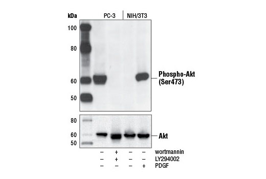
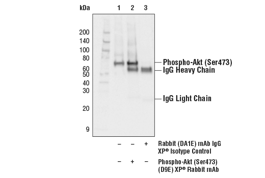
| Phospho-Akt (Ser473) (D9E) XP® Rabbit mAb 4060 | 20 µl |
|
H M R Hm Mk Dm Z B | 60 | Rabbit IgG |
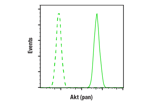
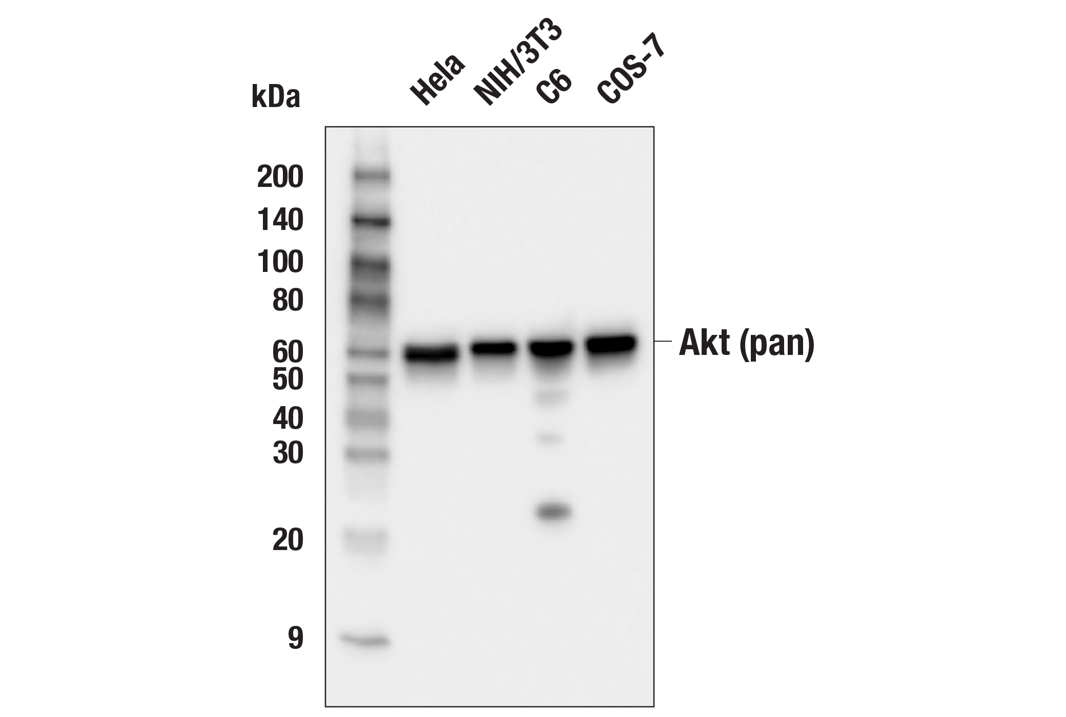
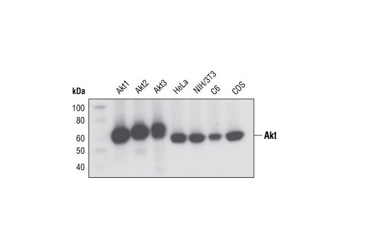
| Akt (pan) (C67E7) Rabbit mAb 4691 | 20 µl |
|
H M R Mk Dm | 60 | Rabbit IgG |
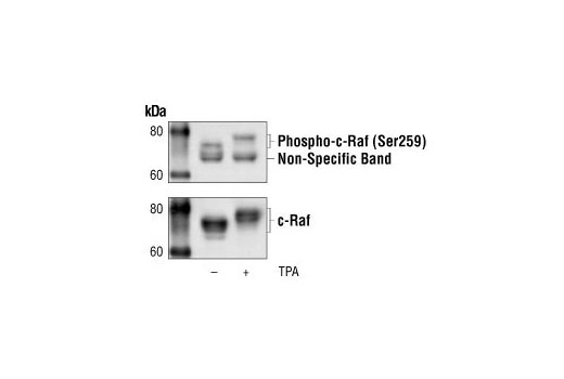
| Phospho-c-Raf (Ser259) Antibody 9421 | 20 µl |
|
H M R Mk X | 74 | Rabbit |
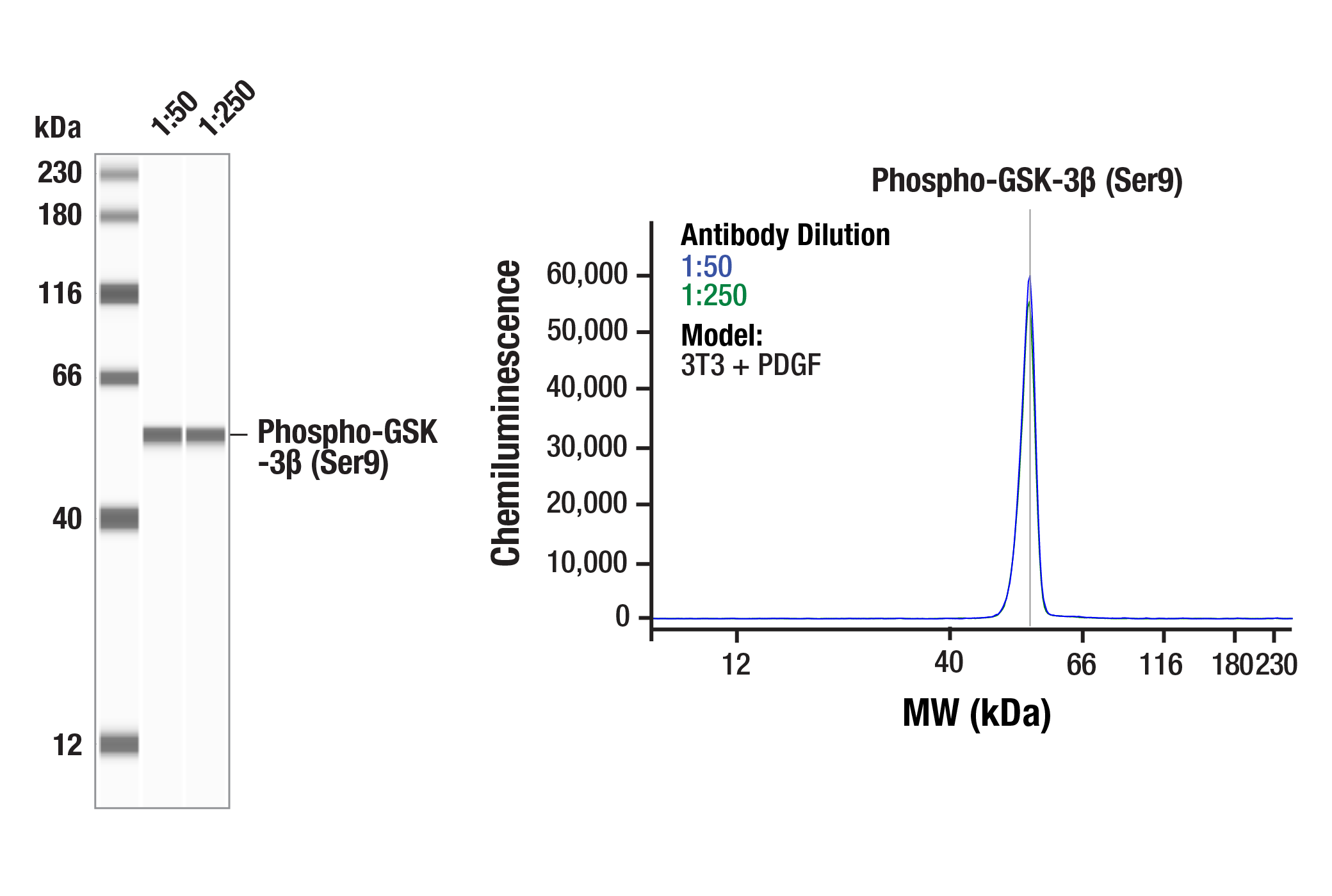
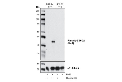
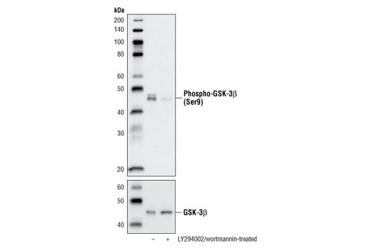
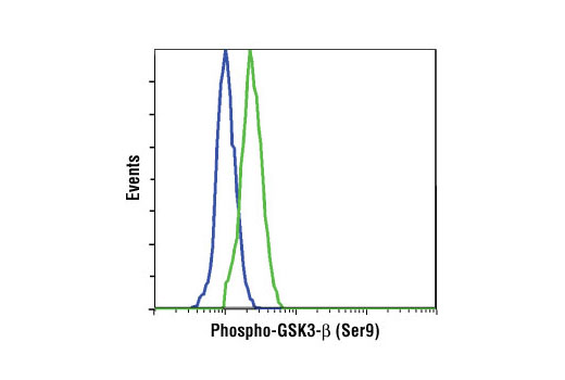
| Phospho-GSK-3β (Ser9) (D85E12) XP® Rabbit mAb 5558 | 20 µl |
|
H M R Hm | 46 | Rabbit IgG |
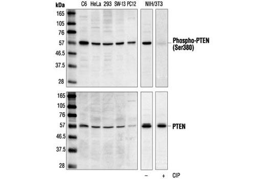
| Phospho-PTEN (Ser380) Antibody 9551 | 20 µl |
|
H M R | 54 | Rabbit |
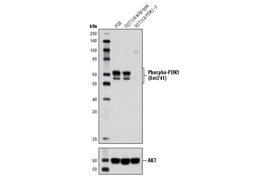
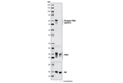
| Phospho-PDK1 (Ser241) (C49H2) Rabbit mAb 3438 | 20 µl |
|
H M R | 58 to 68 | Rabbit IgG |
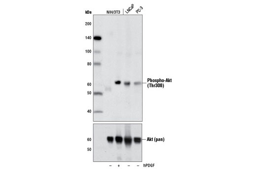
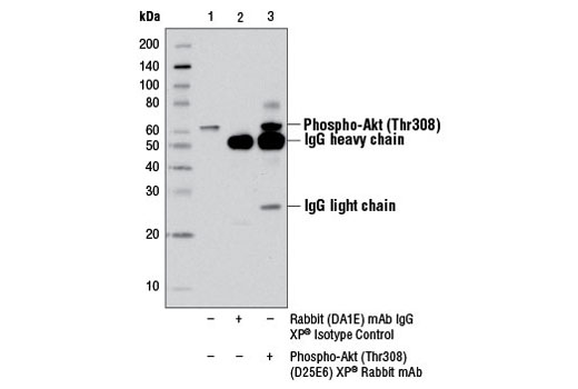
| Phospho-Akt (Thr308) (D25E6) XP® Rabbit mAb 13038 | 20 µl |
|
H M R Mk | 60 | Rabbit IgG |
| Anti-rabbit IgG, HRP-linked Antibody 7074 | 100 µl |
|
Goat |
Product Information
Antibodies are produced by immunizing rabbits with synthetic phosphopeptides corresponding to residues surrounding Ser473 or Thr308 of human Akt, Ser9 of human GSK-3ß, Ser259 of human c-Raf, Ser380 of human PTEN or Ser241 of human PDK1. Polyclonal antibodies are purified by protein A and peptide affinity chromatography.
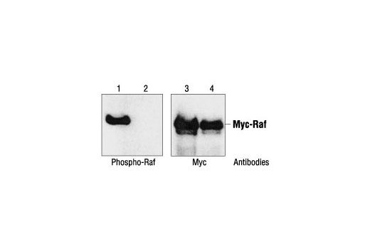
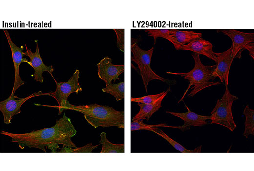
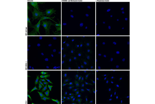
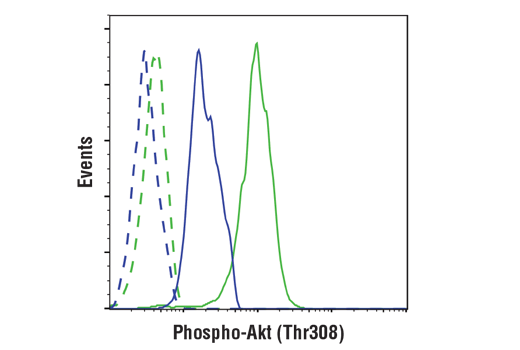
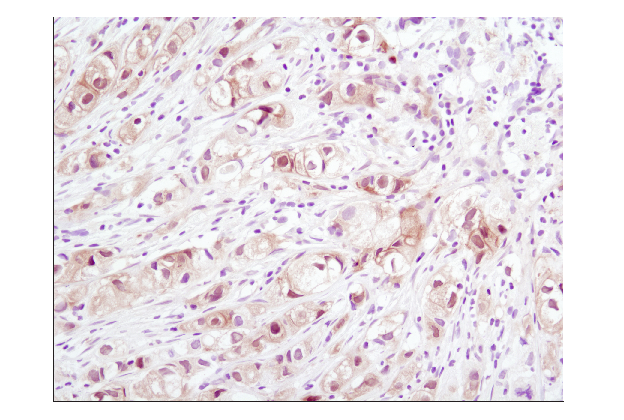
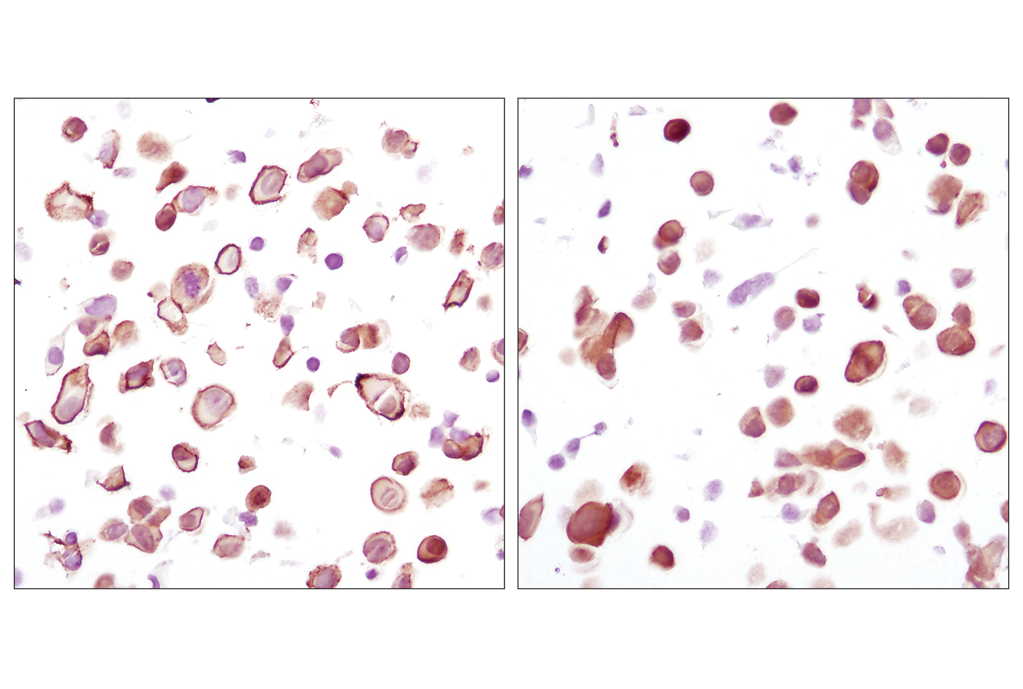
Akt, also referred to as PKB or Rac, plays a critical role in controlling cell survival and apoptosis (1-3). This protein kinase is activated by insulin and various growth and survival factors to function in a wortmannin-sensitive pathway involving PI3 kinase (2,3). Akt is activated by phospholipid binding and activation loop phosphorylation at Thr308 by PDK1 (4) and by phosphorylation within the carboxy terminus at Ser473. The previously elusive PDK2 responsible for phosphorylation of Akt at Ser473 has been identified as mammalian target of rapamycin (mTOR) in a rapamycin-insensitive complex with rictor and Sin1 (5,6). Akt promotes cell survival by inhibiting apoptosis through phosphorylation and inactivation of several targets, including Bad (7), forkhead transcription factors (8), c-Raf (9), and caspase-9. PTEN phosphatase is a major negative regulator of the PI3K/Akt signaling pathway (10). LY294002 is a specific PI3 kinase inhibitor (11). Another essential Akt function is the regulation of glycogen synthesis through phosphorylation and inactivation of GSK-3α and β (12,13). Akt may also play a role in insulin stimulation of glucose transport (12). In addition to its role in survival and glycogen synthesis, Akt is involved in cell cycle regulation by preventing GSK-3β-mediated phosphorylation and degradation of cyclin D1 (14) and by negatively regulating the cyclin-dependent kinase inhibitors p27 Kip1 (15) and p21 Waf1/Cip1 (16). Akt also plays a critical role in cell growth by directly phosphorylating mTOR in a rapamycin-sensitive complex containing raptor (17). More importantly, Akt phosphorylates and inactivates tuberin (TSC2), an inhibitor of mTOR within the mTOR-raptor complex (18,19).
Explore pathways related to this product.
STRING - Known and Predicted Protein-Protein Interactions.
Except as otherwise expressly agreed in a writing signed by a legally authorized representative of CST, the following terms apply to Products provided by CST, its affiliates or its distributors. Any Customer's terms and conditions that are in addition to, or different from, those contained herein, unless separately accepted in writing by a legally authorized representative of CST, are rejected and are of no force or effect.
Products are labeled with For Research Use Only or a similar labeling statement and have not been approved, cleared, or licensed by the FDA or other regulatory foreign or domestic entity, for any purpose. Customer shall not use any Product for any diagnostic or therapeutic purpose, or otherwise in any manner that conflicts with its labeling statement. Products sold or licensed by CST are provided for Customer as the end-user and solely for research and development uses. Any use of Product for diagnostic, prophylactic or therapeutic purposes, or any purchase of Product for resale (alone or as a component) or other commercial purpose, requires a separate license from CST. Customer shall (a) not sell, license, loan, donate or otherwise transfer or make available any Product to any third party, whether alone or in combination with other materials, or use the Products to manufacture any commercial products, (b) not copy, modify, reverse engineer, decompile, disassemble or otherwise attempt to discover the underlying structure or technology of the Products, or use the Products for the purpose of developing any products or services that would compete with CST products or services, (c) not alter or remove from the Products any trademarks, trade names, logos, patent or copyright notices or markings, (d) use the Products solely in accordance with CST Product Terms of Sale and any applicable documentation, and (e) comply with any license, terms of service or similar agreement with respect to any third party products or services used by Customer in connection with the Products.