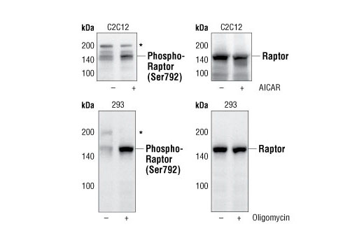
*Cross-reacting bands at 200 kDa.
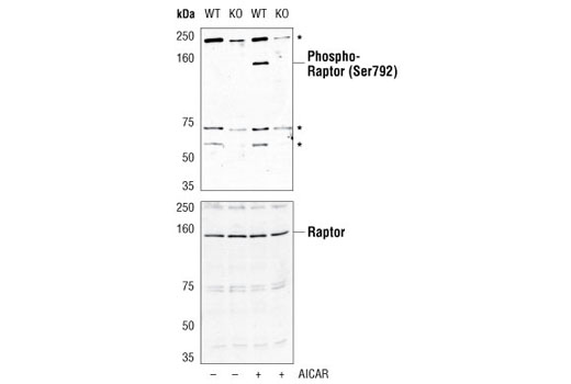
*Cross-reacting bands at 60, 70 and 240 kDa
| Cat. # | Size | Qty. | Price |
|---|---|---|---|
| 8359T | 1 Kit (7 x 20 microliters) |
|
| Product Includes | Quantity | Applications | Reactivity | MW(kDa) | Isotype |
|---|---|---|---|---|---|
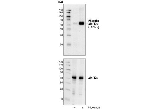
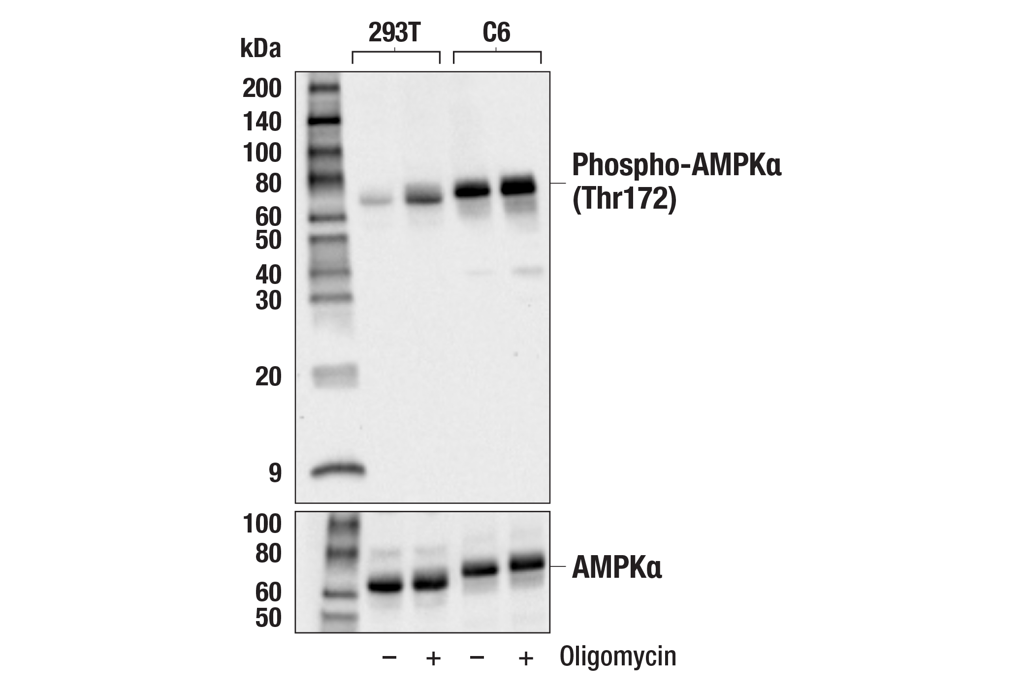
| Phospho-AMPKα (Thr172) (40H9) Rabbit mAb 2535 | 20 µl |
|
H M R Hm Mk Dm Sc | 62 | Rabbit IgG |
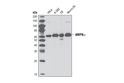
| AMPKα (D63G4) Rabbit mAb 5832 | 20 µl |
|
H M R Mk B | 62 | Rabbit |
| Phospho-Raptor (Ser792) Antibody 2083 | 20 µl |
|
H M R | 150 | Rabbit |
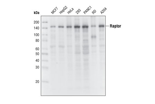
| Raptor (24C12) Rabbit mAb 2280 | 20 µl |
|
H M R Mk | 150 | Rabbit |
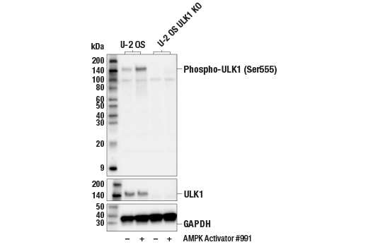
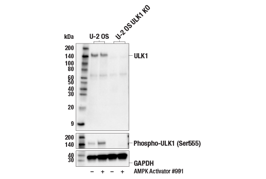
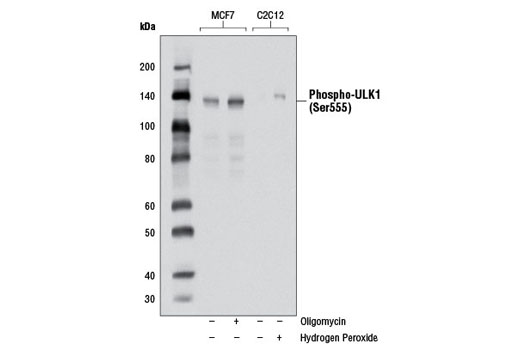
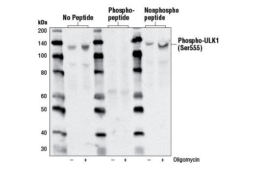
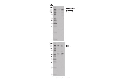
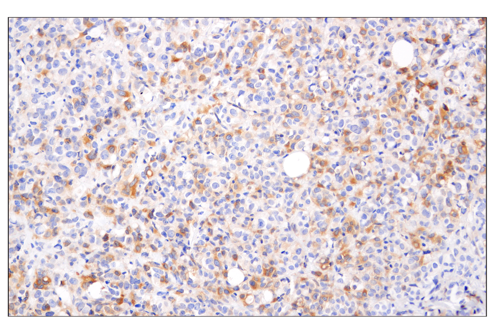
| Phospho-ULK1 (Ser555) (D1H4) Rabbit mAb 5869 | 20 µl |
|
H M | 140-150 | Rabbit IgG |
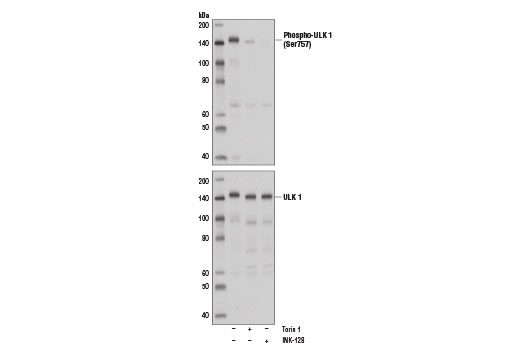
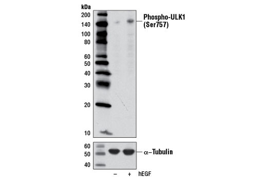
| Phospho-ULK1 (Ser757) Antibody 6888 | 20 µl |
|
H M Mk | 140-150 | Rabbit |
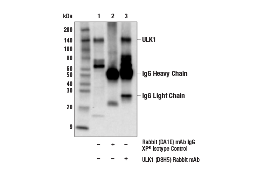
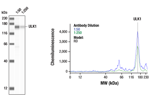
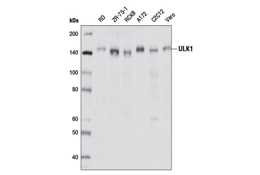
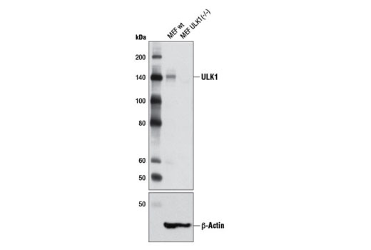
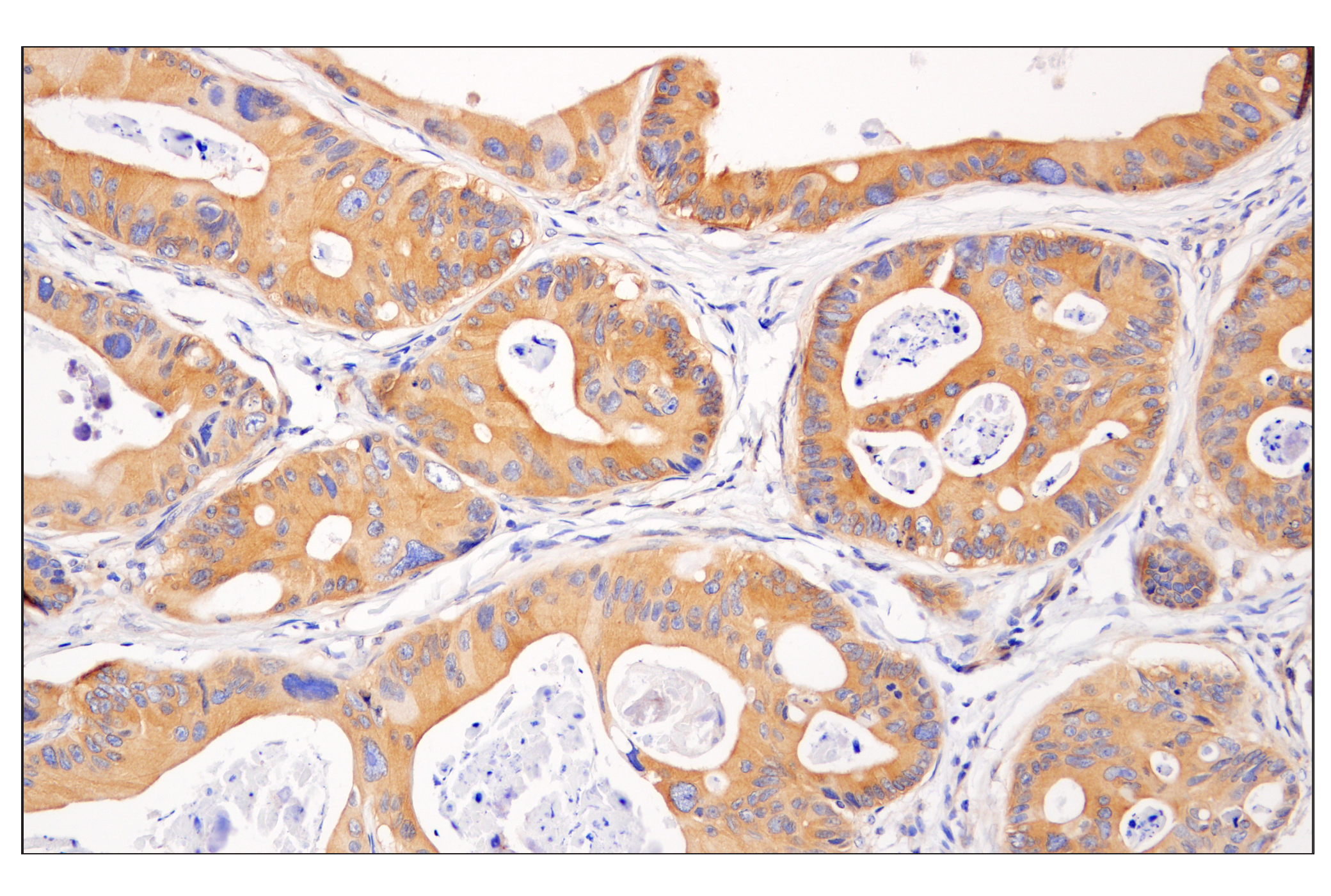
| ULK1 (D8H5) Rabbit mAb 8054 | 20 µl |
|
H M R Mk | 150 | Rabbit IgG |
| Anti-rabbit IgG, HRP-linked Antibody 7074 | 100 µl |
|
Goat |
Product Information
Activation-state specific monoclonal antibodies are produced by immunizing animals with a synthetic phosphopeptides corresponding to residues surrounding Ser555 of human ULK1 protein or residues surrounding Thr172 of human AMPKα protein. Monoclonal antibodies are produced by immunizing animals with a synthetic peptides corresponding to human raptor protein, residues surrounding Lys40 of human AMPKα protein, or residues surrounding Arg600 of human ULK1 protein. Activation-state specific polyclonal antibodies are produced by immunizing animals with synthetic phosphopeptides corresponding to residues surrounding Ser757 of human ULK1 protein or residues surrounding Ser792 of human raptor protein. Polyclonal antibodies are purified by protein A and peptide affinity chromatography.
Two related serine/threonine kinases, UNC-51-like kinase 1 and 2 (ULK1, ULK2), were discovered as mammalian homologs of the C. elegans gene unc-51 in which mutants exhibited abnormal axonal extension and growth (1-4). Both proteins are widely expressed and contain an amino-terminal kinase domain followed by a central proline/serine rich domain and a highly conserved carboxy-terminal domain. The roles of ULK1 and ULK2 in axon growth have been linked to studies showing that the kinases are localized to neuronal growth cones and are involved in endocytosis of critical growth factors, such as NGF (5). Yeast two-hybrid studies found ULK1/2 associated with modulators of the endocytic pathway, SynGAP, and syntenin (6). Structural similarity of ULK1/2 has also been recognized with the yeast autophagy protein Atg1/Apg1 (7). Knockdown experiments using siRNA demonstrated that ULK1 is essential for autophagy (8), a catabolic process for the degradation of bulk cytoplasmic contents (9,10). It appears that Atg1/ULK1 can act as a convergence point for multiple signals that control autophagy (11), and can bind to several autophagy-related (Atg) proteins, regulating phosphorylation states and protein trafficking (12-16).
Raptor mediates the binding of mTORC1 to ULK1, which phosphorylates and inhibits ULK1 under nutrient rich conditions. AMPK also associates directly with ULK1 and, upon nutrient deprivation, can readily reverse the inhibitory effect of mTORC1 by phosphorylating raptor and initiating autophagy (17,18).
Explore pathways related to this product.
STRING - Known and Predicted Protein-Protein Interactions.
Except as otherwise expressly agreed in a writing signed by a legally authorized representative of CST, the following terms apply to Products provided by CST, its affiliates or its distributors. Any Customer's terms and conditions that are in addition to, or different from, those contained herein, unless separately accepted in writing by a legally authorized representative of CST, are rejected and are of no force or effect.
Products are labeled with For Research Use Only or a similar labeling statement and have not been approved, cleared, or licensed by the FDA or other regulatory foreign or domestic entity, for any purpose. Customer shall not use any Product for any diagnostic or therapeutic purpose, or otherwise in any manner that conflicts with its labeling statement. Products sold or licensed by CST are provided for Customer as the end-user and solely for research and development uses. Any use of Product for diagnostic, prophylactic or therapeutic purposes, or any purchase of Product for resale (alone or as a component) or other commercial purpose, requires a separate license from CST. Customer shall (a) not sell, license, loan, donate or otherwise transfer or make available any Product to any third party, whether alone or in combination with other materials, or use the Products to manufacture any commercial products, (b) not copy, modify, reverse engineer, decompile, disassemble or otherwise attempt to discover the underlying structure or technology of the Products, or use the Products for the purpose of developing any products or services that would compete with CST products or services, (c) not alter or remove from the Products any trademarks, trade names, logos, patent or copyright notices or markings, (d) use the Products solely in accordance with CST Product Terms of Sale and any applicable documentation, and (e) comply with any license, terms of service or similar agreement with respect to any third party products or services used by Customer in connection with the Products.