Would you like to visit your country specific website?
Antibodies, reagents, and resources, such as pathways, protocols & videos, to help you accelerate your research.
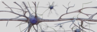
Learn about key cellular mechanisms that drive neurodegenerative diseases.
Important epi biomarkers, regulators, and histone modifications for several types of cancer.
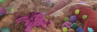
Amplify multiple biomarkers simultaneously in FFPE tissue and generate images in 2 days.
Download “Solutions for Advancing Neurodenerative Therapeutics Discovery” and discover more!
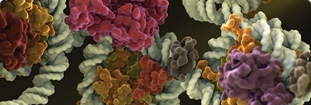
How a collaboration between Cornell, MIT, Yale, and CST is elucidating cellular pathways.