| Cat. # | Size | Qty. | Price |
|---|---|---|---|
| 29530T | 1 Kit (5 x 20 microliters) |
|
| Product Includes | Quantity | Applications | Reactivity | MW(kDa) | Isotype |
|---|---|---|---|---|---|
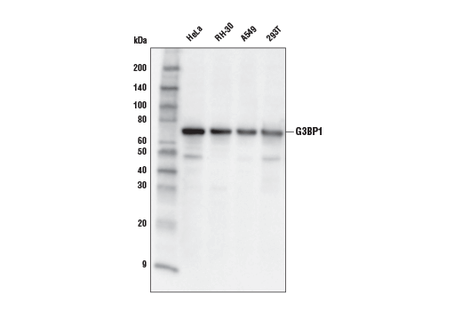
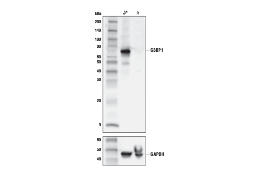
| G3BP1 (E9G1M) XP® Rabbit mAb 61559 | 20 µl |
|
H Mk | 68 | Rabbit IgG |
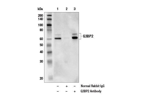
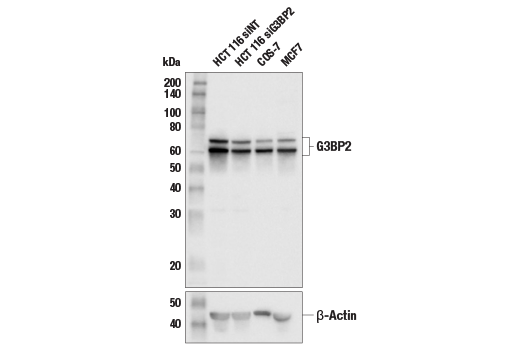
| G3BP2 Antibody 31799 | 20 µl |
|
H M R Mk | 60, 70 | Rabbit |
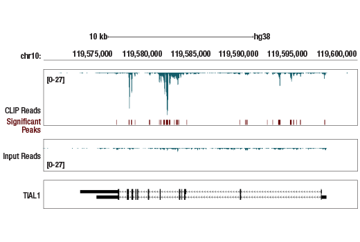
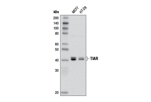
| TIAR (D32D3) XP® Rabbit mAb 8509 | 20 µl |
|
H M R Mk | 42 | Rabbit IgG |
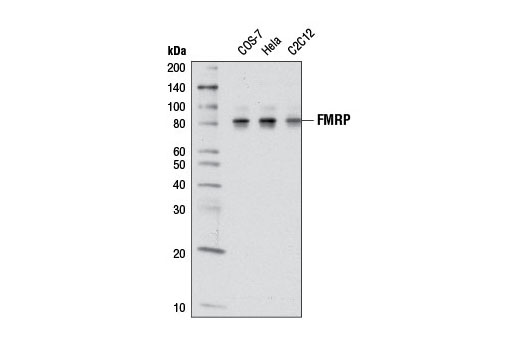
| FMRP (D14F4) Rabbit mAb 7104 | 20 µl |
|
H M R Mk | 80 | Rabbit IgG |
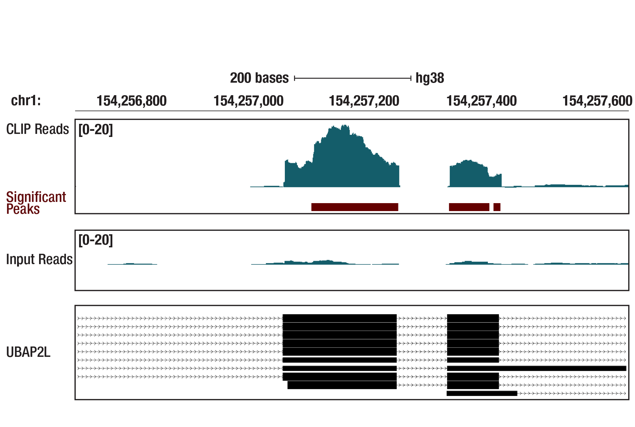
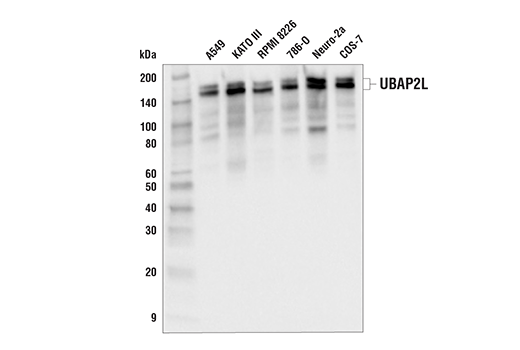
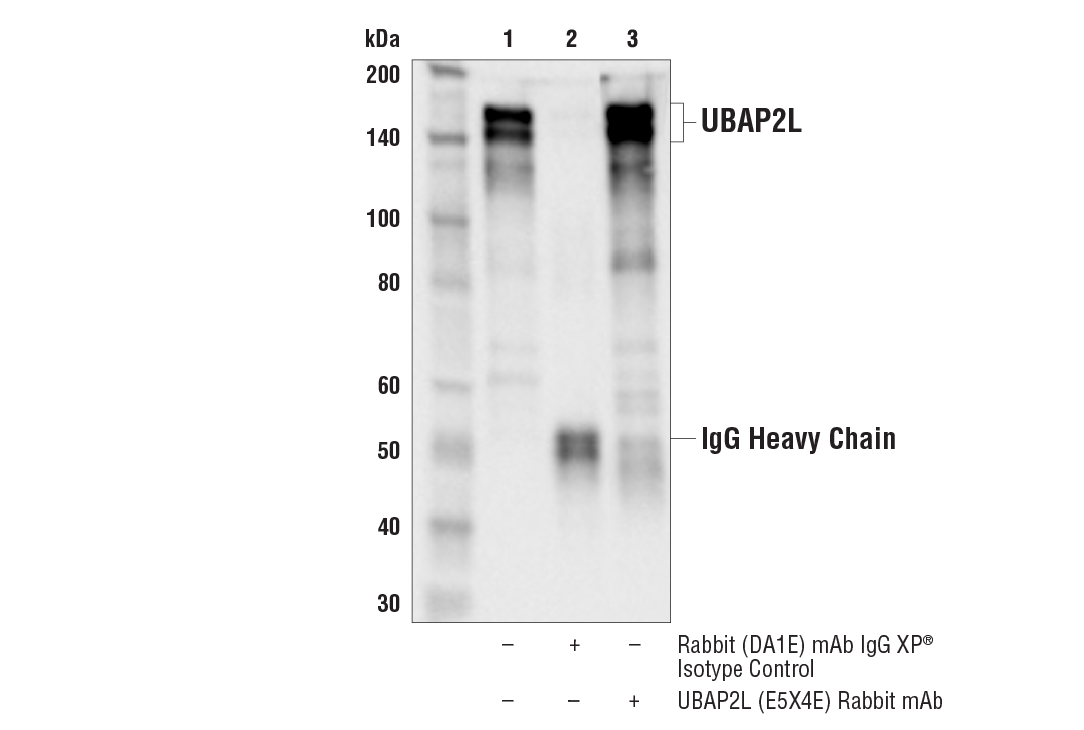
| UBAP2L (E5X4E) Rabbit mAb 40199 | 20 µl |
|
H M Mk | 150, 160 | Rabbit IgG |
| Anti-rabbit IgG, HRP-linked Antibody 7074 | 100 µl |
|
Goat |
Product Information
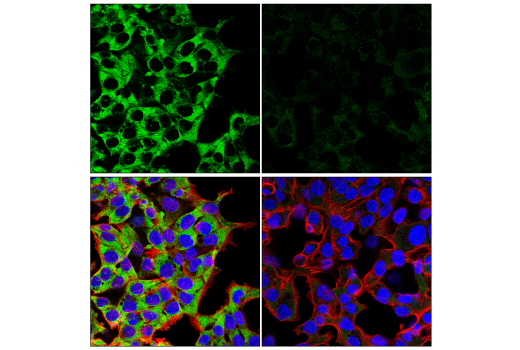
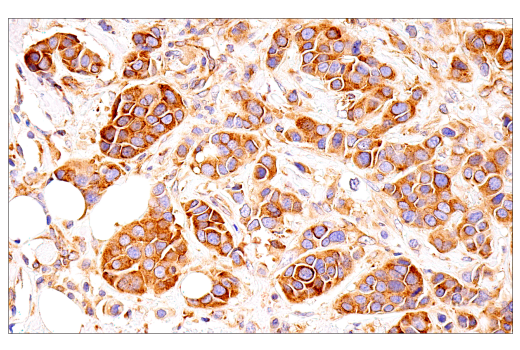
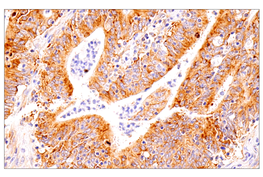
Monoclonal antibodies are produced by immunizing animals with synthetic peptides corresponding to residues surrounding Val218 of human G3BP1 protein, Gly552 of human FMRP protein, residues near the carboxy terminus of human TIAR protein, or recombinant protein specific to the amino terminus of human UBAP2L. Polyclonal antibodies are produced by immunizing animals with a synthetic peptide corresponding to residues near the carboxy terminus of human G3BP2 protein, and are purified by peptide affinity chromatography.
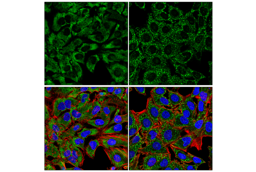
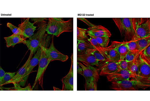
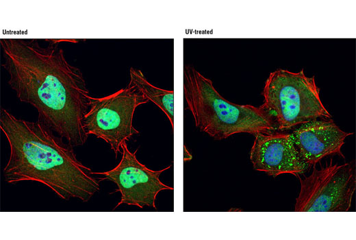
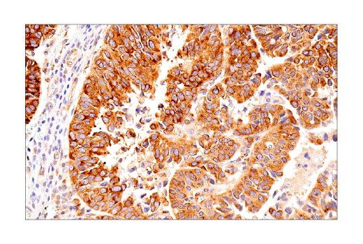
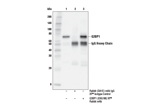
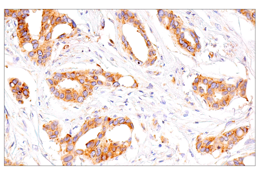
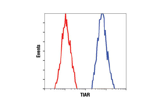
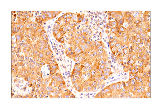
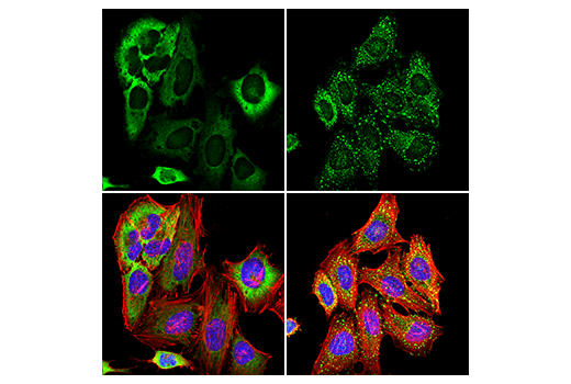
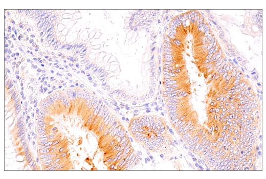
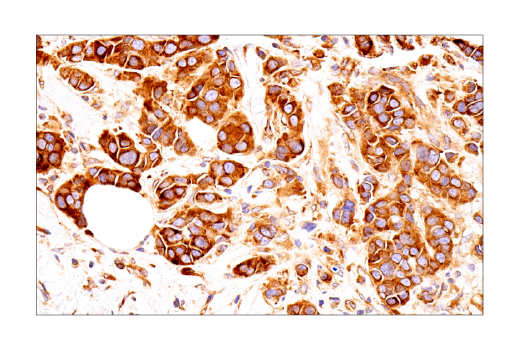
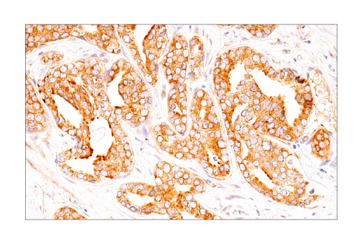
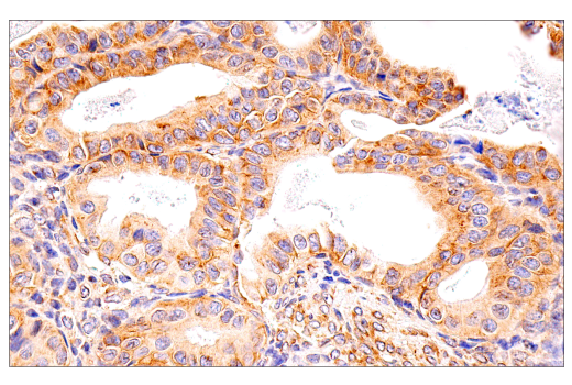
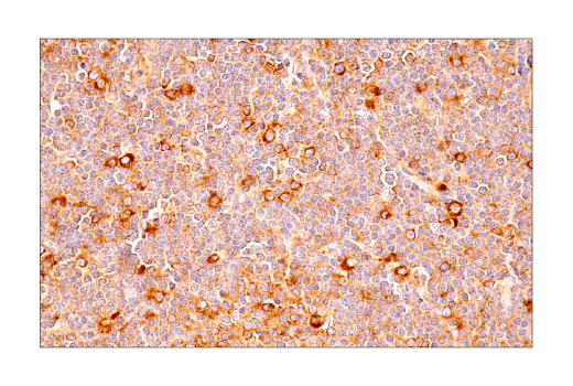
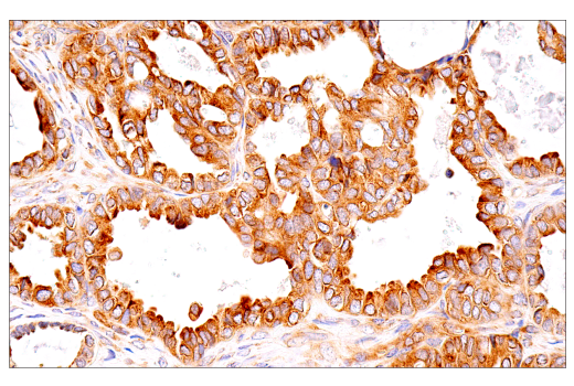
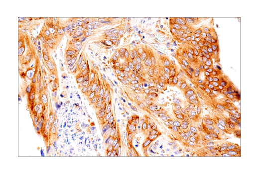
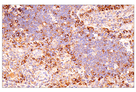
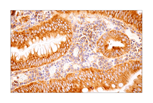
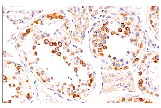
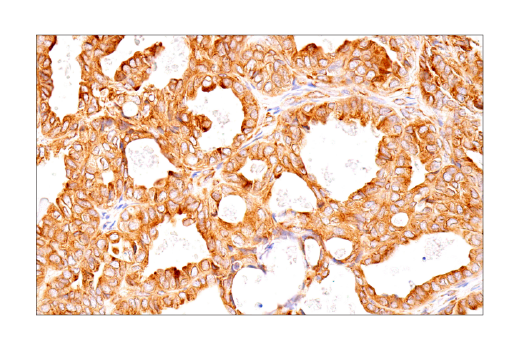
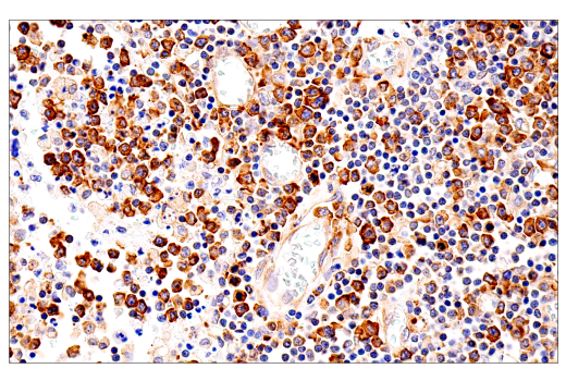
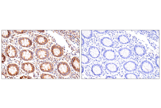
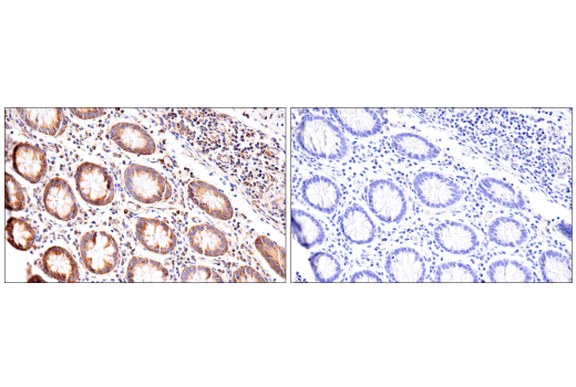
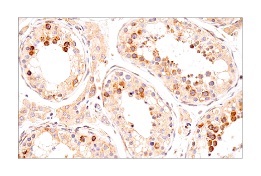
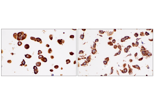
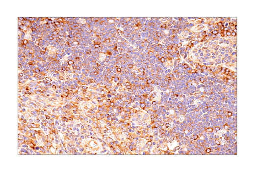
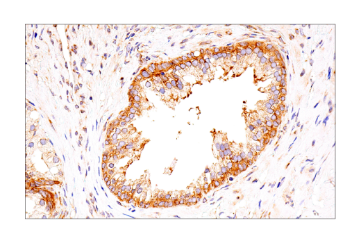
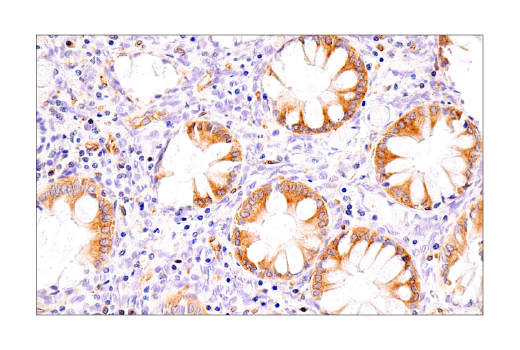
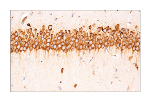
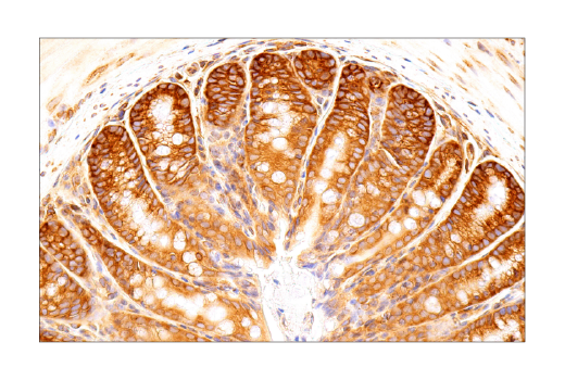
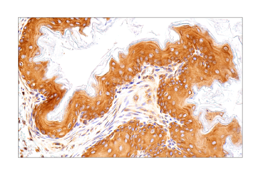
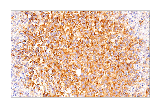
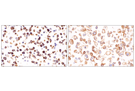
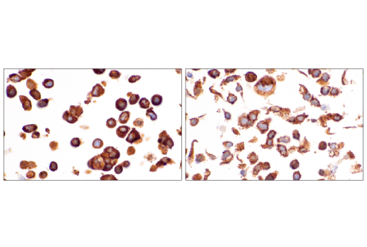
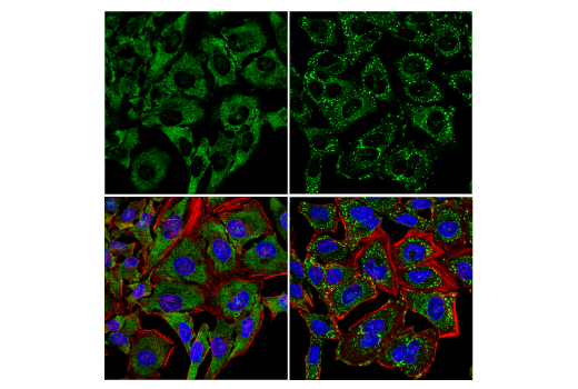
Stress granules (SGs) are cytoplasmic condensates that form at sites of stalled mRNA translation in response to various cellular stressors. SGs are composed of translationally inactive 48S preinitiation complexes (PICs), untranslated mRNA, and a complex mixture of RNA-binding proteins (RBPs). Two key mediators of SG assembly are the RBPs Ras-GTPase-activating protein-binding proteins 1 and 2 (G3BP1/2), which bind to excess mRNA and recruit additional SG-associated proteins. Overexpression of either G3BP1 or G3BP2 is enough to induce SG formation even in the absence of stress, and reduced levels of G3BP1 or G3BP2 protein severely inhibit SG formation, highlighting their crucial role in this process (1-4). Additional SG-associated proteins include TIA-1-related protein (TIAR), fragile X mental retardation protein (FMRP), and ubiquitin-associated protein 2-like (UBAP2L). TIAR is a member of the RNA-recognition motif (RRM) family of RBPs (5,6). In response to cellular stress, TIAR associates with G3BP1/2 and its family member TIA-1 to form SG condensates (7,8). The two major isoforms of TIAR are the products of alternative mRNA splicing (9,10). FMRP (also known as FMR1) and its two autosomal homologs (FXR1 and FXR2) all bind RNA and play a role in the pathogenesis of fragile X syndrome (11-13). Each of these related proteins can associate with one another as well as form homodimers (13). FMRP can act as a translation regulator and is a component of RNAi effector complexes (RISC), suggesting a role in gene silencing (14). In addition, FMRP, FXR1, and FXR2 are components of SGs and have been implicated in the translational regulation of mRNAs (15). UBAP2L is a ubiquitous and highly conserved protein containing an N-terminal ubiquitin-associated (UBA) domain involved in the ubiquitin-proteasome system (UPS) and aggregate formation induced by proteasome inhibitors (16). It can also interact with the Polycomb group protein BMI1 to form a Polycomb subcomplex and regulate hematopoietic stem cell activity (17). UBAP2L is essential for the formation of SGs, and some studies suggest that arginine methylation by PRMT1 inhibits UBAP2L interaction with SG elements and overall SG assembly (18-20).
Except as otherwise expressly agreed in a writing signed by a legally authorized representative of CST, the following terms apply to Products provided by CST, its affiliates or its distributors. Any Customer's terms and conditions that are in addition to, or different from, those contained herein, unless separately accepted in writing by a legally authorized representative of CST, are rejected and are of no force or effect.
Products are labeled with For Research Use Only or a similar labeling statement and have not been approved, cleared, or licensed by the FDA or other regulatory foreign or domestic entity, for any purpose. Customer shall not use any Product for any diagnostic or therapeutic purpose, or otherwise in any manner that conflicts with its labeling statement. Products sold or licensed by CST are provided for Customer as the end-user and solely for research and development uses. Any use of Product for diagnostic, prophylactic or therapeutic purposes, or any purchase of Product for resale (alone or as a component) or other commercial purpose, requires a separate license from CST. Customer shall (a) not sell, license, loan, donate or otherwise transfer or make available any Product to any third party, whether alone or in combination with other materials, or use the Products to manufacture any commercial products, (b) not copy, modify, reverse engineer, decompile, disassemble or otherwise attempt to discover the underlying structure or technology of the Products, or use the Products for the purpose of developing any products or services that would compete with CST products or services, (c) not alter or remove from the Products any trademarks, trade names, logos, patent or copyright notices or markings, (d) use the Products solely in accordance with CST Product Terms of Sale and any applicable documentation, and (e) comply with any license, terms of service or similar agreement with respect to any third party products or services used by Customer in connection with the Products.