View Featured Offers >>
35302
Senescence β-Galactosidase Activity Assay Kit (Fluorescence, Flow Cytometry)
Cellular Assay Kits
Assay Kit

Senescence β-Galactosidase Activity Assay Kit (Fluorescence, Flow Cytometry) #35302

Citations (0)
Flow cytometric analysis of MCF7 cells, untreated (blue) or treated with Etoposide #2200 (12.5 μM, 24 hr; green) and allowed to recover for 3 days, using SA-β-Gal Fluorescent Substrate (solid lines) compared to unlabeled MCF7 cells (dashed line).
To Purchase # 35302
Cat. # Size Qty. Price
35302S
1 Kit  (100 tests)

Product Includes Quantity (with Count)
Bafilomycin A1 54645 1 x 33 µg
SA-β-Gal Fluorescent Substrate 1 x 2 mg

Protocol

PRINT

View >Collapse >

#35302 Senescence β-Galactosidase Activity Assay Kit (Fluorescence, Flow Cytometry)

A. Solutions and Reagents

Supplied Reagents:

  1. Bafilomycin A1 (#54645): Prepare a 100 µM stock (1000X) by adding 530 µl DMSO. Once reconstituted, use within 3 months. Due to production methods, excess bafilomycin is provided. This material can be retained for use in other experiments or discarded.
  2. SA-beta-Gal Fluorescent Substrate (#38154): Prepare a 6.6 mM stock (200X) by adding 352 µl DMSO. Once reconstituted, use within 1 month.

Additional Reagents (Not Supplied):

  1. Dimethyl Sulfoxide (DMSO) (#12611)
  2. Phosphate Buffered Saline (PBS) (#12528)
  3. Bovine Serum Albumin (BSA) (#9998)
  4. 4% Formaldehyde, Methanol-Free (#47746) (optional)

B. Detection of β-Galactosidase Activity

NOTE: Suggested dilutions and volumes are provided for reference. Optimal conditions for SA-beta-Gal Fluorescent Substrate performance may vary by cell type and container type.

NOTE: Cell treatments to induce senescence should be completed before initiating staining with this kit. Ensure that an untreated control is included, to provide a baseline measurement of fluorescence for comparison.

NOTE: Formaldehyde fixation may be performed at the completion of the incubation step with the SA-beta-Gal Fluorescent Substrate #38154. Fixation with up to 4% formaldehyde for up to 15 minutes will not significantly reduce the fluorescence of the substrate. However, permeabilization with methanol or Triton X-100 will cause a significant reduction in fluorescence. If permeabilization is required for immunolabeling of intracellular targets, conduct an experiment to determine whether this will significantly compromise detection of the fluorescent substrate in that cell model.

Analysis of Suspension Cells

  1. Dilute the prepared 100 µM Bafilomycin A1 stock 1:1000 in the desired volume of culture medium, to produce a 100 nM solution. Use within 4 hrs.
  2. Remove culture medium from cells through centrifugation. Add sufficient volume of Bafilomycin A1-containing medium to resuspend cells (roughly 1 mL per 1x10⁶ cells). Incubate at 37°C for 1 hr.
  3. Add sufficient volume of the prepared 200X SA-β-Gal Substrate Solution directly to the cells in Bafilomycin A1-containing medium to produce a 1:200 dilution (33 µM). Gently mix cells to ensure even distribution of SA-β-Gal Substrate and incubate at 37°C for 2-4 hours.
  4. Wash the stained cells three times with 3 mL of 1X PBS, through centrifugation.
  5. Resuspend cells in cold PBS containing 2% FBS or 0.5% BSA.
  6. (Optional) Perform immunostaining of live cells with antibodies against extracellular targets.
  7. (Optional) Fix and permeabilize cells to perform immunostaining with antibodies against intracellular targets. See cautionary note above.
  8. If necessary, obtain a cell count using a hemocytometer or other method, and collect an aliquot of the desired number of cells. Analyze the cells by flow cytometry.

Analysis of Adherent Cells

  1. Dilute the prepared 100 µM Bafilomycin A1 stock 1:1000 in the desired volume of culture medium, to produce a 100 nM solution. Use within 4 hrs.
  2. Remove culture medium from cells via aspiration. Add sufficient volume of Bafilomycin A1-containing medium to cover cells. Incubate at 37°C for 1 hr.
  3. Add sufficient volume of the prepared 200X SA-β-Gal Substrate Solution directly to the cells in Bafilomycin A1-containing medium to produce a 1:200 dilution (33 µM). Gently mix cells to ensure even distribution of SA-β-Gal Substrate and incubate at 37°C for 2-4 hours.
  4. Wash the stained cells three times with 3 mL of 1X PBS.
  5. Remove PBS and trypsinize cells to remove from culture dish, per normal cell culture methods. Add media containing serum to inactivate trypsin. Collect cells, centrifuge and remove supernatant, and resuspend cells in cold PBS containing 2% FBS or 0.5% BSA.
  6. (Optional) Perform immunostaining of live cells with antibodies against extracellular targets.
  7. (Optional) Fix and permeabilize cells to perform immunostaining with antibodies against intracellular targets. See cautionary note above.
  8. If necessary, obtain a cell count using a hemocytometer or other method, and collect an aliquot of the desired number of cells. Analyze the cells by flow cytometry.

posted April 2010

Protocol Id: 1986

Product Description

The Senescence β-Galactosidase Activity Assay Kit (Fluorescence, Flow Cytometry) allows the quantitative measure of senescence-associated β-galactosidase activity in living cells via flow cytometry using a cell permeable fluorogenic substrate, which becomes fluorescent upon hydrolysis by β-galactosidase. The fluorescent molecule is optimally excited at 490 nm (blue laser) and peak emission occurs at 514 nm (usually FL1). Each kit contains sufficient quantities of reagents to perform up to 100 assays.

Background

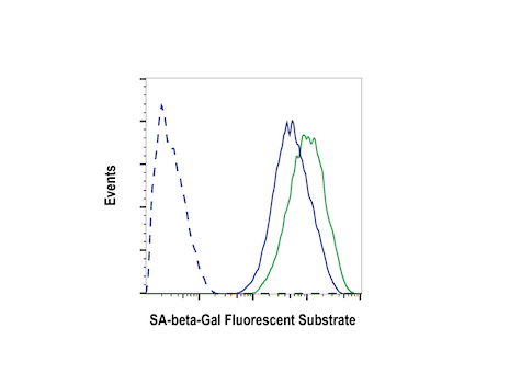
Limited capacity to replicate is a defining characteristic of most normal cells and culminates in senescence, an arrested state in which the cell remains viable (1). Senescent cells are not stimulated to divide by serum or passage in culture, and senescence invokes a specific cell cycle profile that differs from most damage-induced arrest processes or contact inhibition (2). An enlarged cell size, expression of pH-dependent β-galactosidase activity (3), and an altered pattern of gene expression (4,5) further characterize senescent cells.

Limited Uses

Except as otherwise expressly agreed in a writing signed by a legally authorized representative of CST, the following terms apply to Products provided by CST, its affiliates or its distributors. Any Customer's terms and conditions that are in addition to, or different from, those contained herein, unless separately accepted in writing by a legally authorized representative of CST, are rejected and are of no force or effect.

Products are labeled with For Research Use Only or a similar labeling statement and have not been approved, cleared, or licensed by the FDA or other regulatory foreign or domestic entity, for any purpose. Customer shall not use any Product for any diagnostic or therapeutic purpose, or otherwise in any manner that conflicts with its labeling statement. Products sold or licensed by CST are provided for Customer as the end-user and solely for research and development uses. Any use of Product for diagnostic, prophylactic or therapeutic purposes, or any purchase of Product for resale (alone or as a component) or other commercial purpose, requires a separate license from CST. Customer shall (a) not sell, license, loan, donate or otherwise transfer or make available any Product to any third party, whether alone or in combination with other materials, or use the Products to manufacture any commercial products, (b) not copy, modify, reverse engineer, decompile, disassemble or otherwise attempt to discover the underlying structure or technology of the Products, or use the Products for the purpose of developing any products or services that would compete with CST products or services, (c) not alter or remove from the Products any trademarks, trade names, logos, patent or copyright notices or markings, (d) use the Products solely in accordance with CST Product Terms of Sale and any applicable documentation, and (e) comply with any license, terms of service or similar agreement with respect to any third party products or services used by Customer in connection with the Products.

For Research Use Only. Not for Use in Diagnostic Procedures.
Cell Signaling Technology is a trademark of Cell Signaling Technology, Inc.
All other trademarks are the property of their respective owners. Visit our Trademark Information page.