| Cat. # | Size | Qty. | Price |
|---|---|---|---|
| 29650T | 1 Kit (8 x 20 microliters) |
|
| Product Includes | Quantity | Applications | Reactivity | MW(kDa) | Isotype |
|---|---|---|---|---|---|
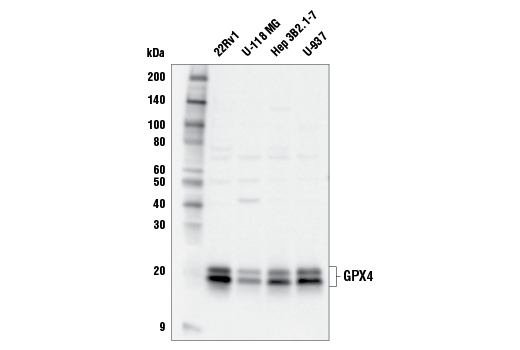
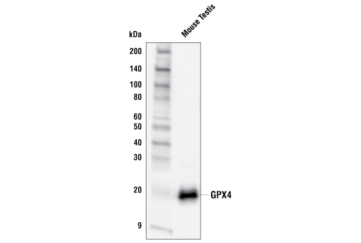
| GPX4 Antibody 52455 | 20 µl |
|
H M Mk | 20, 22 | Rabbit |
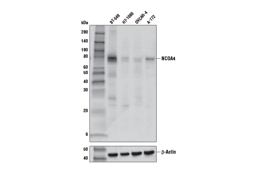
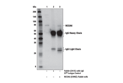
| NCOA4 (E8H8Z) Rabbit mAb 66849 | 20 µl |
|
H | 80 | Rabbit IgG |
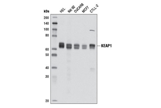
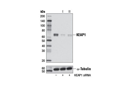
| KEAP1 (D6B12) Rabbit mAb 8047 | 20 µl |
|
H M R | 60-64 | Rabbit IgG |
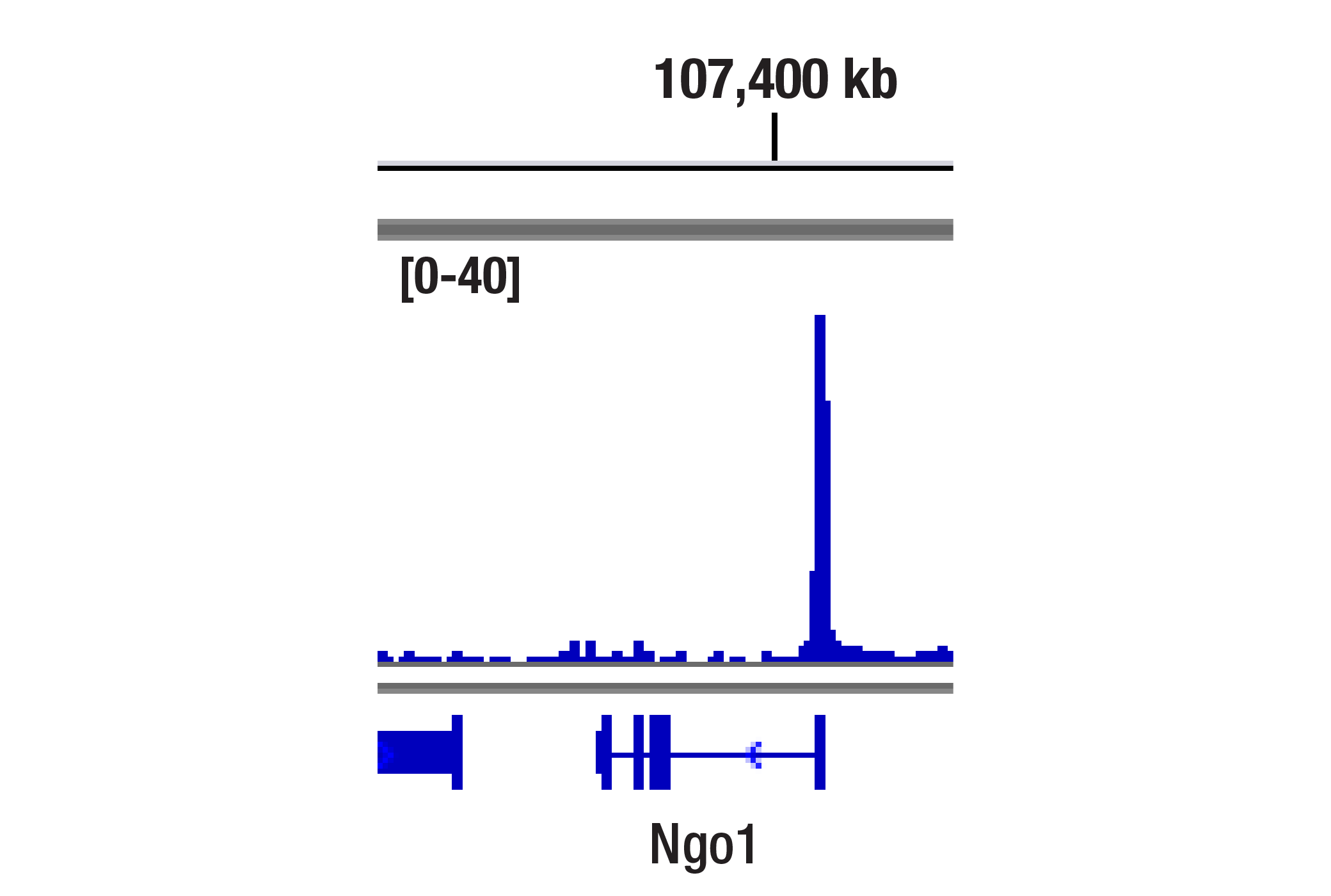
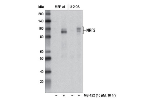
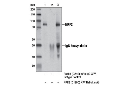
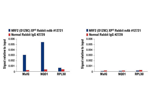
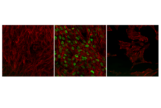
| NRF2 (D1Z9C) XP® Rabbit mAb 12721 | 20 µl |
|
H M Mk | 97-100 | Rabbit IgG |
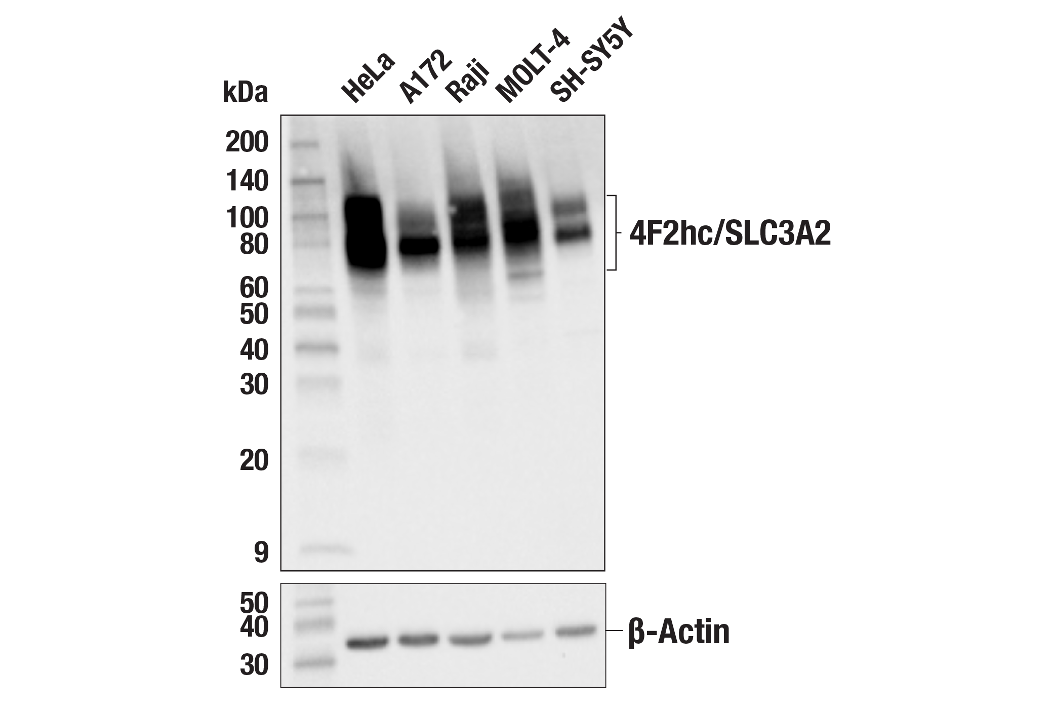
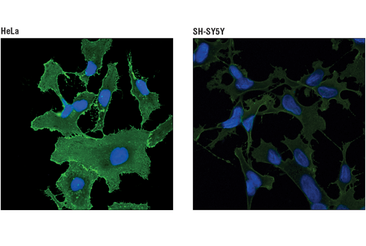
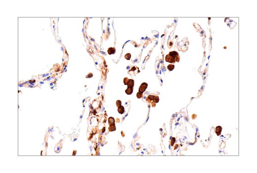
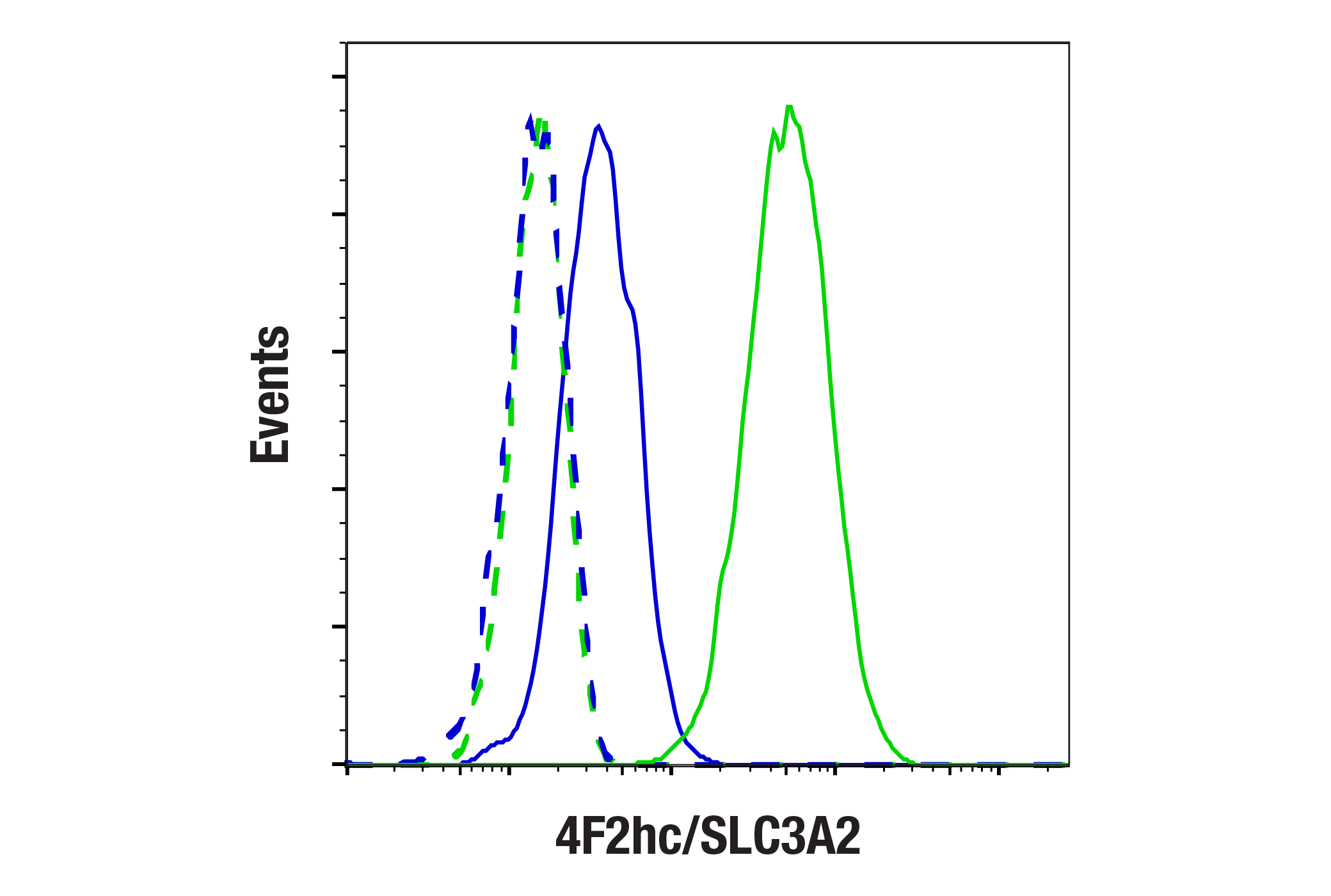
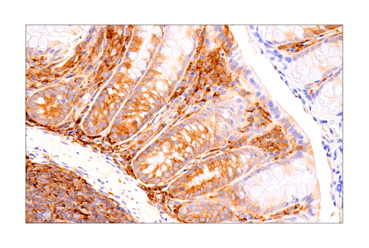
| 4F2hc/SLC3A2 (D3F9D) XP® Rabbit mAb 47213 | 20 µl |
|
H | 75-120 | Rabbit IgG |
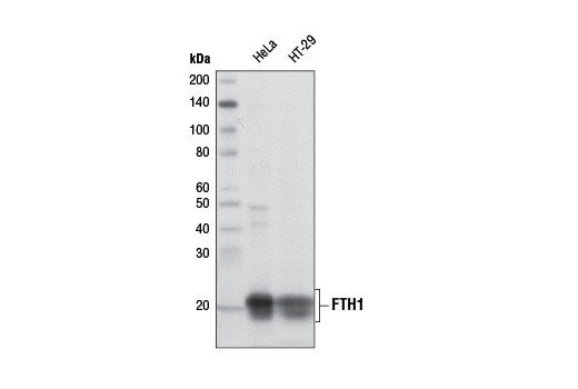
| FTH1 (D1D4) Rabbit mAb 4393 | 20 µl |
|
H M R Mk | 21 | Rabbit IgG |
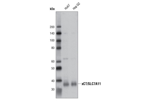
| xCT/SLC7A11 (D2M7A) Rabbit mAb 12691 | 20 µl |
|
H | 35 | Rabbit IgG |
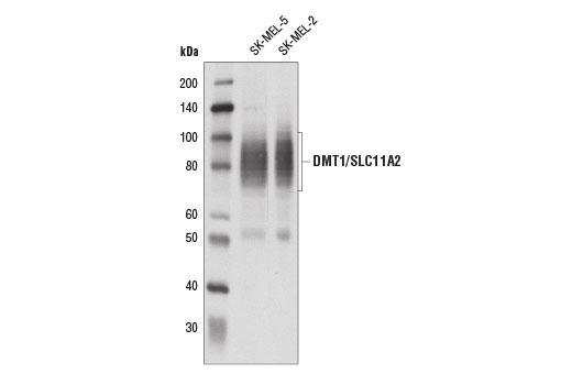
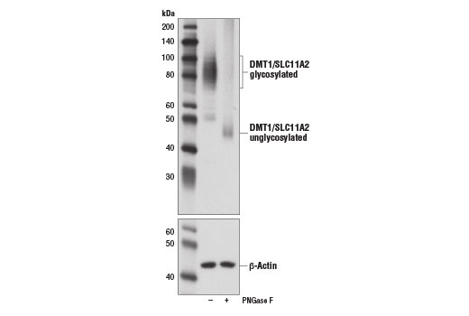
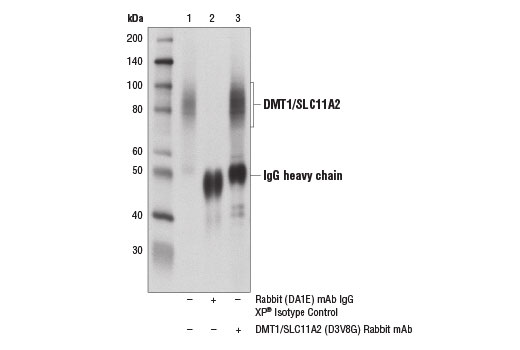
| DMT1/SLC11A2 (D3V8G) Rabbit mAb 15083 | 20 µl |
|
H | 55, 70-100 | Rabbit IgG |
| Anti-rabbit IgG, HRP-linked Antibody 7074 | 100 µl |
|
Goat |
Product Information
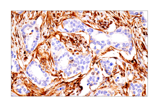
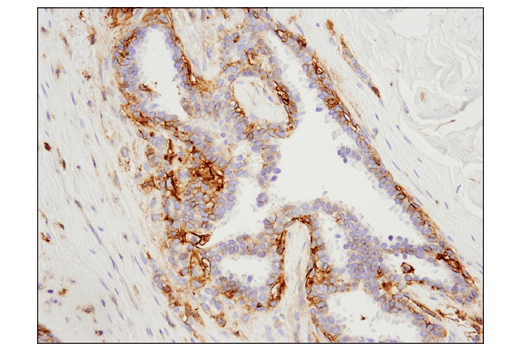
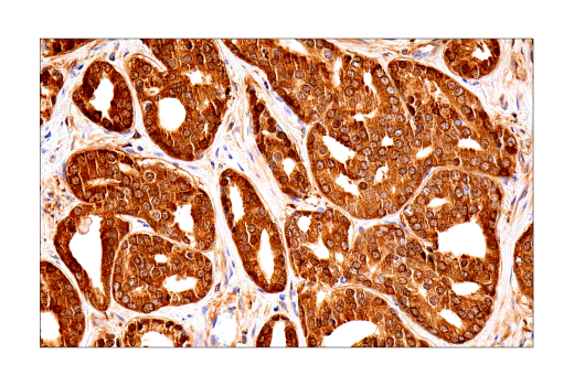
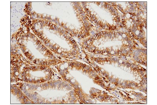
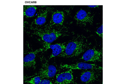
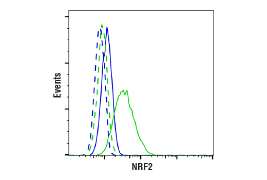
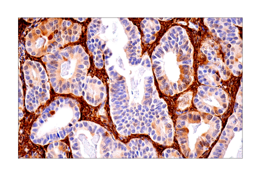
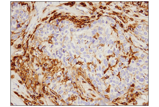
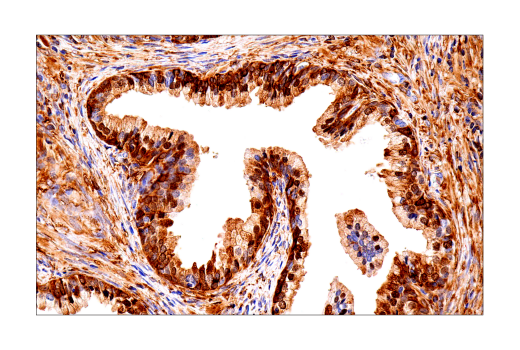
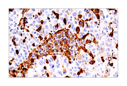
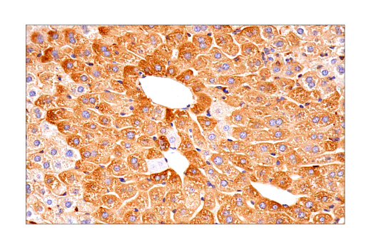
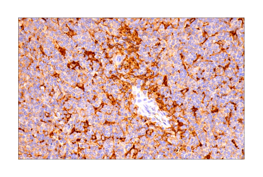
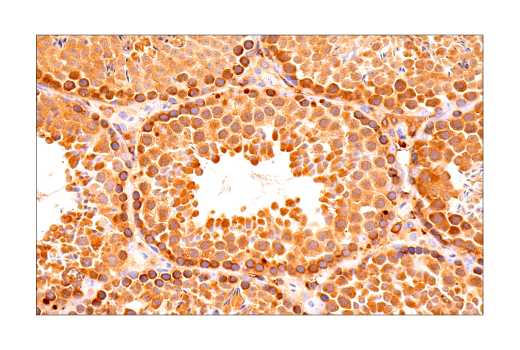
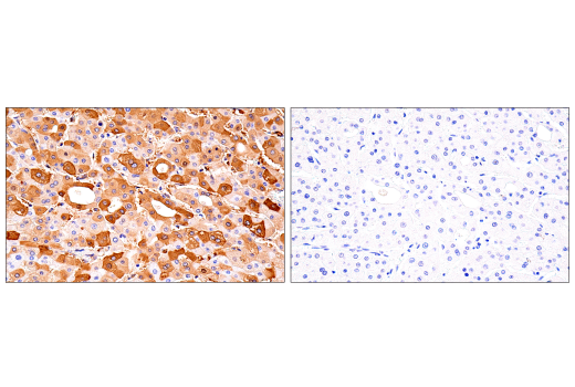
Monoclonal antibodies are produced by immunizing animals with synthetic peptides corresponding to residues surrounding Gly213 of human NCOA4, Ala275 of human NRF2, Val509 of human 4F2hc/CD98, Ala224 of human xCT/SLC7A11, residues near the carboxy terminus of human KEAP1 and human FTH1, and residues near the amino terminus of human DMT1. GPX4 Antibody is a polyclonal antibody produced by immunizing animals with a synthetic peptide corresponding to residues near the carboxy terminus of human GPX4 protein. Antibodies are purified by peptide affinity chromatography.
Ferroptosis is an iron-dependent form of regulated cell death associated with an increase in lipid peroxides (reviewed in 1,2). Free divalent iron (Fe2+) can lead to spontaneous lipid peroxidation through a Fenton reaction. Ferroptosis is regulated by signaling pathways that control iron storage and oxidative stress. Iron homeostasis is controlled, in part, by ferritin, an iron storage protein consisting of a complex of heavy (FTH1) and light (FTL) chains. Levels of ferritin may be regulated by a selective autophagy process targeting ferritin, termed ferritinophagy. This pathway is mediated by nuclear receptor coactivator 4 (NCOA4), a selective cargo receptor for ferritin (3,4). The divalent metal transporter SLC11A2/DMT1/NRAMP2 regulates iron homeostasis through non-heme absorption in the intestine (5). The glutathione peroxidase pathway has been identified as a key antioxidant defense pathway triggering ferroptosis. The compound RSL3, which directly inhibits GPX4, was identified as an activator of ferroptosis (6). GPX4 converts GSH into oxidized glutathione (GSSH) and reduces cytotoxic lipid peroxides. The glutathione peroxidase pathway is further regulated by System Xc-, an amino acid antiporter consisting of a disulfide-linked heterodimer of SLC7A11/xCT and SLC3A2/4F2hc/CD98, and is inhibited by the ferroptosis inducer erastin (7). Regulation of genes involved in oxidative stress, including GPX4, are largely controlled by the transcription factor NRF2 and serves as a defense against ferroptosis (8). Under normal conditions, expression of NRF2 is inhibited through interaction with KEAP1, part of a ubiquitin E3 ligase complex that leads to NRF2 proteasomal degradation. Oxidative stress leads to conformational changes in KEAP1 that disrupts this interaction, resulting in stabilization of NRF2. This process is further regulated through the autophagy pathway in which the autophagy cargo receptor p62/SQSTM1 can competitively inhibit the KEAP1-NRF2 complex, leading to upregulation of NRF2.
Except as otherwise expressly agreed in a writing signed by a legally authorized representative of CST, the following terms apply to Products provided by CST, its affiliates or its distributors. Any Customer's terms and conditions that are in addition to, or different from, those contained herein, unless separately accepted in writing by a legally authorized representative of CST, are rejected and are of no force or effect.
Products are labeled with For Research Use Only or a similar labeling statement and have not been approved, cleared, or licensed by the FDA or other regulatory foreign or domestic entity, for any purpose. Customer shall not use any Product for any diagnostic or therapeutic purpose, or otherwise in any manner that conflicts with its labeling statement. Products sold or licensed by CST are provided for Customer as the end-user and solely for research and development uses. Any use of Product for diagnostic, prophylactic or therapeutic purposes, or any purchase of Product for resale (alone or as a component) or other commercial purpose, requires a separate license from CST. Customer shall (a) not sell, license, loan, donate or otherwise transfer or make available any Product to any third party, whether alone or in combination with other materials, or use the Products to manufacture any commercial products, (b) not copy, modify, reverse engineer, decompile, disassemble or otherwise attempt to discover the underlying structure or technology of the Products, or use the Products for the purpose of developing any products or services that would compete with CST products or services, (c) not alter or remove from the Products any trademarks, trade names, logos, patent or copyright notices or markings, (d) use the Products solely in accordance with CST Product Terms of Sale and any applicable documentation, and (e) comply with any license, terms of service or similar agreement with respect to any third party products or services used by Customer in connection with the Products.