
High-Content Imaging Resource Center
Reliable CST® Solutions to Drive Your Drug Discovery Forward
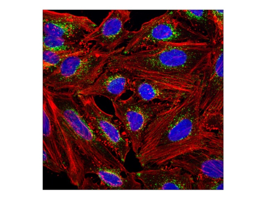
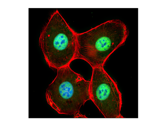
Confocal immunofluorescent analysis of untreated SK-MEL-2 cells using SQSTM1/p62 (D5L7G) Mouse Monoclonal Antibody #88588, DyLight® 554 Phalloidin #13054, and DAPI #4083.
In the high-stakes world of drug development, reproducible data that propels your screening forward with confidence is essential. High-content imaging (HCI) unlocks deep insights into complex cellular processes at the scale required to meet demanding timelines, but only when supported by antibodies you can trust. HCI-ready immunofluorescence-immunocytochemistry (IF-IC)-validated antibodies from CST are rigorously tested so you can:
- Generate data you trust: Eliminate false positives due to non-specific binding, or false negatives due to poor target specificity. Using highly specific antibodies is essential for image segmentation and accurate data quantification.
- Save valuable time: HCI-ready IF-IC-validated antibodies are guaranteed to work in your assay, eliminating the need to identify or test them yourself.
- Avoid failed experiments: HCI workflows are challenging enough. Remove reagent reliability as a variable so you can focus on the insights, not the troubleshooting.
Precision HCI Reagents and Custom Solutions Enable Accurate Image Analysis at Scale
 | High-Content Imaging-Ready Antibodies Select from a broad catalog of validated antibodies guaranteed to deliver reliable results. |
| |
 | Bulk Orders & Lot Reservations Future-proof your HCI assay with a consistent supply throughout your project. |
| |
Catalog & Custom Antibody Conjugation Solutions Leverage CST conjugation expertise. Flexible options seamlessly fit into your assay. | |
| |
 | Application Workflow Reagents Utilize high-quality reagents from CST throughout your HCI-workflow. |
| |
Common Markers for High-Content Imaging
The following is just a small selection of HCI-ready antibodies offered. Browse our comprehensive portfolio for more HCI-ready antibodies.
Organelle & Morphological
Confocal immunofluorescent analysis of HeLa cells using LAMP1 (D2D11) Rabbit Monoclonal Antibody #9091 (green), DyLight® 554 Phalloidin #13054, and DRAQ5 #4084 (blue).
Proliferation & Cell Cycle
- Ki-67 (Universal proliferation marker)
- Phospho-Histone H3 (Ser10) (Mitotic cell marker)
- Cyclin D1 (G1/S transition regulator)
- PCNA (DNA replication marker)
- p21 Waf1/Cip1 (Cell cycle inhibitor)
- MCM2 (Robust proliferation marker)
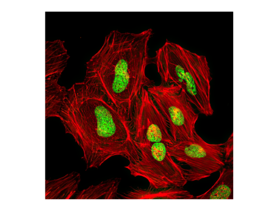
Confocal immunofluorescent analysis of HeLa cells using Ki-67 (8D5) Mouse Monoclonal Antibody #9449 (green), DyLight® 554 Phalloidin #13054, and DRAQ5 #4084 (blue).
Cell Death & Stress Response
- Cleaved Caspase-3 (Apoptosis induction)
- Phospho-Histone H2A.X (Ser139) (DNA double-strand breaks)
- LC3B (Autophagy marker)
- SQSTM1/p62 (Autophagic Flux Marker)
- CHOP (ER stress-induced apoptosis)
- HIF-1 alpha (Hypoxic response)
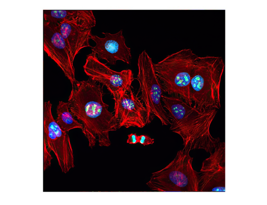
Confocal immunofluorescence of HeLa cells following UV irradiation. Double-strand DNA breaks were detected using Phospho-Histone H2A.X (Ser139) (D7T2V) Mouse Monoclonal Antibody #80312 (green), and the actin cytoskeleton was counterstained with DyLight® 554 Phalloidin #13054 (red).
Signal Transduction & Translocation
- NF-kappaB p65 (Key mediator of immune activity)
- Phospho-Akt (Ser473) (Central node in survival signaling)
- Phospho-STAT3 (Tyr705) (Cytokine signaling)
- Phospho-ERK1/2 (Thr202/Tyr204) (MAP kinase signaling)
- YAP (Hippo pathway effector)
- Phospho-S6 (Ser235/236) (mTORC1 signaling activity)
Confocal immunofluorescence analysis of HT-1080 cells stimulated with hTNF-α #8902 (20 ng/ml, 20 min). Cells were probed with NF-κB p65 (D14E12) Rabbit Monoclonal Antibody #8242 (green), demonstrating robust nuclear translocation. Actin filaments were labeled with DyLight® 554 Phalloidin #13054 (red), and DNA was stained with DRAQ5 #4084 (blue).
Can’t find an HCI-ready antibody for your target of interest? Submit a Product Development Request.
High-Content Imaging FAQs
DyLight is a registered trademark of Thermo Fisher Scientific, Inc. and its subsidiaries.