| Cat. # | Size | Qty. | Price |
|---|---|---|---|
| 90298T | 1 Kit (9 x 20 microliters) |
|
| Product Includes | Quantity | Applications | Reactivity | MW(kDa) | Isotype |
|---|---|---|---|---|---|
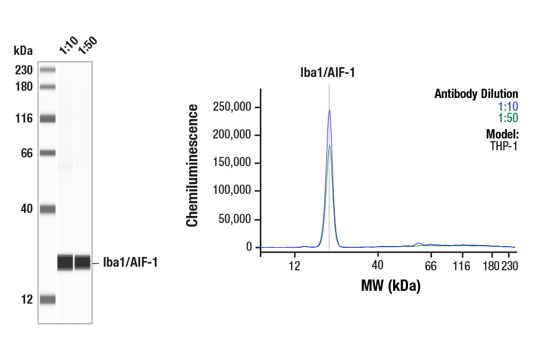
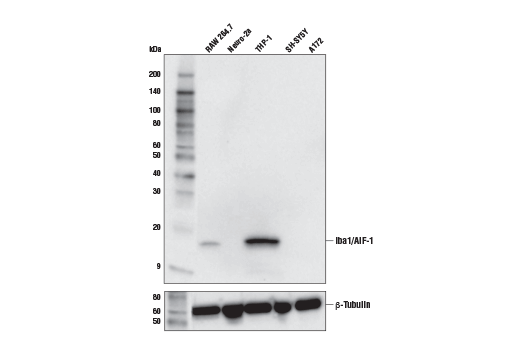
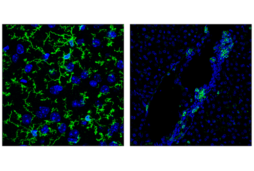
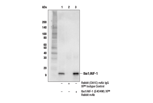
| Iba1/AIF-1 (E4O4W) XP® Rabbit mAb 17198 | 20 µl |
|
H M R Hm Mk | 17 | Rabbit IgG |
| TMEM119 (E3E1O) Rabbit mAb 90840 | 20 µl |
|
M | Rabbit IgG | |
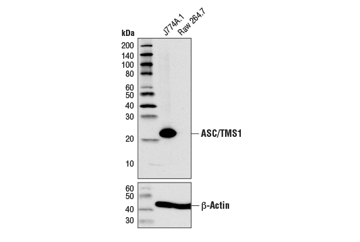
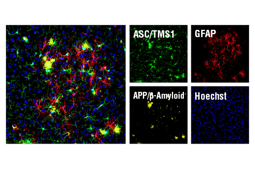
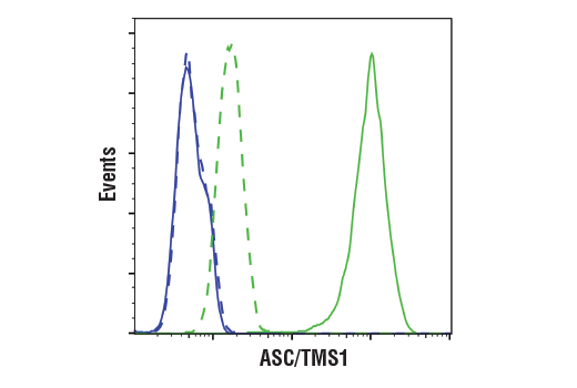
| ASC/TMS1 (D2W8U) Rabbit mAb 67824 | 20 µl |
|
M | 22 | Rabbit IgG |
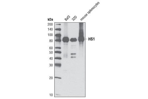
| HS1 (D5A9) XP® Rabbit mAb 3892 | 20 µl |
|
M | 80 | Rabbit IgG |
| CD11b/ITGAM (M1/70) Rat mAb 46512 | 20 µl |
|
M | Rat IgG2b kappa | |
| CD45 (30-F11) Rat mAb 55307 | 20 µl |
|
M | Rat IgG2b kappa | |
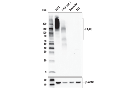
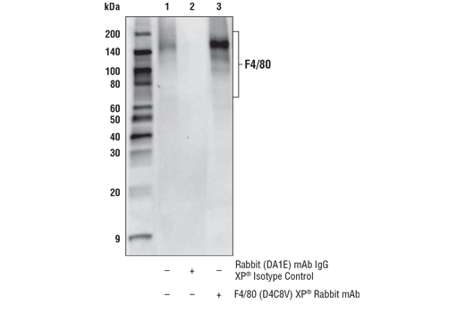
| F4/80 (D4C8V) XP® Rabbit mAb 30325 | 20 µl |
|
M | 65-250 | Rabbit IgG |
| Ki-67 (D3B5) Rabbit mAb 9129 | 20 µl |
|
H M R | Rabbit IgG | |
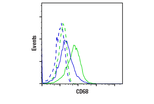
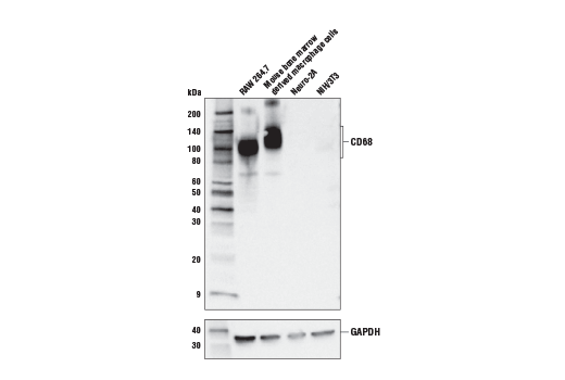
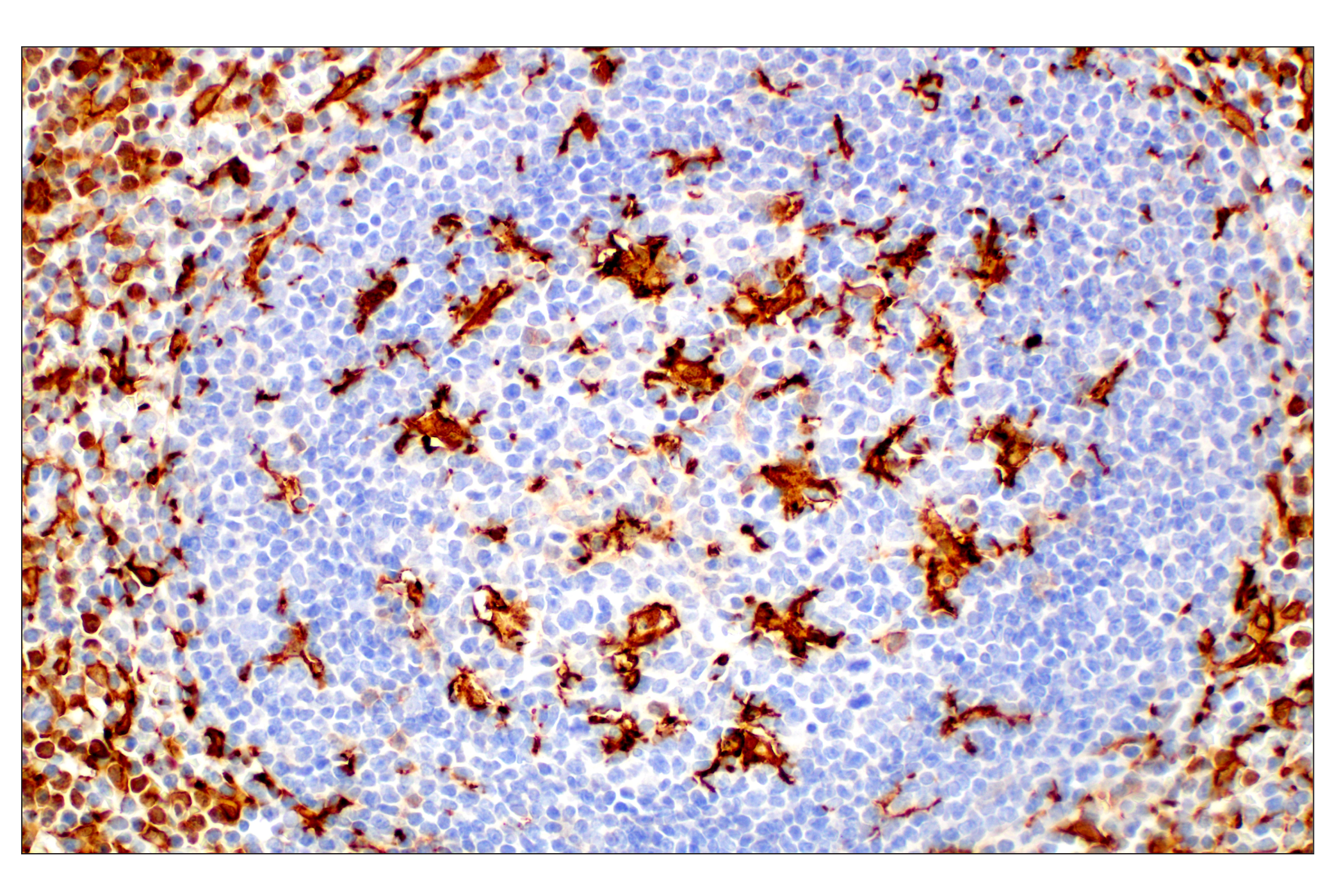
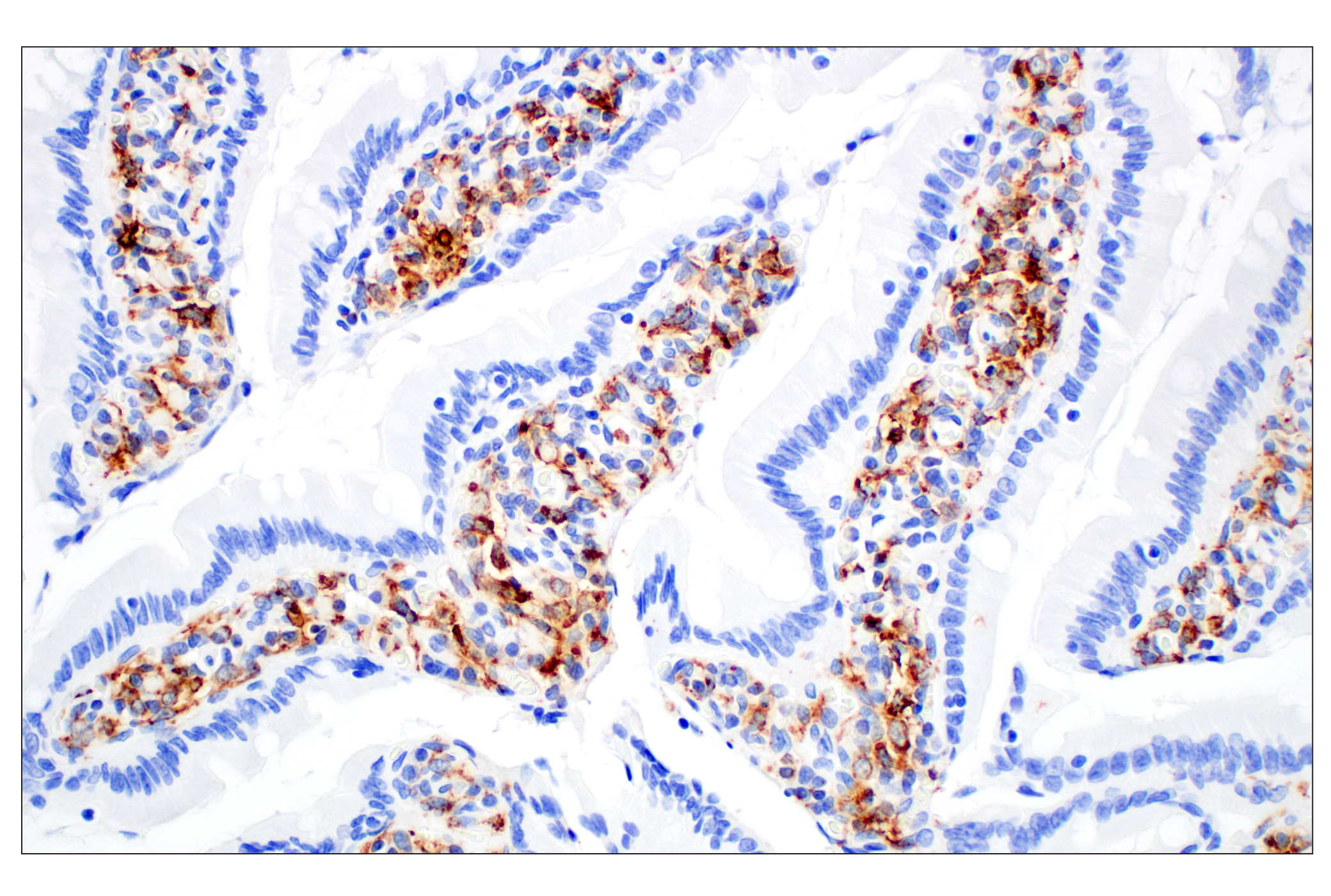
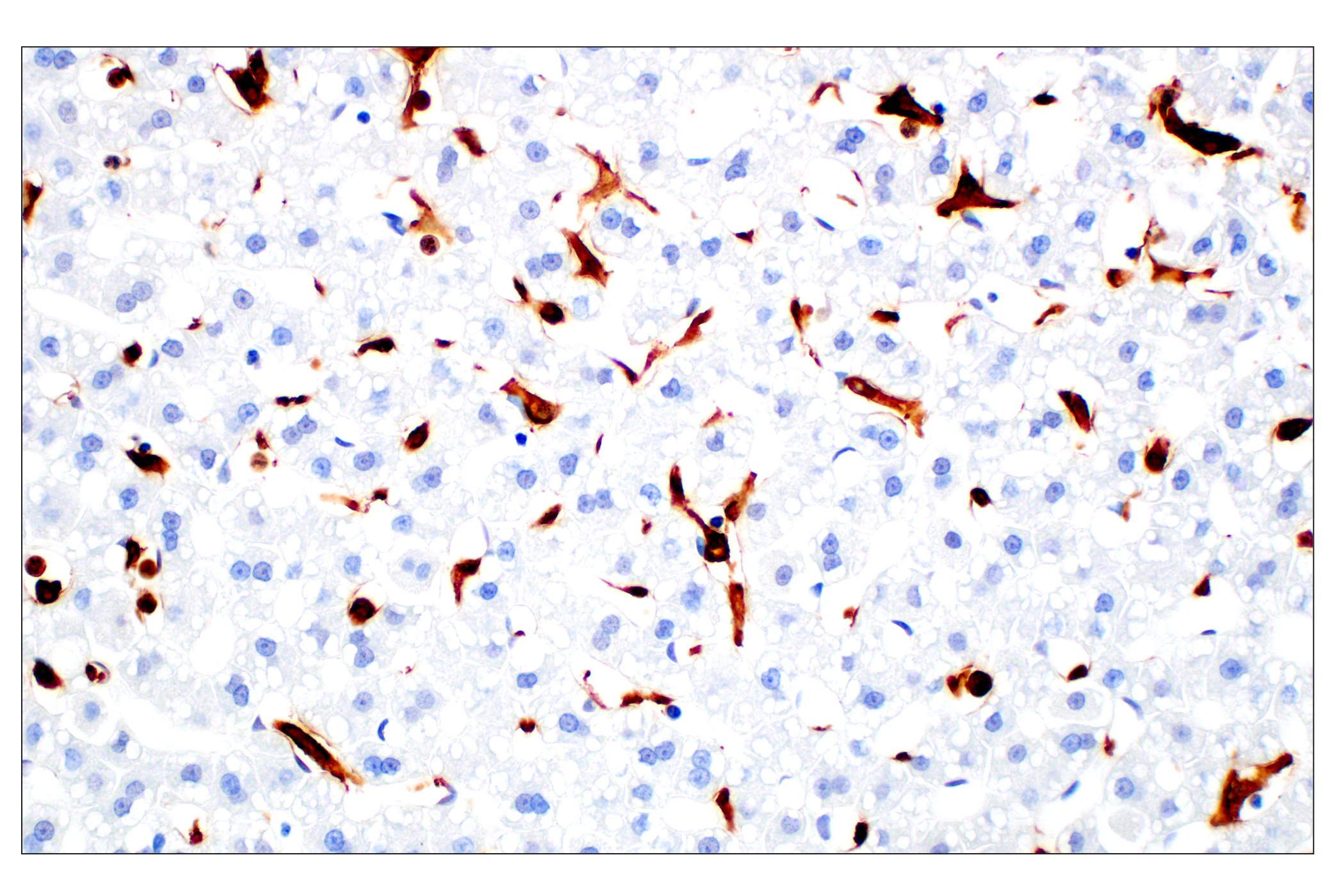
| CD68 (E3O7V) Rabbit mAb 97778 | 20 µl |
|
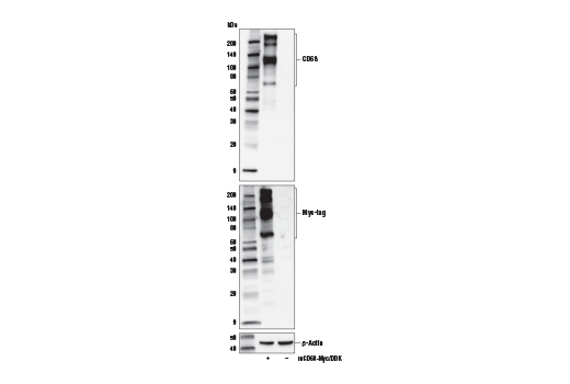
M R Hm | 70-80, 130-140, 200 | Rabbit IgG |
| Anti-rabbit IgG, HRP-linked Antibody 7074 | 100 µl |
|
Goat |
Product Information
Monoclonal antibodies are produced by immunizing animals with synthetic peptides corresponding to residues surrounding Ala139 of human Iba1/AIF-1 protein, Leu310 of mouse HS1 protein, Gln260 of mouse F4/80 protein, the amino terminus of human TMEM119 and Ki-67 proteins, and recombinant mouse ASC/TMS1 protein and mouse CD68 that reacts with an epitope surrounding Gly113. CD11b/ITGAM (M1/70) Rat mAb and CD45 (30-F11) Rat mAb were purified from tissue culture supernatant via affinity chromatography.
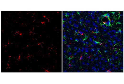
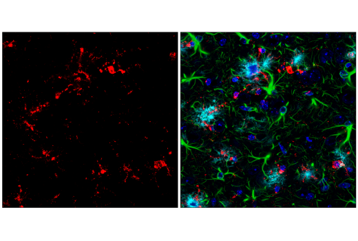
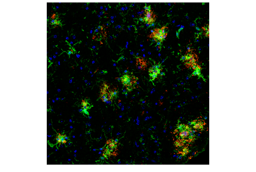
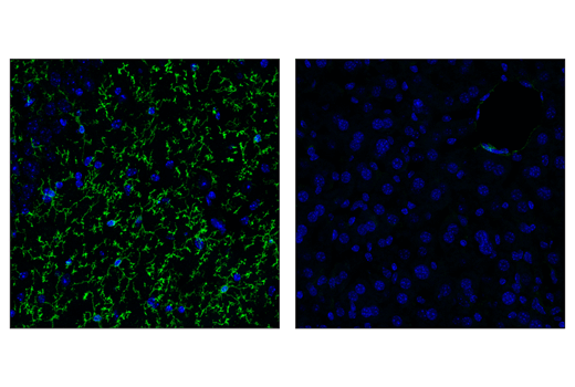
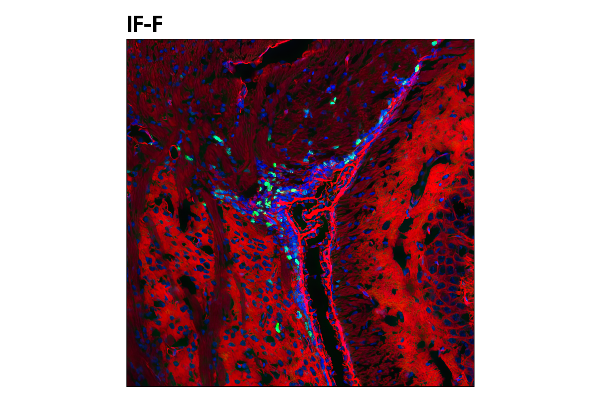
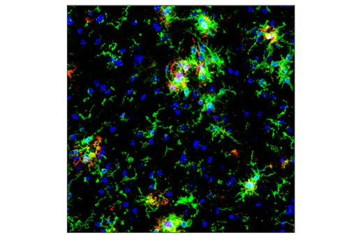
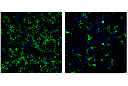
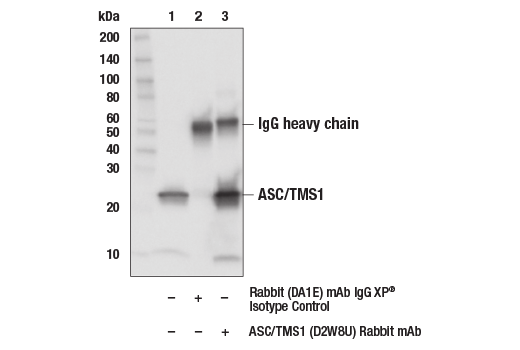
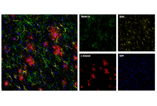
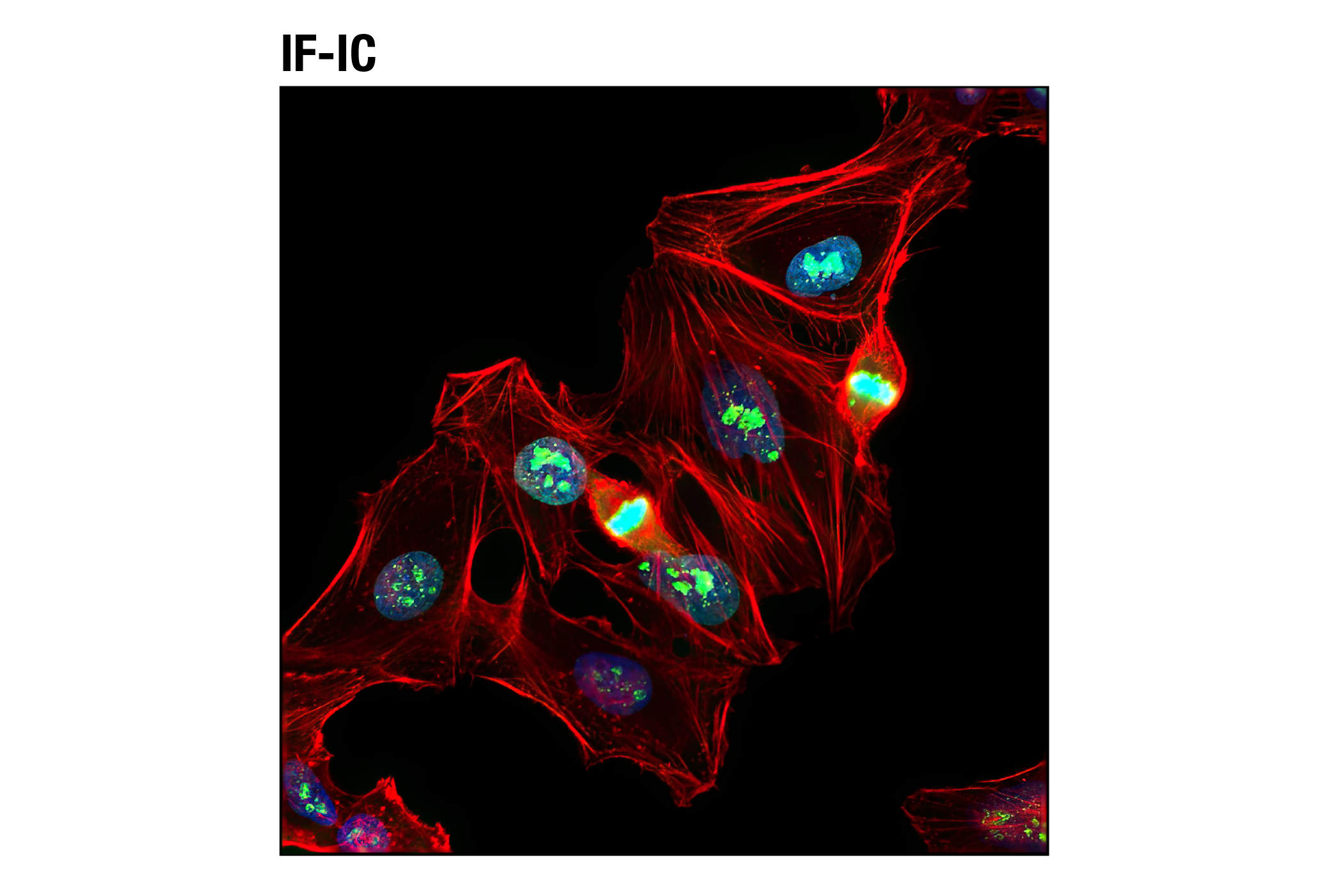
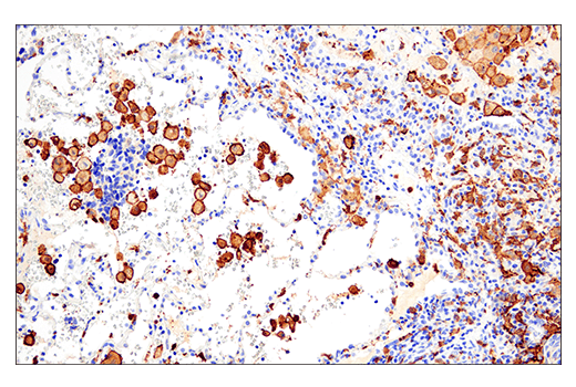
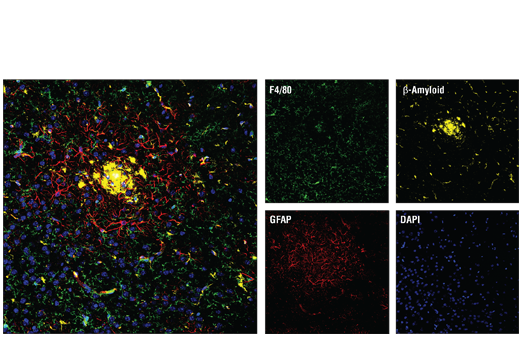
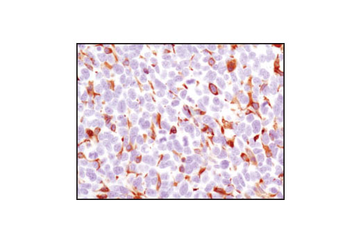
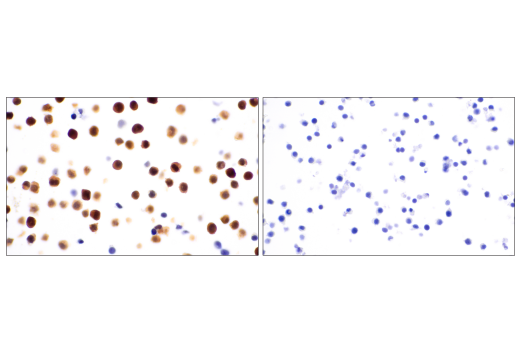
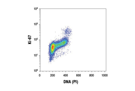
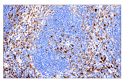
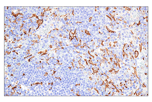
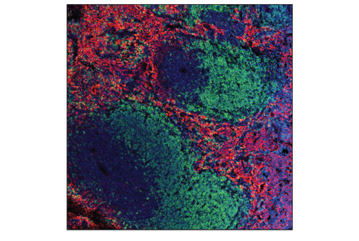
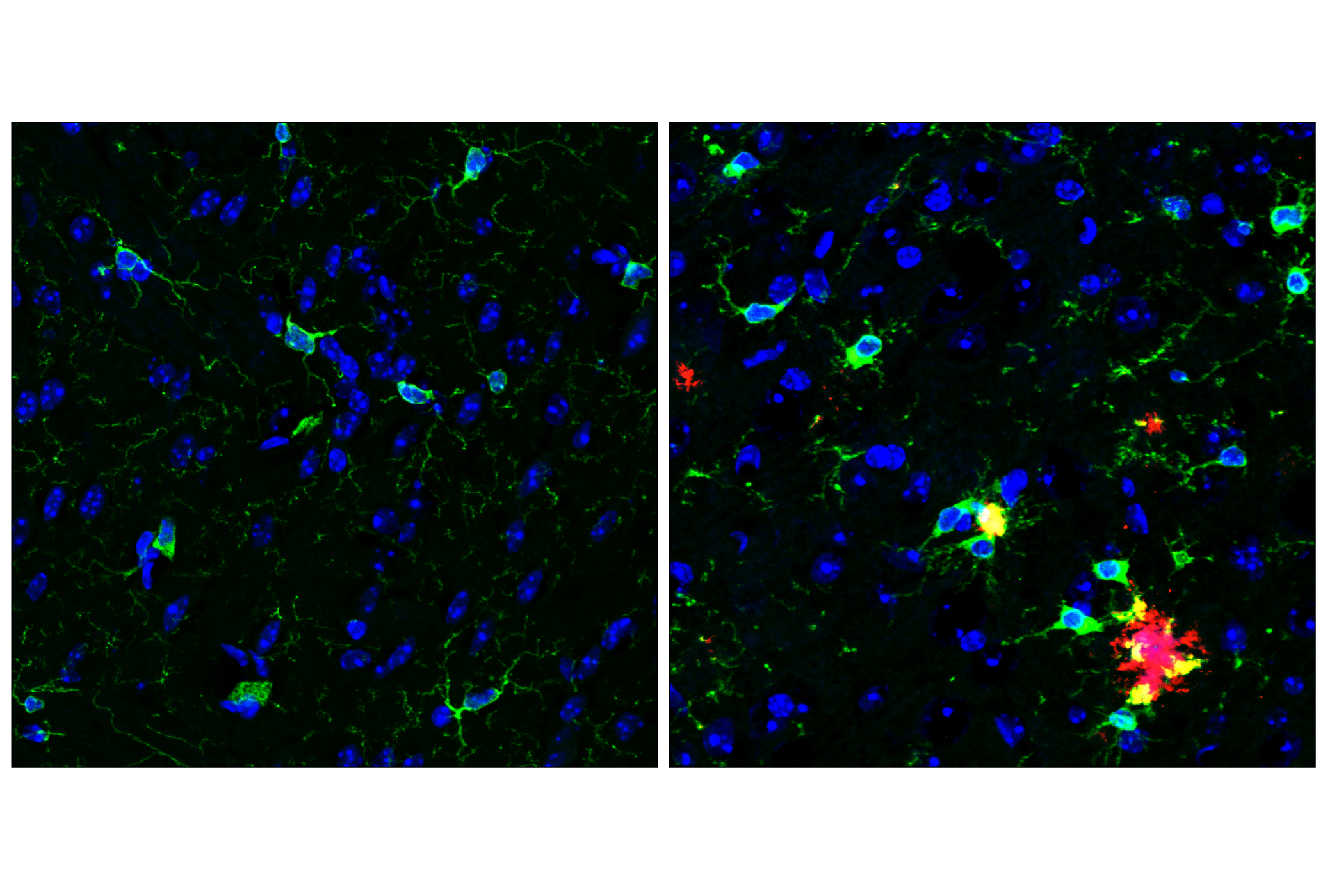
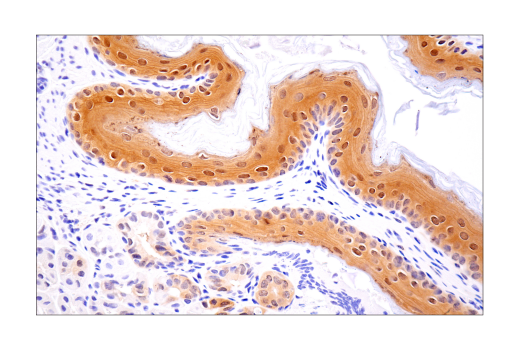
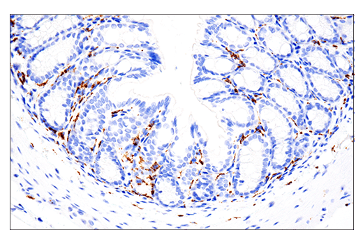
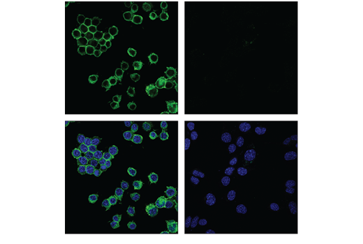
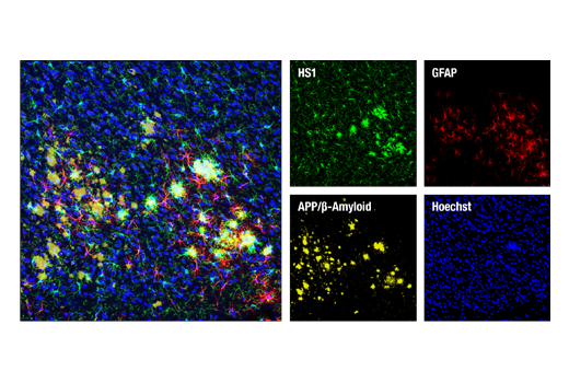
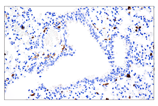
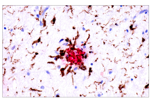
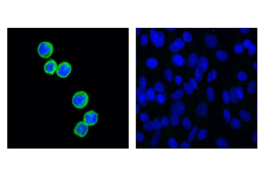
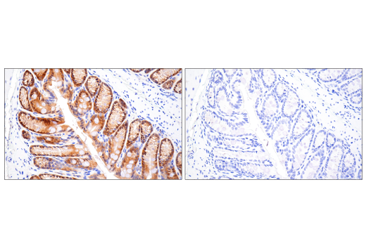
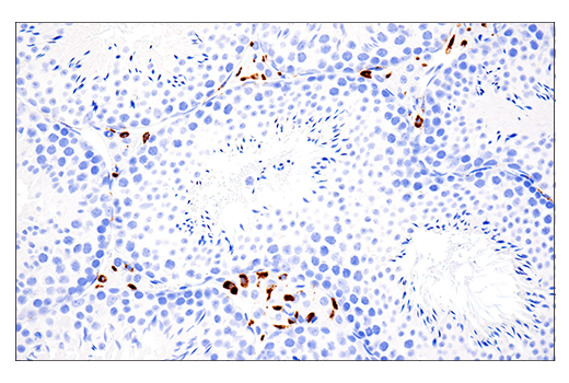
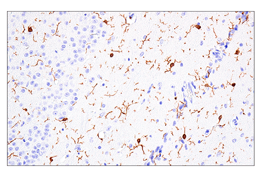
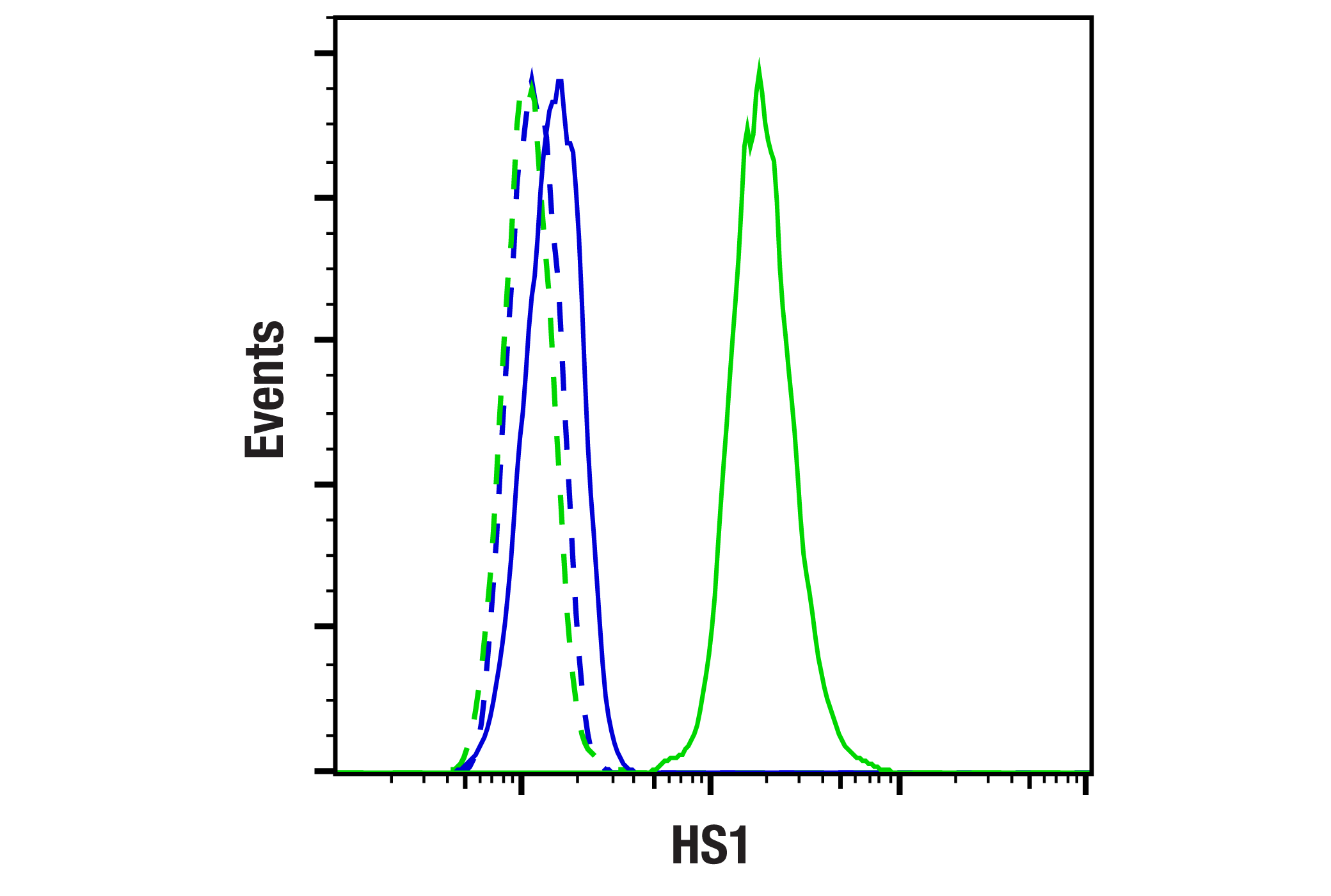
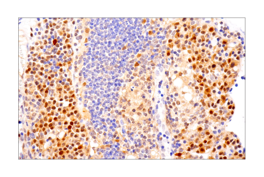
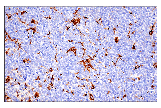
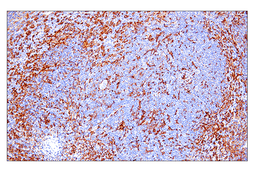
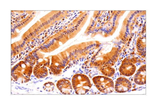
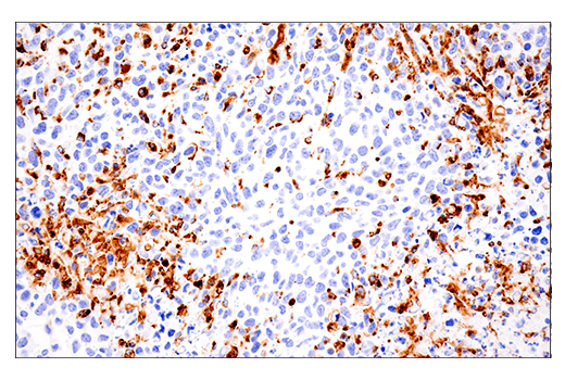
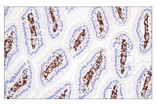
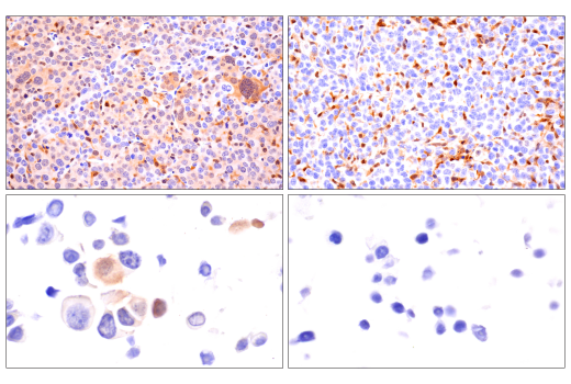
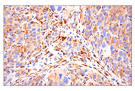
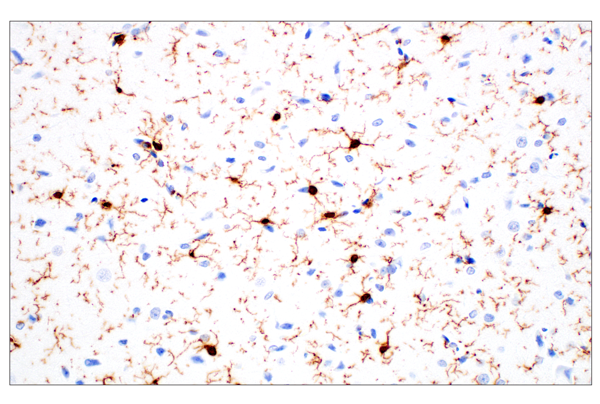
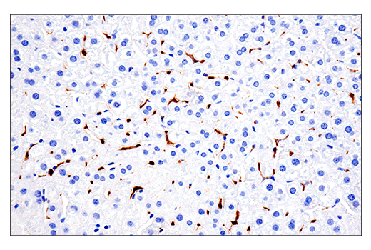
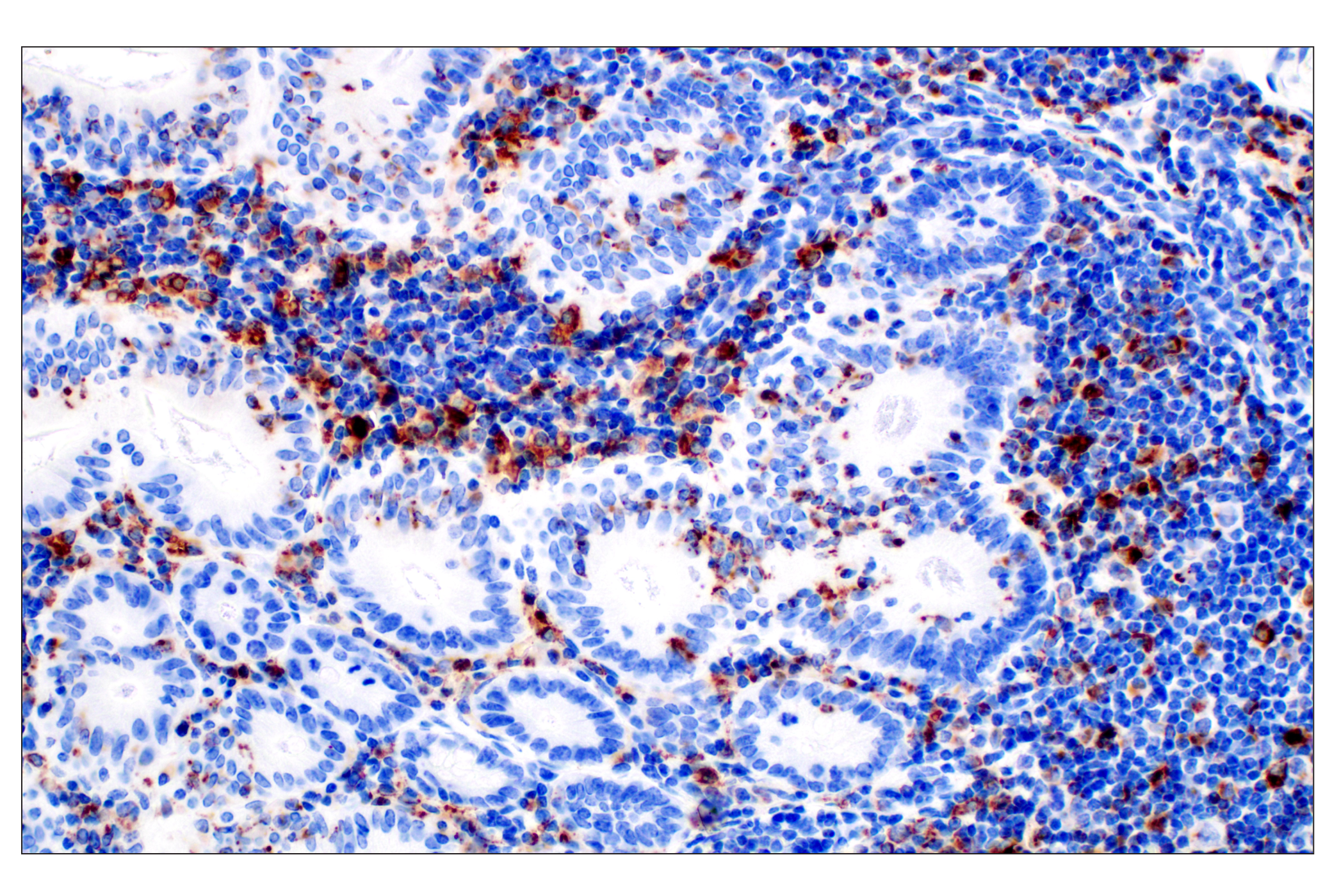
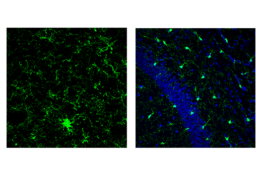
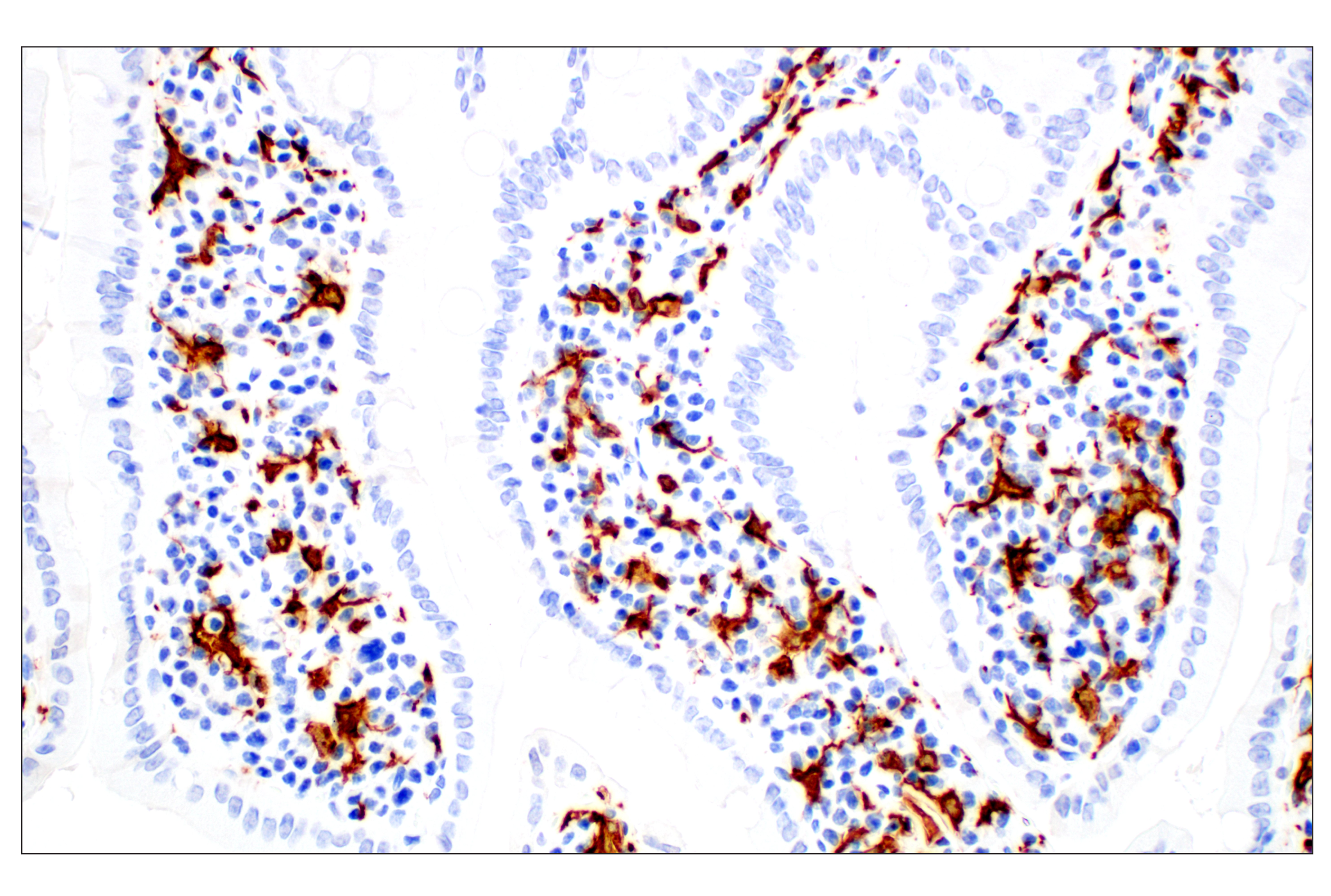
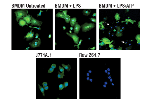
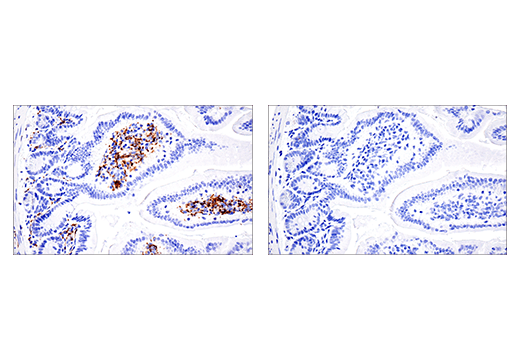
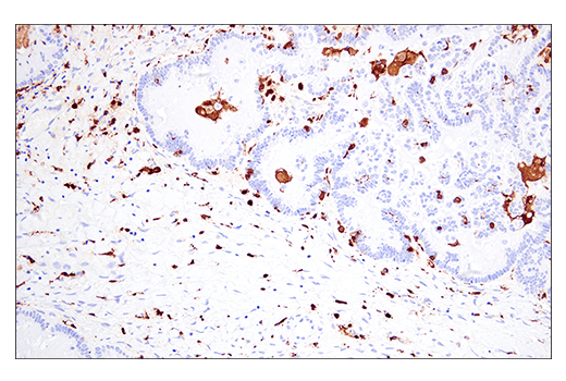
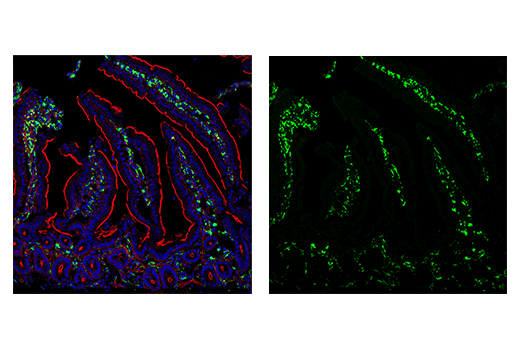
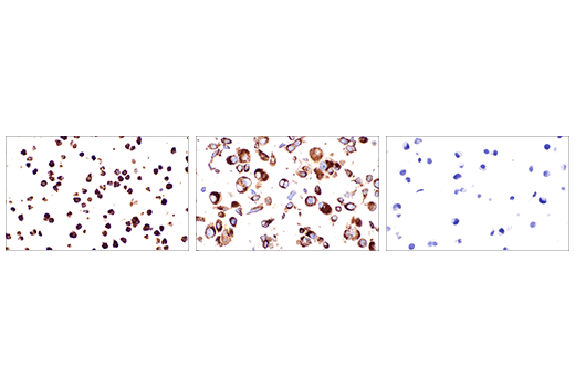
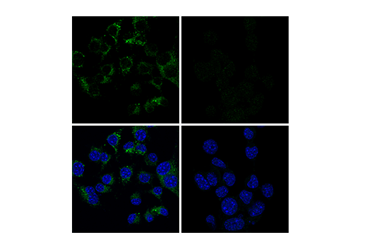
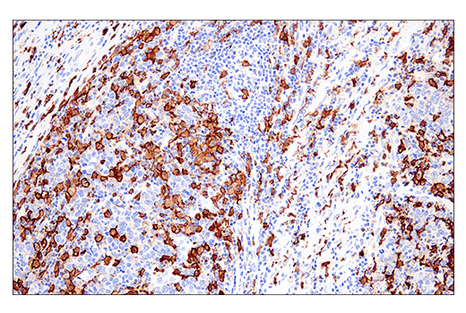
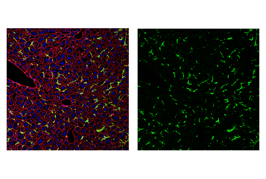
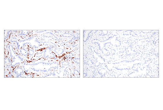
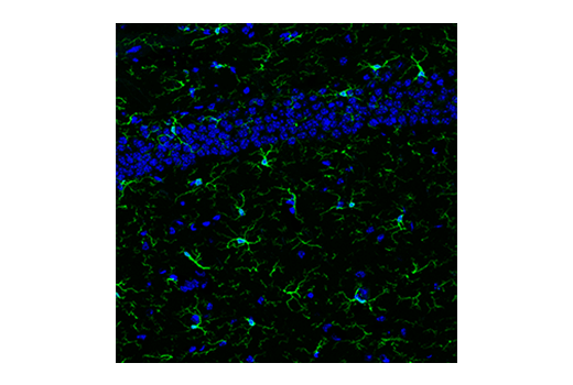
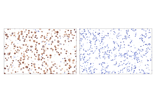
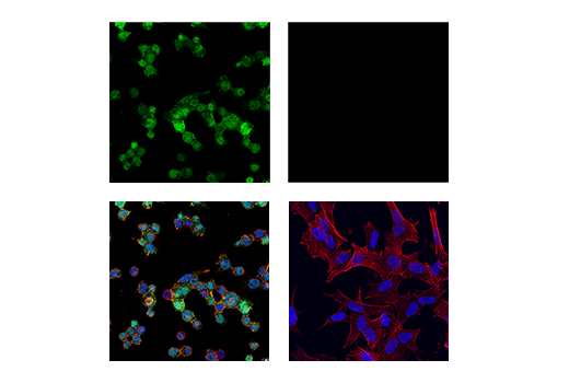
Microglia are the resident macrophages of the central nervous system, responsible for immune response and maintenance of CNS homeostasis (1). Ionized calcium-binding adaptor molecule 1 (Iba1), also known as allograft inflammatory factor 1 (AIF-1), is uniquely expressed in cells of monocytic lineage and is, therefore, widely used as a marker for microglia/macrophages in the brain and other tissue (2,3). Transmembrane protein 119 (TMEM119) is a cell-surface protein of unknown function, expressed exclusively by the microglia subset of myeloid and neural cells (4). Iba1+ microglia with both ramified and amoeboid morphologies express TMEM119, while Iba1+ macrophages are TMEM119 negative (5). TMEM119 and other homeostatic genes have been shown to be downregulated in disease-associated microglia (DAM) (6). Cluster of differentiation molecule 11b (CD11b)/Integrin alpha M (ITGAM) is a transmembrane protein expressed by, and commonly used as a marker for, myeloid lineage cells, including neutrophils, monocytes, macrophages, and microglia (7). F4/80 (EMR1) is a heavily glycosylated G-protein-coupled receptor and is a well-established marker for mouse macrophages (8-10). Expression of F4/80 has been observed in microglia and subset populations of dendritic cells (11). The protein phosphatase (PTP) receptor CD45 is a type I transmembrane protein expressed in all nucleated hematopoietic cells (12). Studies suggest CD45 plays a role in regulation of microglial activation (13,14). CD68 (macrosialin) is a heavily glycosylated transmembrane protein that is expressed by and commonly used as a marker for monocytes and macrophages (15,16). It localizes to the lysosomal membrane and is upregulated during microglial activation (17,18). Ki-67 is a nuclear nonhistone protein (19) universally expressed among proliferating cells and absent in quiescent cells (20). Previous work identifying markers of specific brain cell types using RNA-seq has shown HS1 and ASC/TMS1 to be useful and specific tools to study microglia (21). HS1 is a protein kinase substrate that is expressed only in tissues and cells of hematopoietic origin (22) and ASC/TMS1 has been found to be a critical component of inflammatory signaling where it associates with and activates caspase-1 in response to pro-inflammatory signals (23).
Explore pathways related to this product.
STRING - Known and Predicted Protein-Protein Interactions.
Except as otherwise expressly agreed in a writing signed by a legally authorized representative of CST, the following terms apply to Products provided by CST, its affiliates or its distributors. Any Customer's terms and conditions that are in addition to, or different from, those contained herein, unless separately accepted in writing by a legally authorized representative of CST, are rejected and are of no force or effect.
Products are labeled with For Research Use Only or a similar labeling statement and have not been approved, cleared, or licensed by the FDA or other regulatory foreign or domestic entity, for any purpose. Customer shall not use any Product for any diagnostic or therapeutic purpose, or otherwise in any manner that conflicts with its labeling statement. Products sold or licensed by CST are provided for Customer as the end-user and solely for research and development uses. Any use of Product for diagnostic, prophylactic or therapeutic purposes, or any purchase of Product for resale (alone or as a component) or other commercial purpose, requires a separate license from CST. Customer shall (a) not sell, license, loan, donate or otherwise transfer or make available any Product to any third party, whether alone or in combination with other materials, or use the Products to manufacture any commercial products, (b) not copy, modify, reverse engineer, decompile, disassemble or otherwise attempt to discover the underlying structure or technology of the Products, or use the Products for the purpose of developing any products or services that would compete with CST products or services, (c) not alter or remove from the Products any trademarks, trade names, logos, patent or copyright notices or markings, (d) use the Products solely in accordance with CST Product Terms of Sale and any applicable documentation, and (e) comply with any license, terms of service or similar agreement with respect to any third party products or services used by Customer in connection with the Products.