

- Home /
- Research /
- Neuroscience /
- Neurodegeneration Drug Development
Neurodegeneration Drug Development
Put our experience to work for you. Cell Signaling Technology (CST) scientists are biology, application, and therapeutic area experts and are here to streamline your discovery. We are passionate about science and keep up with all the latest neurodegenerative disease research, allowing us to offer a broad antibody portfolio against targets that could lead to new, more efficacious therapies. CST also offers an extensive post-translational, modification-specific antibody portfolio as well as resources like the PhosphoSitePlus® PTM Database.
Solutions Across the Drug Development Continuum
Target ID & Validation | Screening & Lead Optimization | Preclinical Safety & Validation |
|---|---|---|
Target ID and MOA StudiesTarget ValidationFlexible Packaging | Identify Primary and Secondary Endpoints
Platform compatibility
Custom
|
|
Antibodies That Work on YOUR Platform
Customized Formulations for Immunoassay Development
Resources and Guides for Antibodies Compatible With Your Assay Platform
| Platforms | Application | Conjugate Type* | Product Format |
|---|---|---|---|
|
|
|
|
|
|
|
|
|
|
| |
|
|
| |
|
|
| |
|
|
|
|
*Some conjugates directly available for purchase from CST (other conjugate types may be proprietary).
** Growing list of select antibodies
Cellular Targets in Therapeutic Development
Chronic neuroinflammation is associated with the progression of neurodegenerative diseases. Microglia are the resident macrophages of the central nervous system and play key roles in mediating neuronal signaling and neuroinflammatory responses. Cell Signaling Technology offers a wide variety of cell signaling markers associated with chronic neurodegenerative diseases that can be reliably used to determine disease onset and progression.
TREM2 Signaling
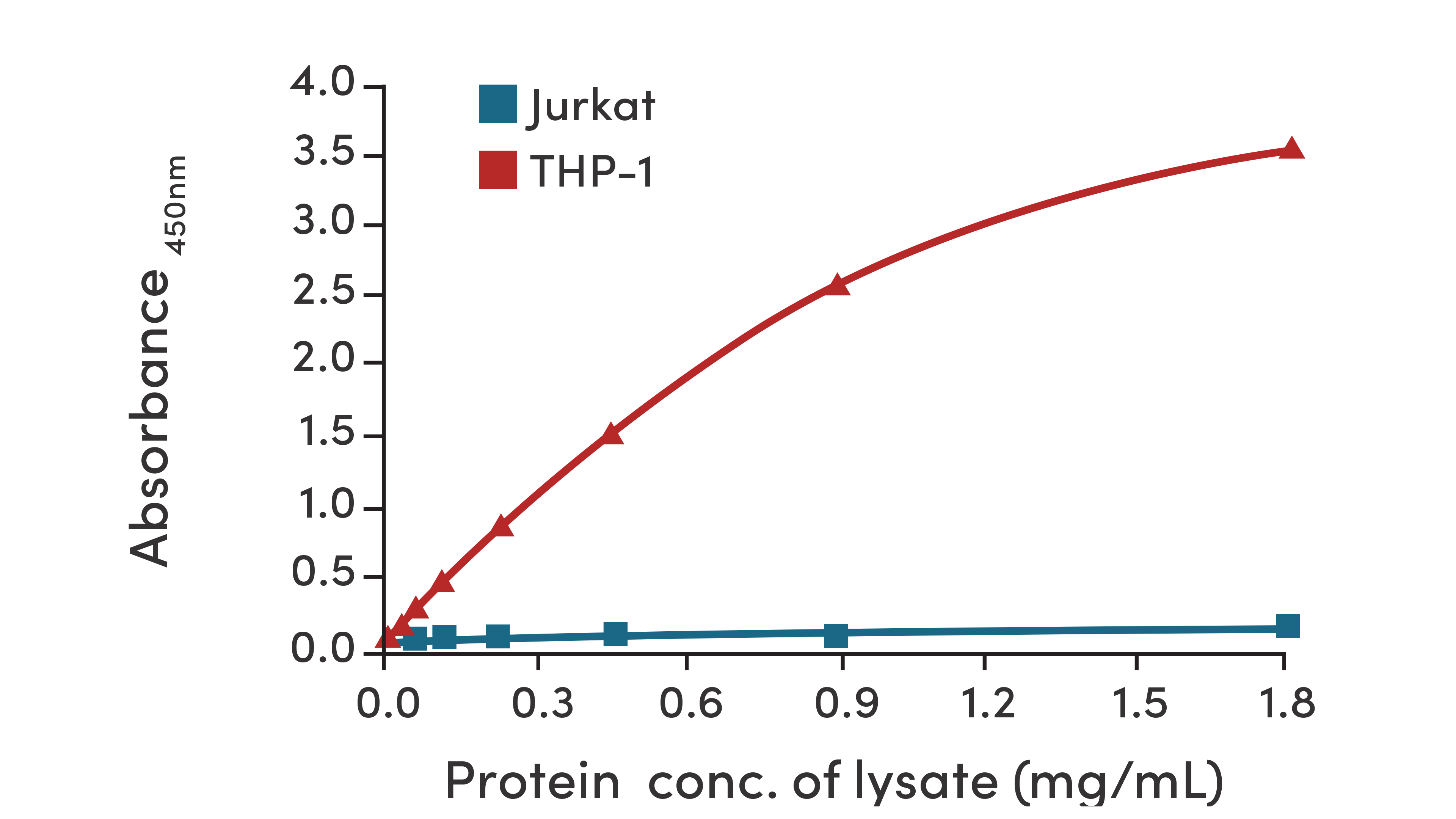
FastScan™ Total TREM2 ELISA Kit #23831: TREM2 protein is detected in THP-1 cells (positive control) but absent in Jurkat cells (negative control). The relationship between lysate protein concentration and the absorbance at 450 nm is shown.
Inflammasome
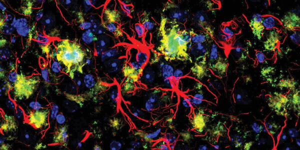
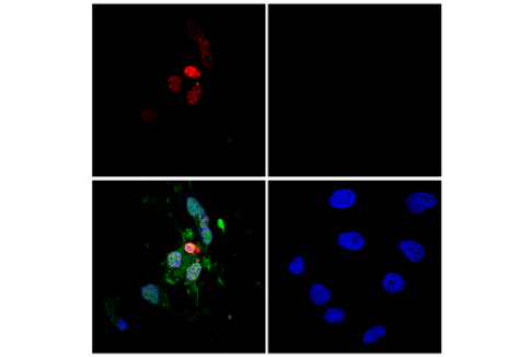
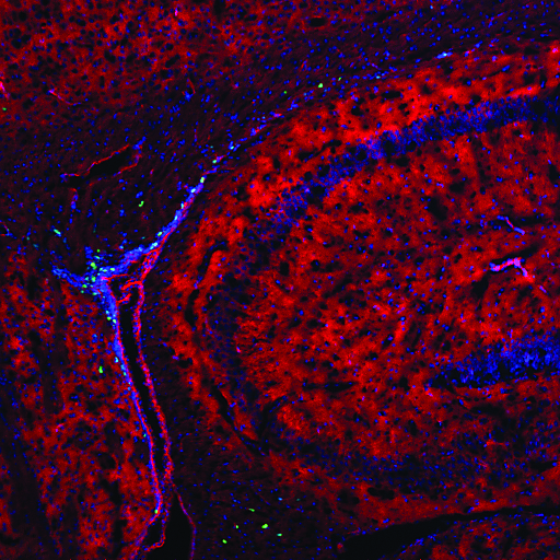
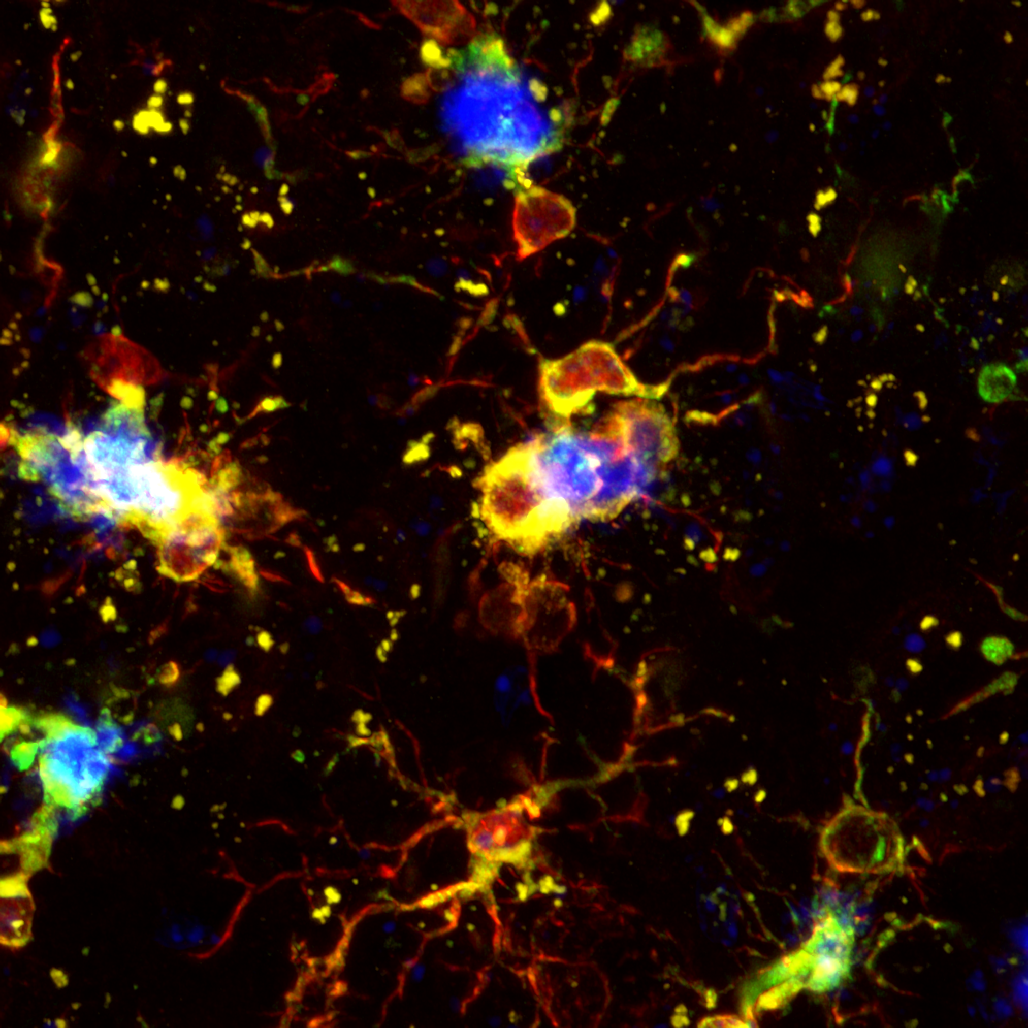
ASC/TMS1 (D2W8U) Rabbit mAb (Mouse Specific) (Alexa Fluor® 647 Conjugate) #23640: Confocal IF analysis of mouse Tg2576 brain that overexpresses mutant human APP695 using #23640 (red), GFAP (Alexa Fluor® 555 Conjugate) #3656 (pseudocolor yellow), β-Amyloid (Alexa Fluor® 488 Conjugate) #51374 (green), and DAPI #4083 (blue).
Cellular Readouts for Neuroinflammation
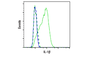
IL-1β (D3U3E) Rabbit mAb #12703: Flow cytometric analysis of THP-1 cells untreated (blue) or treated (green) with LPS #14011 using #12703.
Cell death serves as a readout for neurodegenerative disease progression. Neurons and glia can die and become diseased due to the aberrant regulation of cell death and survival pathways, which may include mitochondrial dysfunction, autophagy, and the activation of both apoptotic and non-apopototic pathways. With validated antibodies, protocols, and sampler kits from Cell Signaling Technology, neurodegenerative disease progression can be reliably assessed by analyzing the different types and activation states of cell death in human and rodent samples.
Necroptosis
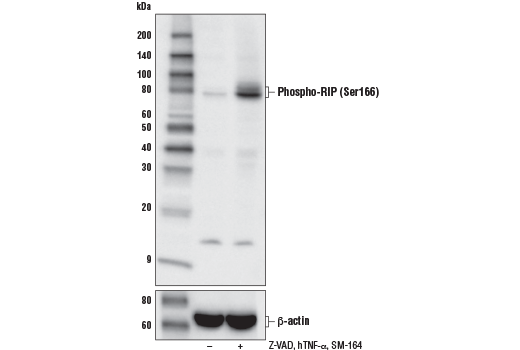
Phospho-RIP (Ser166) (D8I3A) Rabbit mAb #44590: Western blot analysis of HT-29 cells, untreated (-) or treated (+) with Z-VAD (20 μM, added 30 min prior to other compounds), human TNF-α (20 ng/ml, 7 hr), and SM-164 (100 nM, 7 hr), using #44590 (upper) or β-Actin (D6A8) Rabbit mAb #8457 (lower).
Apoptosis
TUNEL Assay Kit (Fluorescence, 640 nm) #64936: Confocal analysis of HeLa cells, treated with Staurosporine #9953 (1 µM, 3 hr; left, positive) or untreated (right, negative), using #64936 (red) followed by staining with Cleaved Caspase-3 (Asp175) Antibody #9661 (green) and DAPI #4083 (blue).
Pyroptosis
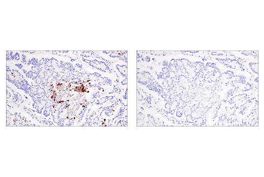
Cleaved Gasdermin D (Asp275) (E7H9G) Rabbit mAb #36425: IHC analysis of paraffin-embedded human colon carcinoma using #36425 in the presence of non-cleaved Gasdermin D peptide (left) or Asp275 cleavage-specific Gasdermin D peptide (right).
The production and processing of proteins that form protein aggregates are associated with frontotemporal lobar degeneration and taupathies. Therapies targeting protein aggregate formation may help slow proteinopathy progression. Protein aggregates found in blood or cerebrospinal fluid may also serve as biomarkers for diagnosing conditions earlier, and for monitoring disease progression and therapeutic response. Antibodies from Cell Signaling Technology are an ideal foundation for biomarker-based assays because they are thoroughly validated for specificity and sensitivity on biologically relevant binary model systems.
Amyloid
β-Amyloid (D54D2) XP® Rabbit mAb #8243: Confocal IF analysis of the subicular cortex from an amyloid mouse model of Alzheimer’s disease mouse using #8243 (green), GFAP #3670 (red), and DAPI #8961 (blue).
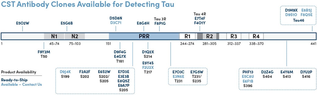
Tau/Phospho-Tau
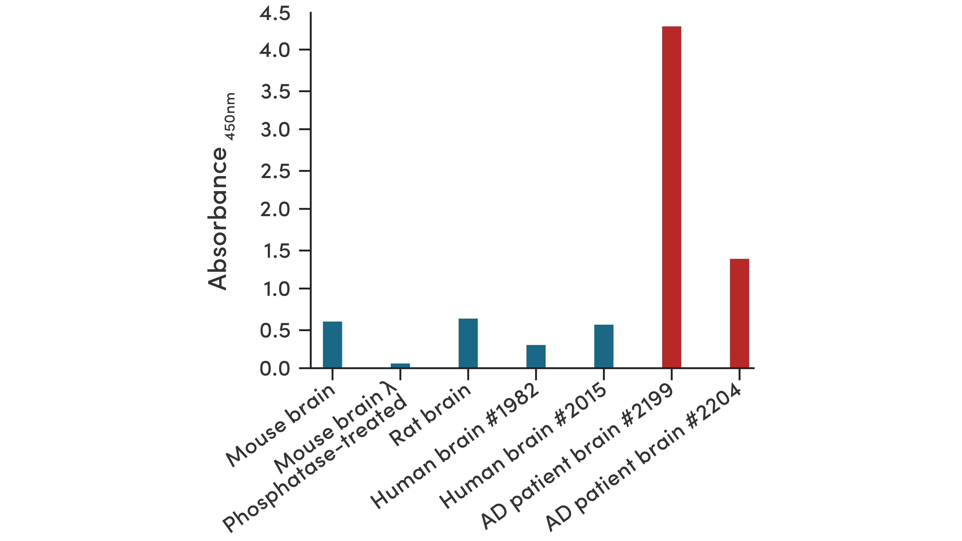
PathScan® Phospho-Tau (Thr217) Sandwich ELISA Kit #59672: #59672 is sensitive enough to detect differences in Thr217 phosphorylation among different Alzheimer’s disease patient samples, which may indicate disease progression.
Additional Neurodegenerative Aggregate Markers
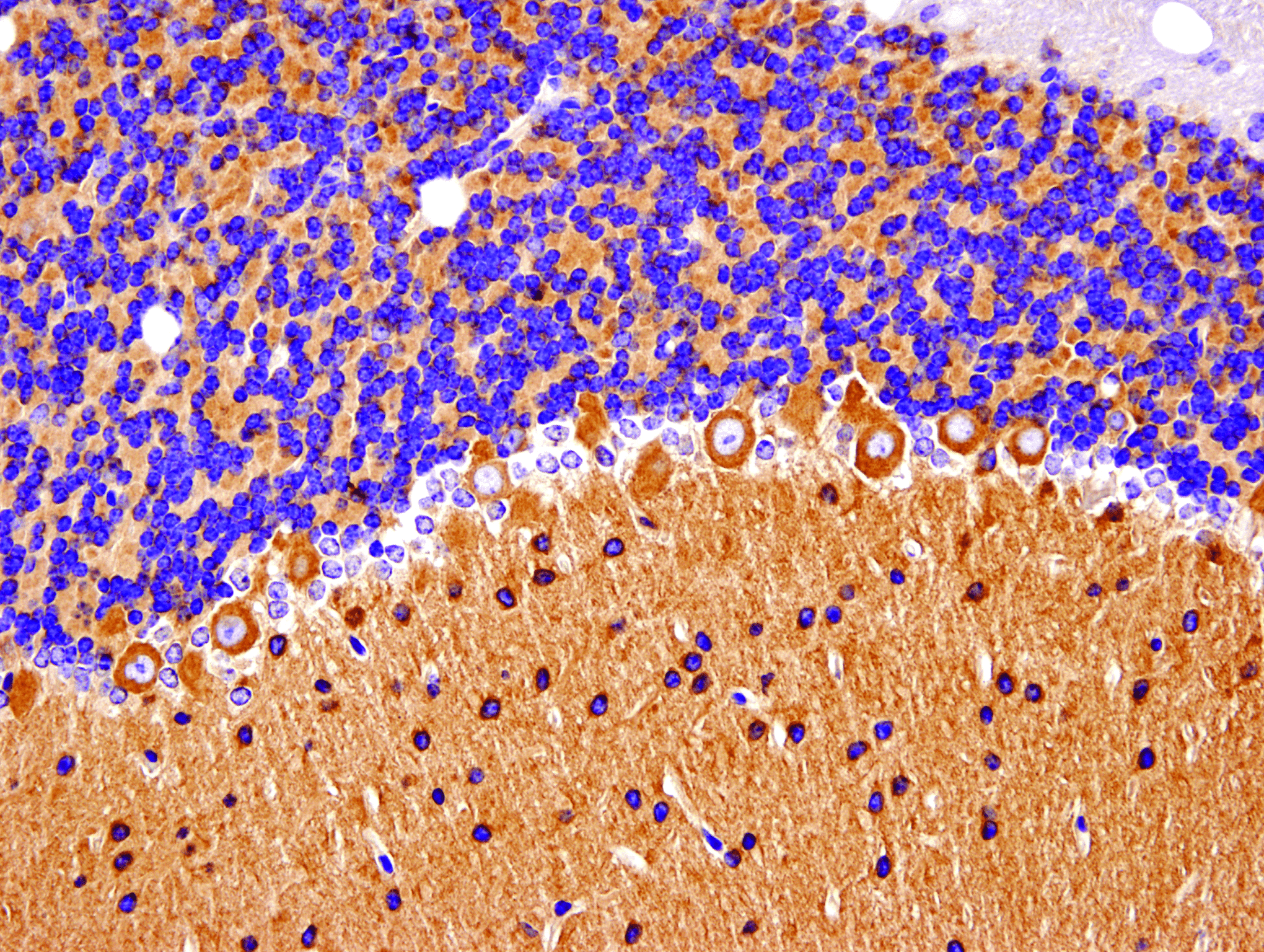
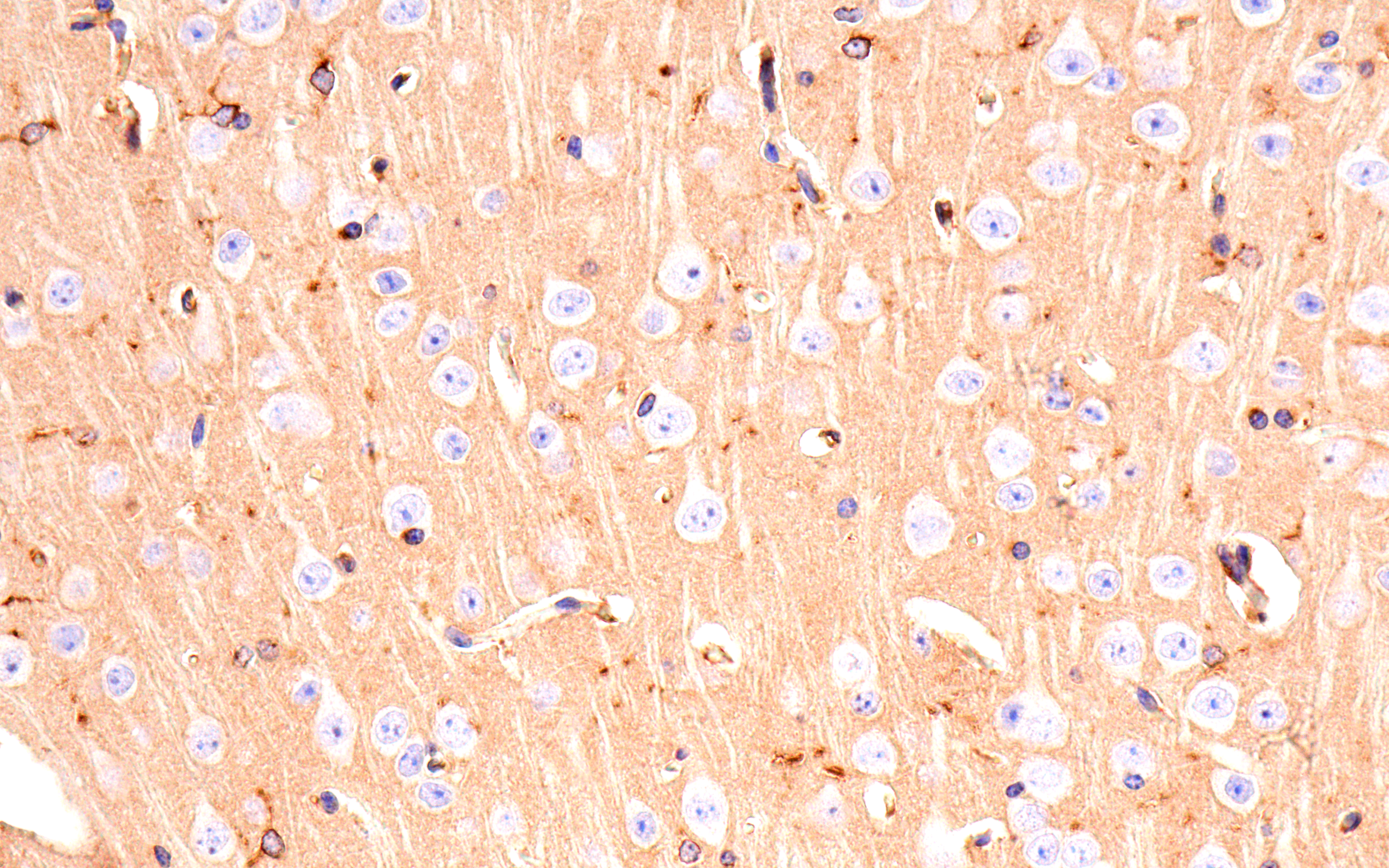
Huntingtin (D7F7) XP Rabbit mAb #5656: IHC analysis of paraffin-embedded mouse cerebellum using #5656.
Measuring cell viability can serve as a readout for determining neuronal disease progression. Aberrant or reduced proliferation, increases in neuronal damage, and the accumulation of senescent cells are all indicators of cell health and neurodegenerative disease status. Cell Signaling Technology offers antibodies and assay kits to determine cell viability efficiently and economically.
Cell Proliferation Markers
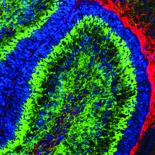
Ki-67 (D3B5) Rabbit mAb #9129: Confocal IF analysis of the ventricular zone in P21 mouse brain using #9129 (green). Actin filaments were labeled with DyLight 554 phalloidin #13054 (red) and DNA labeled with DRAQ5 #4084 (blue).
Neural Damage Markers

PathScan® Total Neurofilament-L Sandwich ELISA Kit #99175: Neurofilament-L protein is detectable in mouse and rat brain, but not HeLa cells (negative control) using #99175, as expected.
Senescence
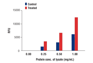
Senescence β-Galactosidase Activity Assay Kit: (Fluorescence, Plate-Based) #23833: β-galactosidase activity in HeLa lysates untreated and treated with Doxorubicin using #23833.
The human brain is responsible for about 20% of basal energy expenditure, despite accounting for only 2% of body weight. Metabolic dysregulation and neurodegeneration are strongly correlated. Metabolic proteins may act as therapeutic targets since abnormal glucose tolerance or insulin resistance is observed in many neurodegenerative conditions. Cell Signaling Technology provides high-quality antibodies and assays to support the interrogation of metabolic pathways and cellular energy homeostasis regulation.
Metabolite Transporters
ApoE (E7X2A) Rabbit mAb #49285: IHC analysis of paraffin-embedded mouse brain using #49285.
Insulin Receptor Signaling
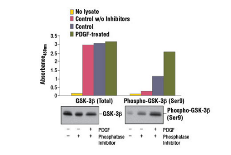
PathScan® Total GSK-3β Sandwich ELISA Kit #7265: Treatment of NIH/3T3 cells with hPDGF-BB #8912 stimulates phosphorylation of GSK-3β at Ser9, as detected by PathScan Phospho-GSK-3β (Ser9) Sandwich ELISA Kit #7311, but does not affect levels of total GSK-3β protein detected by #7265. The absorbance readings at 450 nm are shown in the upper figure, while the corresponding western blots using GSK-3β (27C10) Rabbit mAb #9315 (left panel) or Phospho-GSK-3β (Ser9) (D85E12) XP® Rabbit mAb #5558 (right panel) is shown in the lower figure.
Autophagy
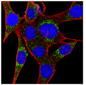
SQSTM1/p62 (D6M5X) Rabbit mAb (Rodent Specific) #23214: Confocal IF analysis of wild-type MEF treated with Chloroquine #14774 (50 μM, 18 hours) using #23214 (green). Actin filaments were labeled with β-Actin (8H10D10) Mouse mAb #3700 (red). Blue pseudocolor = DRAQ5 #4084 (fluorescent DNA dye).
Mitophagy
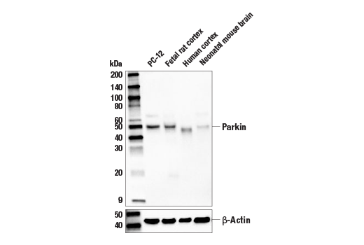
Parkin (D4Z1W) Rabbit mAb #32833: Western blot analysis of extracts from PC-12 cells, and various brain tissues, using #32833 (upper) or β-Actin (D6A8) Rabbit mAb #8457 (lower).
Synaptic plasticity is not only a part of development, learning, and memory, but has also been utilized as a readout for the clinical diagnosis of neurodegenerative diseases, including synaptopathies like Alzheimer’s disease. Cell Signaling Technology provides high-quality antibodies and assays to support the interrogation of synaptic activity and function.
Presynaptic Proteins
Synaptophysin (D8F6H) XP® Rabbit mAb #36406: Confocal IF analysis of mouse brain using #36406 (green), Neurofilament-L #2835 (yellow), Lamin A/C (4C11) Mouse #4777 (red), and DAPI #8961 (blue).
Postsynaptic Proteins
PSD95 (D27E11) XP Rabbit mAb #3450: Confocal IF analysis of rat cerebellum using #3450 (red), Neurofilament-L #2835 (green), DRAQ5 #4084 (pseudocolor blue).
Modulators of Synaptic Plasticity
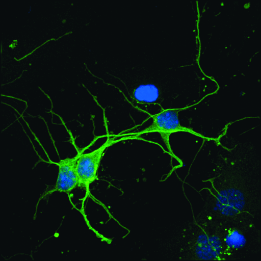
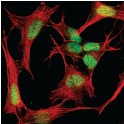
CaMKII-α (6G9) Mouse mAb #50049: Confocal IF analysis of primary neurons using #50049 (green) and DRAQ5 #4084 (pseudocolor blue).
Brain health is heavily reliant on epigenetic mechanisms, and a loss of chromatin dynamics is observed in neurodegenerative diseases. Profiling epigenetic patterns and identifying the chromatin marks associated with disease progression have become increasingly important for the development of therapies against neurodegeneration. Cell Signaling Technology provides a comprehensive and diverse catalog of epigenetics products and an extensive assay portfolio used to measure protein-DNA interactions and histone modifications.
Histone Modifications
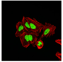
Acetyl-Histone H4 (Lys16) (E2B8W) Rabbit mAb (Alexa Fluor® 488 Conjugate) #56999: Confocal IF analysis of HeLa cells treated with TSA #9950 (1 μM, 4 hr), using #56999 (green). Actin filaments have been labeled with DyLight 554 Phalloidin #13054 (red).
DNA Methylation
MeCP2 (D4F3) XP® Rabbit mAb #3456: Confocal IF analysis of SH-SY5Y cells using #3456 (green) and actin (red)
Immediate Early Genes and Associated Proteins
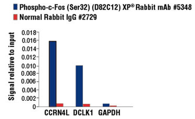
Phospho-c-Fos (Ser32) (D82C12) XP Rabbit mAb #5348: qRT-PCR results for ChIP-enriched DNA from PC-12 cells starved overnight and treated with hβ-NGF #5221 and either #5348 or Normal Rabbit IgG #2729 using SimpleChIP® Enzymatic Chromatin IP Kit (Magnetic Beads) #9003.
RBPs are increasingly becoming therapeutic targets for the treatment of neurodegeneration. Dysregulation of RNA-binding proteins (RBPs) has been linked to amyotrophic lateral sclerosis (ALS), Alzheimer’s disease, and frontotemporal dementia. Mutations in stress granule-associated RBPs lead to the pathological accumulation of protein aggregates that are readily apparent in neurodegenerative disorders. Age-related changes in mRNA methylation and methylation-dependent RBPs have also been associated with neurodegeneration. Cell Signaling Technology offers antibodies against key RBPs, stress granules, and m6A methylators, validated in relevant neuronal systems.
RBPs and Stress Granules
TDP43 (D9R3L) Rabbit mAb (Alexa Fluor® 555 Conjugate) #68779: Confocal IF analysis of mouse cerebellum using #68779 (red), Razs (Alexa Fluor® 647 Conjugate) #37182 (cyan pseudocolor), and DAPI #8961 (blue).
RNA m6A Methylation
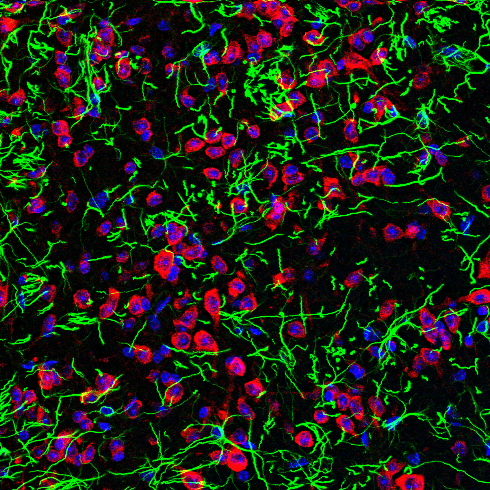
YTHDF2 (E2I2H) Rabbit mAb #71283: Confocal IF analysis of mouse medulla oblongata using #71283 (red), GFAP (Alexa Fluor® 488 Conjugate) #3655 (green), and DAPI #8961 (blue).
Cell Characterization
Studying the brain and nervous system requires examination not only of neurons, but also of microglia, oligodendrocytes, and astrocytes. The key to visualizing and identifying each of these cell types lies in using antibodies that target protein biomarkers specifically expressed and localized within these cells. Cell Signaling Technology provides highly specific, validated antibodies against accepted neuronal markers.
Immature Neuronal Markers
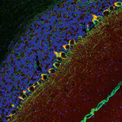
Doublecortin (A8L1U) Rabbit mAb #14802: Confocal IF analysis of P5 mouse cerebellum using #14802 (green), GFAP #3670 (red), and DRAQ5 #4084 (psuedocolor blue).
Mature Neuronal Markers
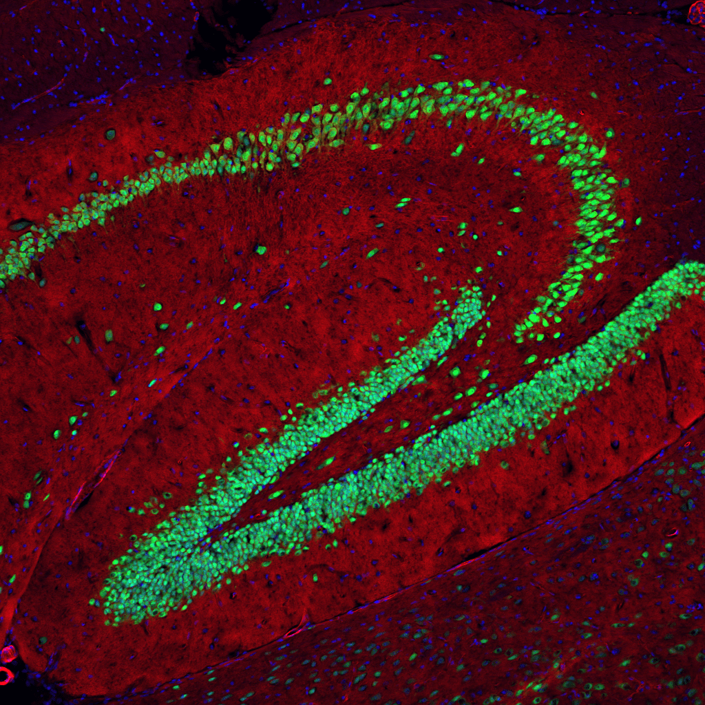
NeuN (D4G4O) XP® Rabbit mAb #24307: Confocal IF analysis of mouse hippocampus using #24307 (green) and actin labeled with DyLight 554 Phalloidin #13054 (red). DR˚AQ5 #4084 (pseudocolor blue).
/browse/?tab=product&search=eaat2Glial cells consist of the non-neuronal supporting cells, including astrocytes, oligodendrocytes, and microglia. Distinct changes in glial cells and their markers are known to be associated with neurodegenerative disease progression. Glial cells also play an important role in overcoming challenges with the blood-brain barrier. Cell Signaling Technology has a wide variety of validated glial marker antibodies to identify and study the different glial cell types.
Astrocytes
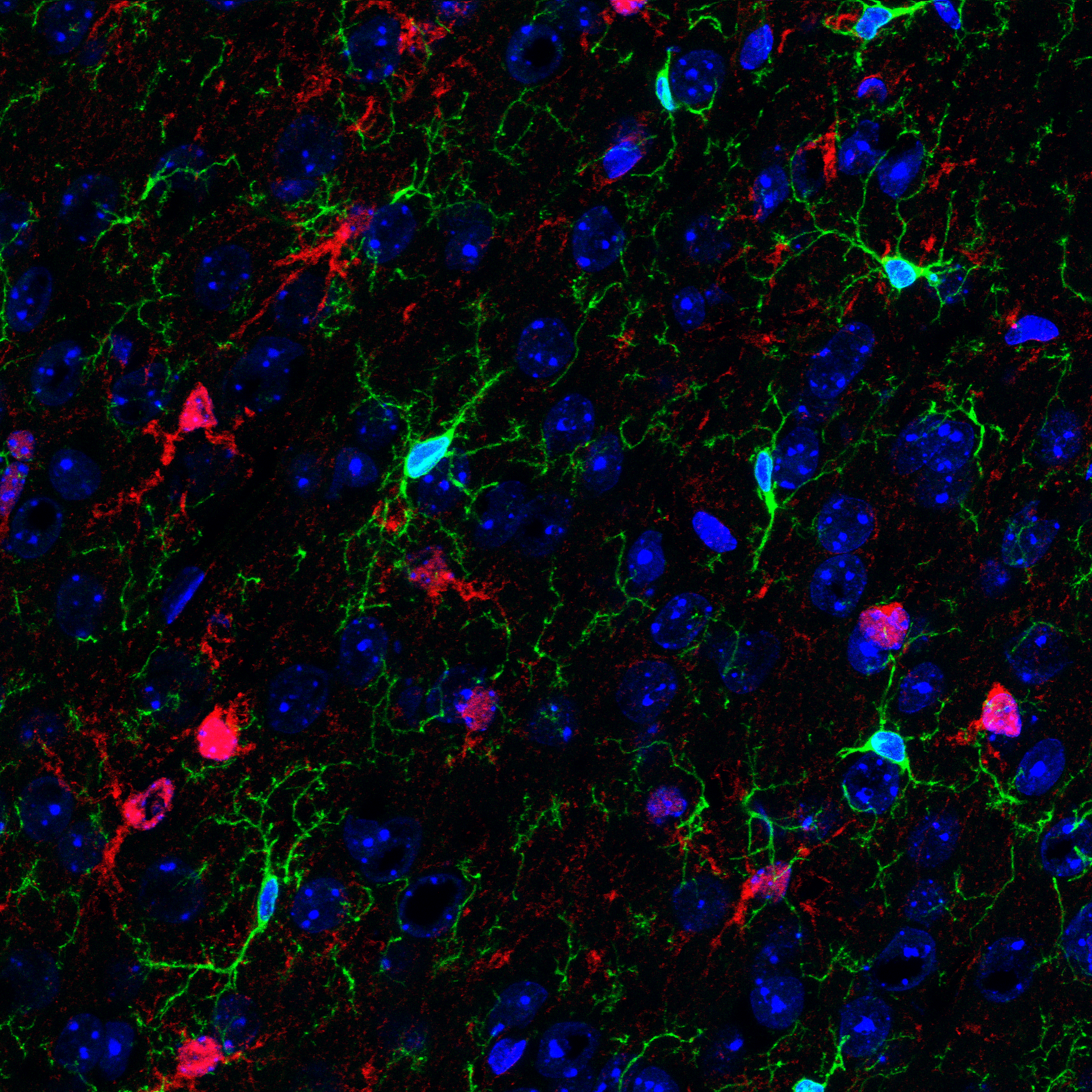
S100B (E7C3A) Rabbit mAb #90393: Confocal IF analysis of fixed frozen mouse cerebral cortex. #90393 (red), HS1 (Alexa Fluor® 488 Conjugate) #68206 (green), and DAPI #8961 (blue).
Oligodendrocytes
Olig2 (E6G6Q) XP® Rabbit mAb #65915: Confocal IF analysis of fixed frozen mouse cerebellum. #65915 (red), GFAP (Alexa Fluor® 488 Conjugate) #3655 (green), and DAPI #4083 (blue).
Microglia
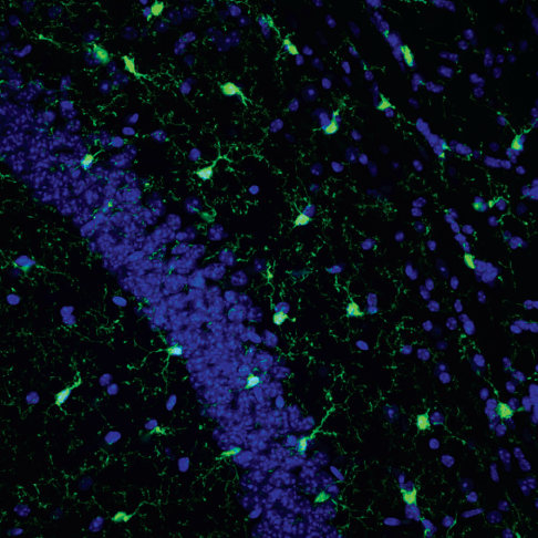
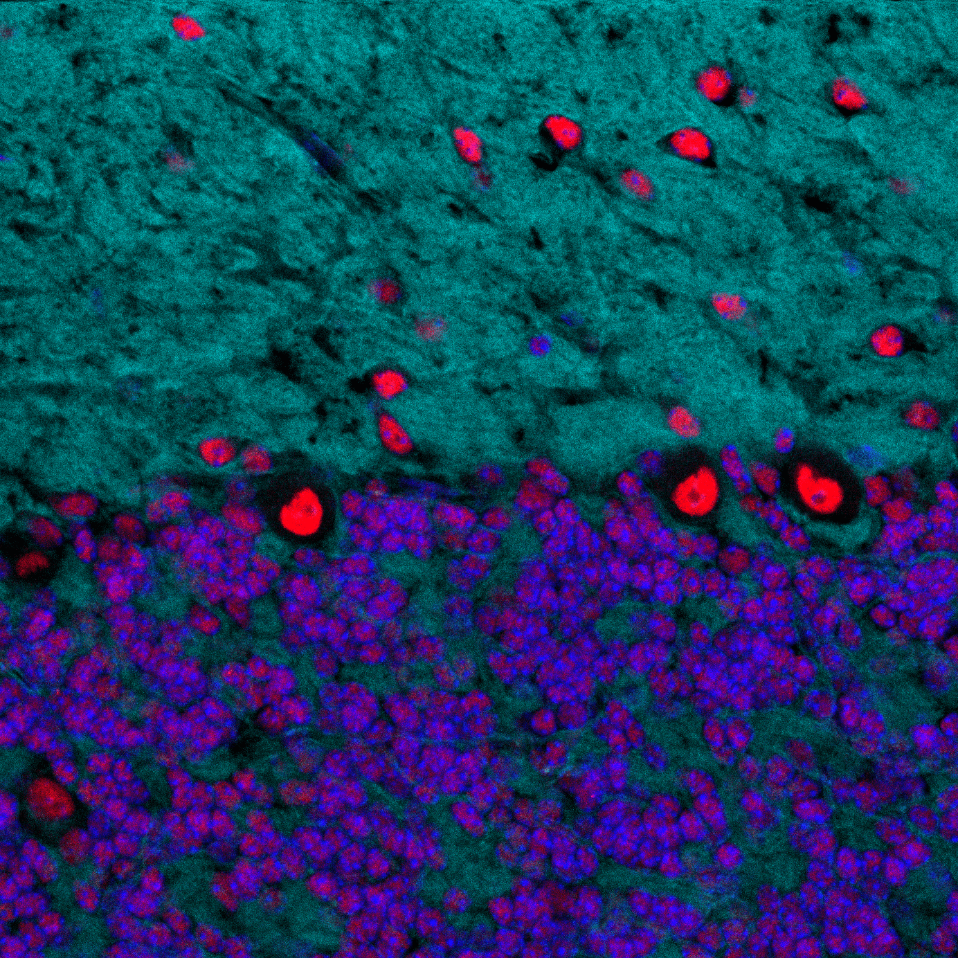
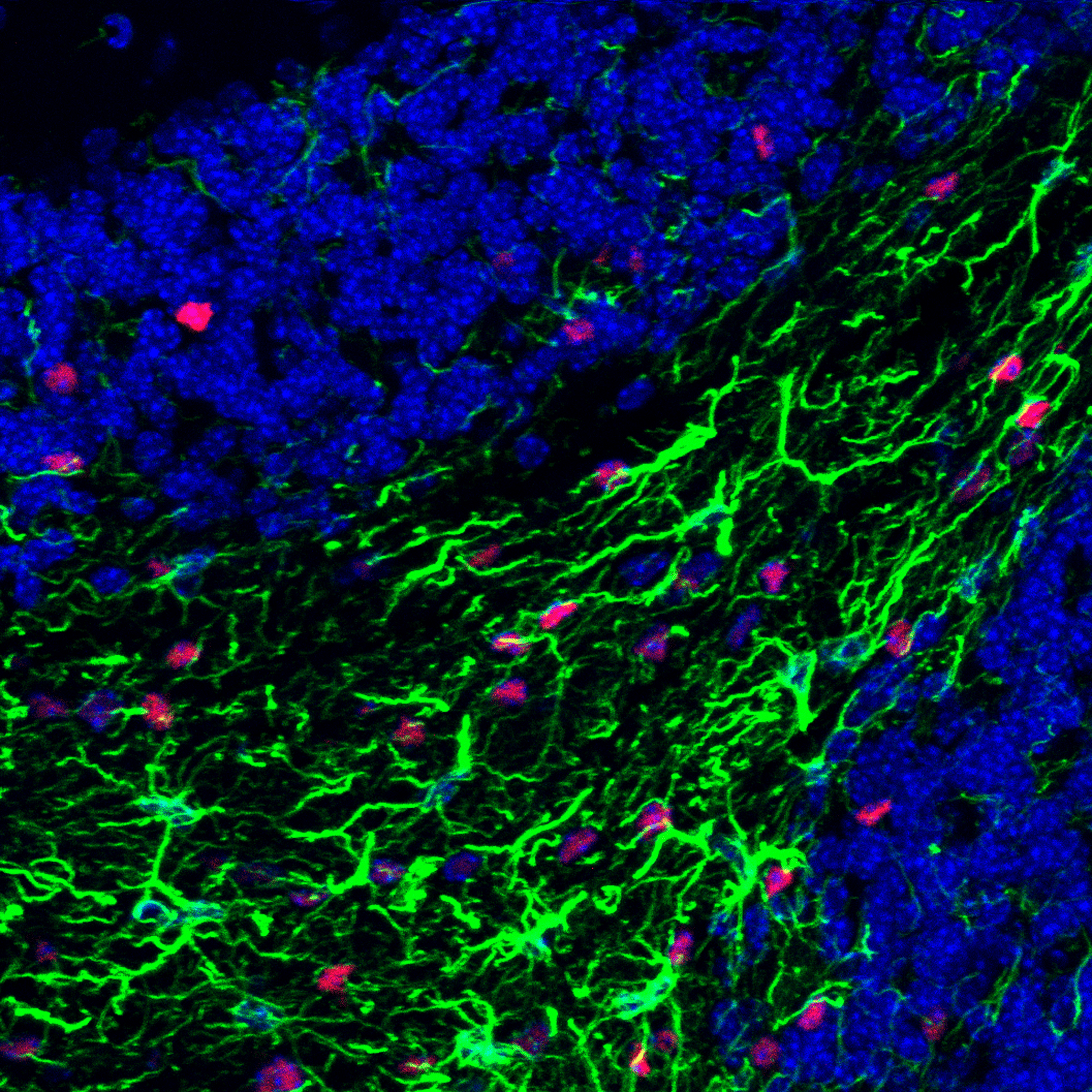
Iba1/AIF-1 (E4O4W) XP Rabbit mAb #17198: Confocal IF analysis of mouse CA1 hippocampus using #17198 (green) and DAPI (blue). Images kindly provided by Dr. Marco Colonna (Washington University) and used with permission.
DAM Markers
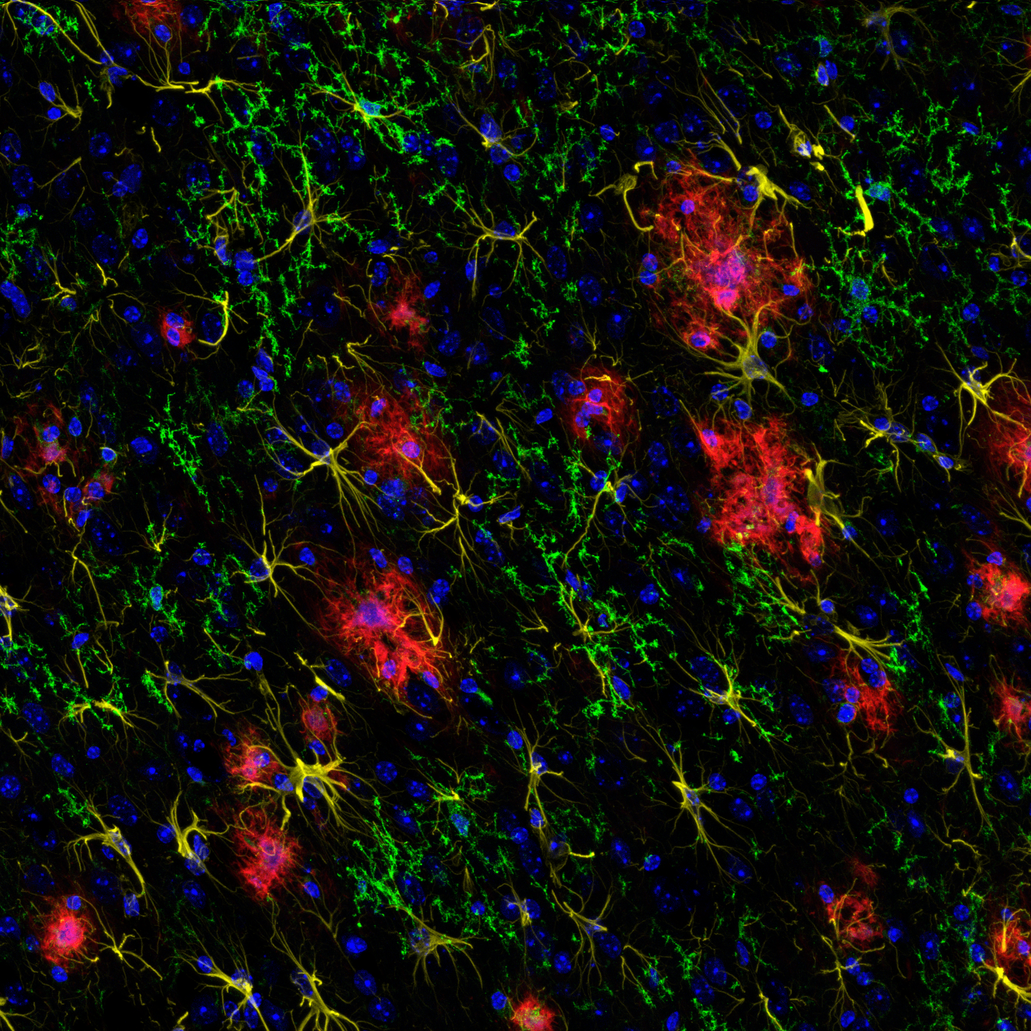
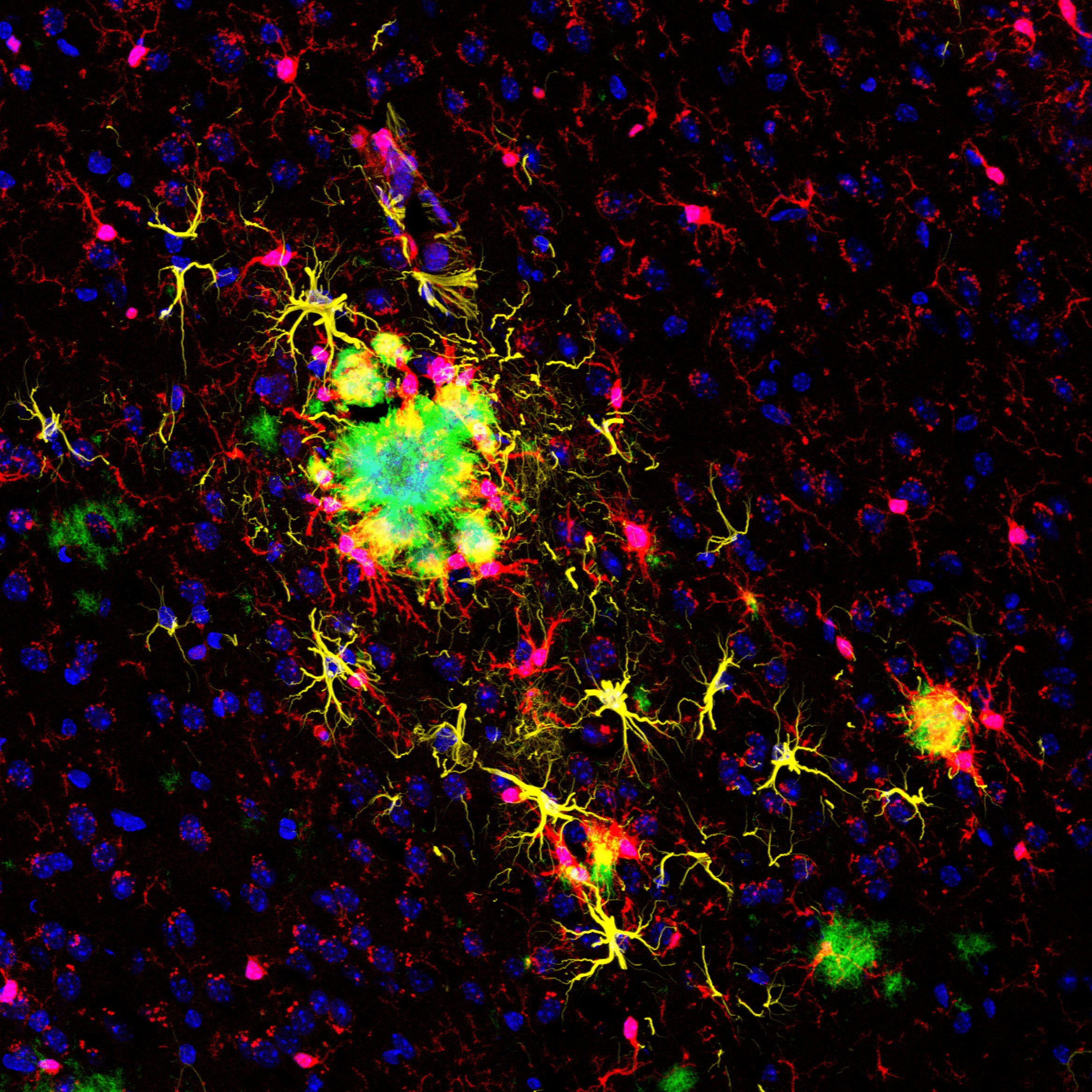
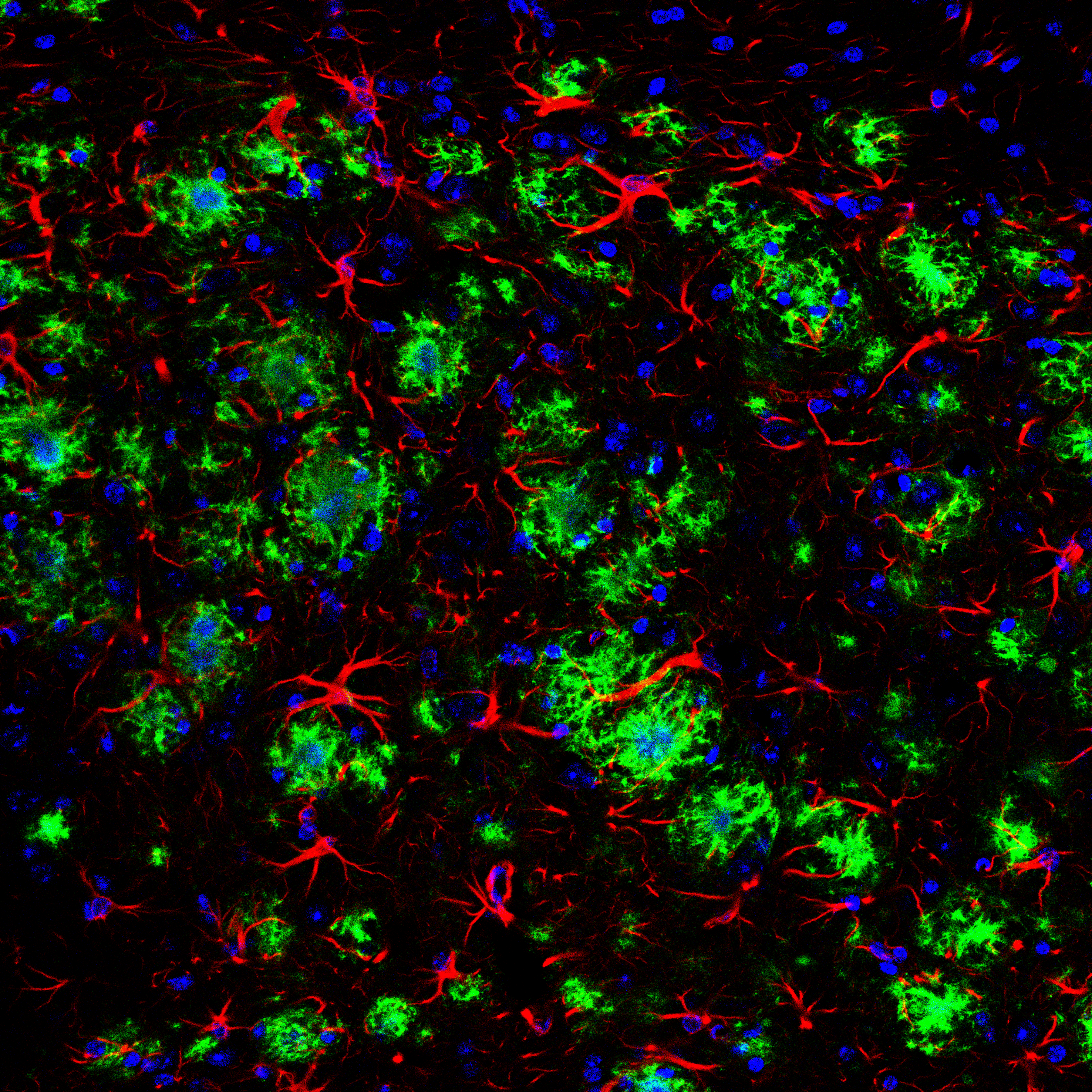
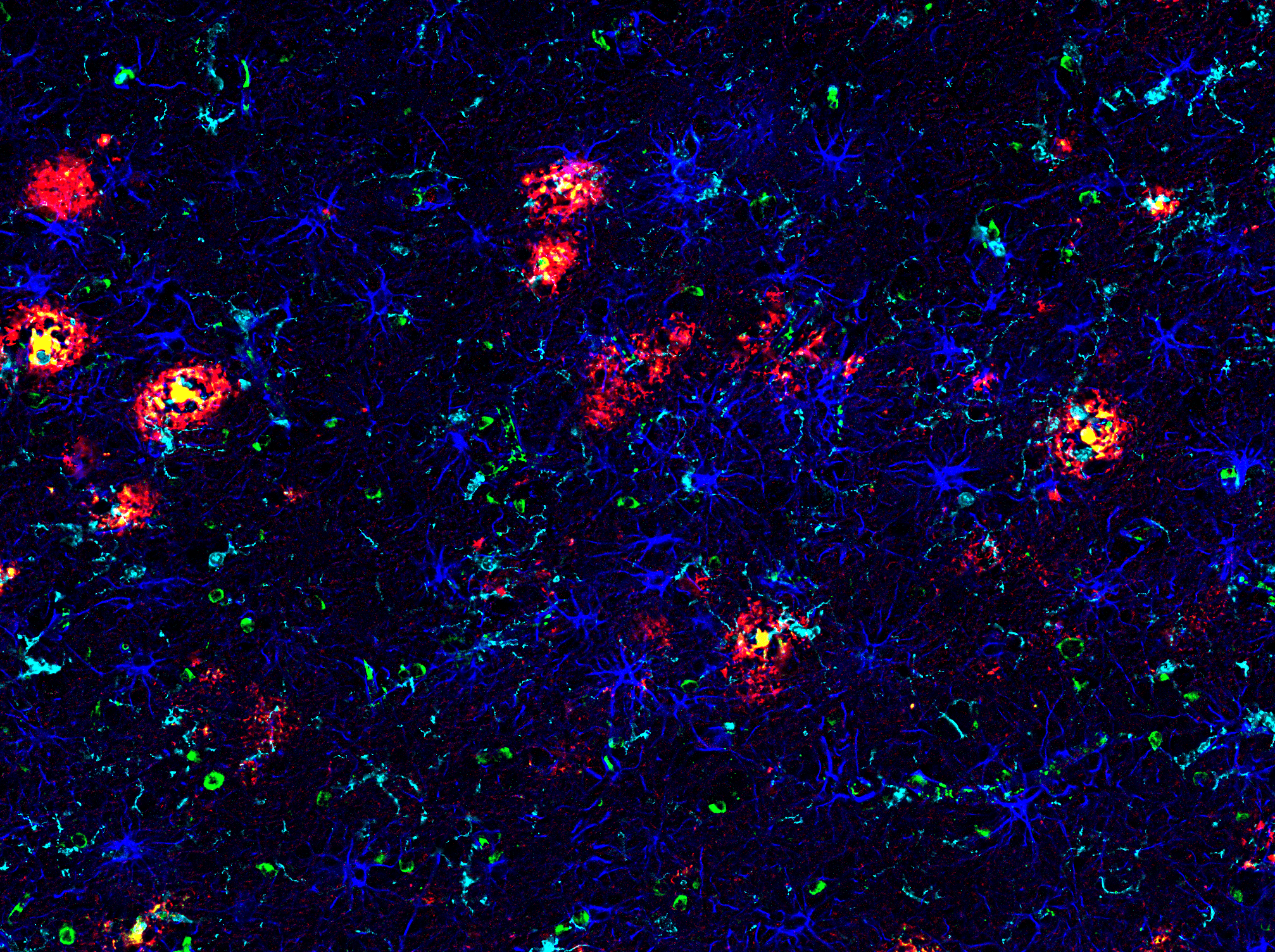
TMEM119 (E3E1O) Rabbit mAb #90840: Confocal IF analysis of an amyloid mouse model of Alzheimer’s disease brain using #90840 (green), GFAP #3670 (yellow), Β-Amyloid (Alexa Fluor® 647 Conjugate) #42284 (red), and DAPI #8961 (blue).
Biofluid Biomarker Assay Tools
Reliable reagents are critical for detecting and analyzing neurodegenerative disease biomarkers in biofluids like blood and CSF for discovery research. CST offers a broad portfolio of top-quality, reliable, and highly specific antibodies for developing pair-based assays that accurately detect low-abundance proteins like phospho-tau (thr217) and beta-amyloid (1-40)/(1-42).
Download the Antibody-Based Assay for Detection Human Alzheimer’s Disease Biomarkers Application Note to learn more about antibody-based assay solutions for neurodegeneration drug development.
Matched Antibody Pairs and ready-to-use ELISA kits in 96- or 384-well formats from CST deliver accurate and reliable Aβ42/40 ratios. Matched Antibody Pairs can be supplied in a carrier-free, conjugation-ready format (BSA-and azide-free) for assay platform-compatibility. Generate data you trust during the compound screening phase of drug development or in translational research studies.
PathScan® RP β-Amyloid (1-42) Sandwich ELISA Kit #27029
FastScan™ β-Amyloid (1-40) ELISA Kit #20882
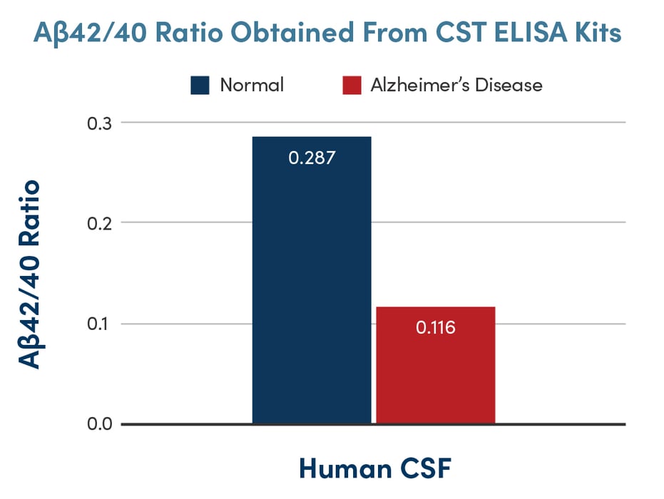
PathScan® RP β-Amyloid (1-42) Sandwich ELISA Kit #27029 and FastScan™ β-Amyloid (1-40) ELISA Kit #20882 are ready-to-use ELISA solutions for measuring Aβ42/40 ratios in human CSF. Aβ42 and Aβ40 were measured using their respective kits and their ratios are indicated.
Compared to a top competitor’s ELISA kit, the CST® FastScan™ Phospho-Tau (Thr181) ELISA Kit #58537 delivers 5-fold greater sensitivity and provides results in 1.5 hours, ~10 times faster than the 15.5–19.5 hours required by the competitor. The antibodies used in the kits are available as an immunoassay platform-compatible matched antibody pair.
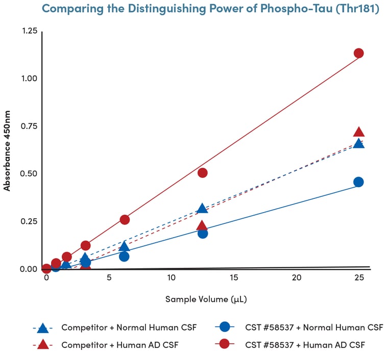
The CST® FastScan™ Phospho-Tau (Thr181) ELISA Kit #58537 outperforms an available ELISA product from a top competitor when measuring phospho-Tau (Thr181) levels in AD CSF.
The binding kinetics of Phospho-Tau (Thr217) (E9Y4S) Rabbit monoclonal antibody #51625 and a commercially available monoclonal antibody from a leading competitor were measured using bio-layer interferometry (BLI) on the Octet RED96 (Sartorius). The higher binding properties, faster association, and lower dissociation rates for the antibody clone from CST demonstrate that it has superior binding characteristics than the competitor antibody.
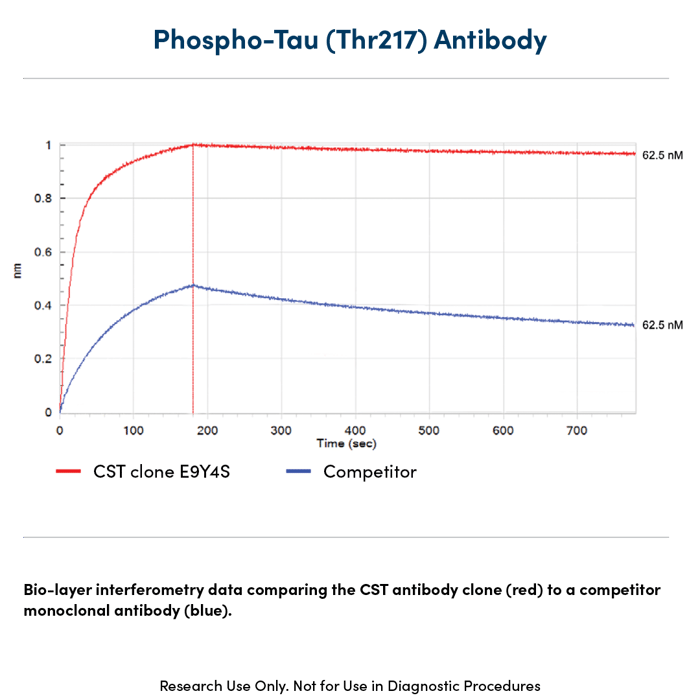
Bio-layer interferometry data comparing the CST® Phospho-Tau (Thr217) (E9Y4S) Rabbit monoclonal antibody #51625 (red) to a leading competitor monoclonal antibody (blue) using bio-layer interferometry on the Octet RED96 (Sartorius).
Matched Antibody Pairs from CST deliver the sensitivity and specificity required to detect biomarkers like phospho-Tau (Thr217) in CSF using your immunoassay platform of choice. Matched Antibody Pairs are supplied in a carrier-free, conjugation-ready format (BSA-and azide-free) to ensure platform compatibility.
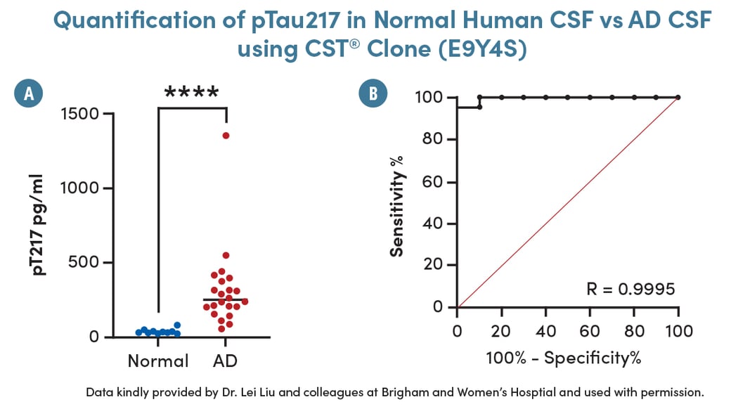
(A) Phopsho-Tau (Thr217) was measured in AD and normal CSF using the SMCxPRO platform. (B) A receiver operating characteristic (ROC) curve was plotted to represent this data with an R score of 0.9995.
CST offers an elite biofluid biomarker portfolio against critical neurodegenerative disease targets, with innovative products being added daily.
| Tau | Amyloid | ALS/FTD |
|---|---|---|
|
| Parkinson's Disease | Synaptic Dysfunction | Neuronal Health |
|---|---|---|
| Microgliosis | Astrogliosis | Huntington's Disease |
|---|---|---|
| Astrocytosis | ||
|---|---|---|
Solutions for Assay Development
Cell Signaling Technology has the breadth of products available to enable several methods of multiplex staining, highlighted below.
Host Species
- One of the simplest ways to multiplex
- Use primary antibodies derived from various host species and isotypes (rabbit IgG, mouse IgG1, rat IgG2a, etc.), then probe with fluorophore-coupled secondaries specific to the host primary.
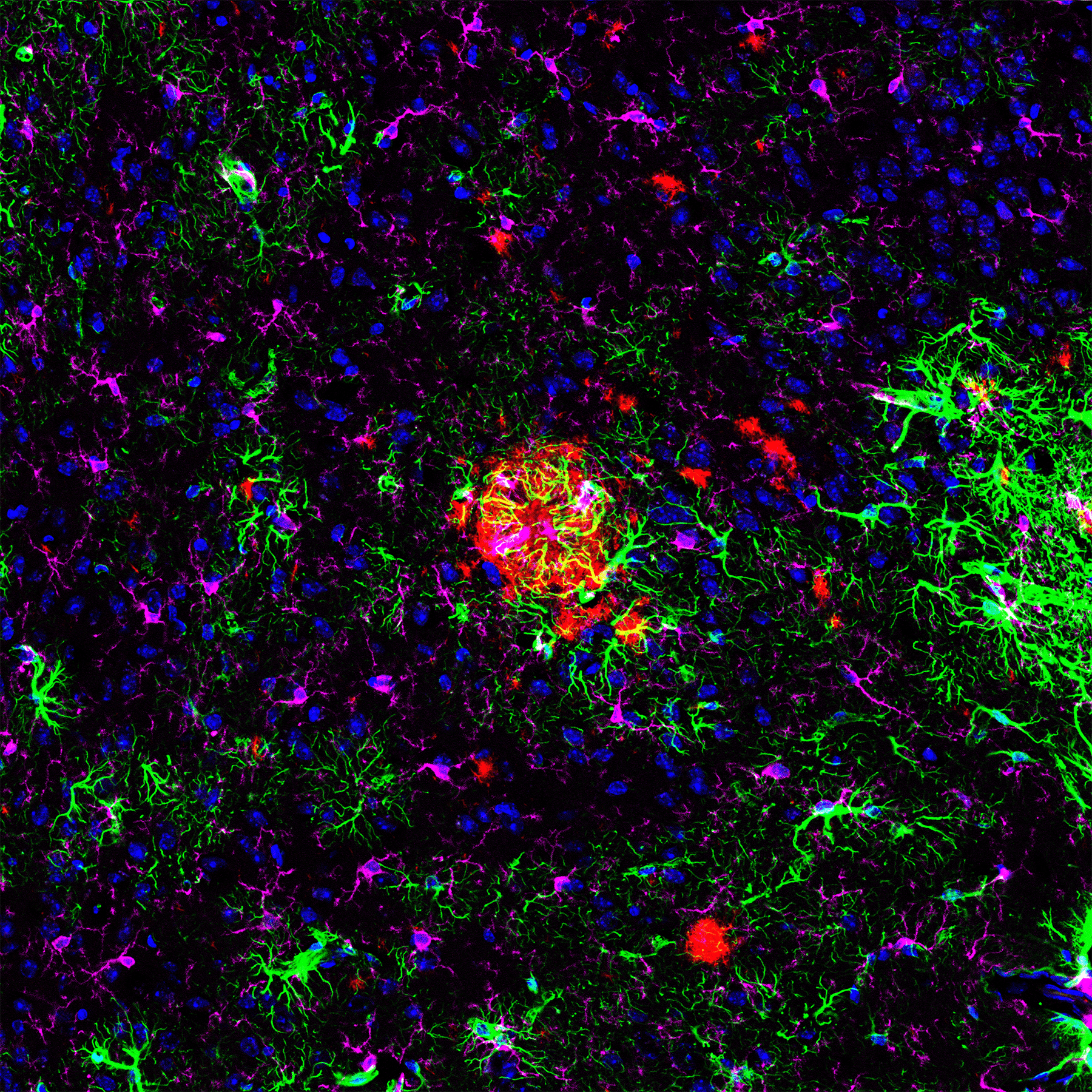
Confocal IF analysis of mouse Tg2576 brain labeled with GFAP #34001 (green), β-Amyloid #15126 (red), HS1 #3892 (magenta), and DAPI #8961 (blue).
Fluorophore Conjugated Antibodies
- Directly conjugate a primary antibody with a specific label, such as a fluorophore or enzyme
- CST offers rigorously validated, ready-to-use antibodies conjugated to the most popular fluorophores.
Confocal IF analysis of an amyloid mouse model of Alzheimer’s disease brain using Alexa Fluor® 647 Conjugated Iba1/AIF-1 #78060 (red), Alexa Fluor® 488 Conjugated HS1 #68206 (green), GPNMB #90205 (yellow), and methoxy XO4 (blue). Image kindly provided by Dr. Simone Briochi of Dr. Marco Colonna’s lab (Washington University) and used with permission.
Conjugation-Ready Carrier-Free Antibodies
- Preferred method when expanding panels or using technologies where a directly conjugated antibody is not available.
- Use carrier-free formulated antibodies. They are conjugation-ready to support platforms requiring labels such as metals, fluorophores, and oligonucleotides.
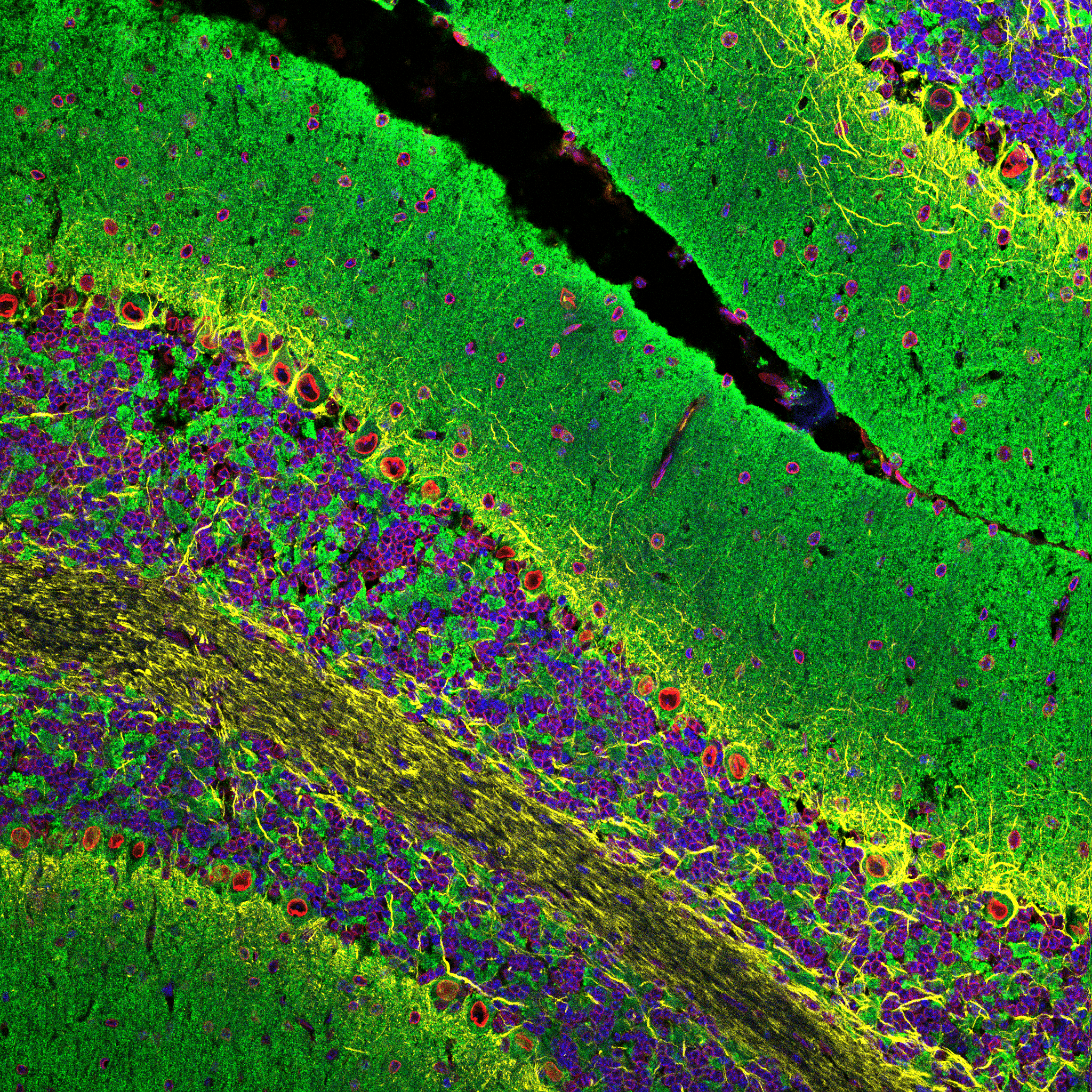
Multiplex IF imaging of the human prefrontal cortex from an Alzheimer’s disease patient stained with GFAP (blue), microglia with Iba-1/AIF-1 #17198 (cyan), ApoE #13366 (green), and β-Amyloid #9888 (red). Imaged with the Akoya PhenoCycler platform. Image kindly provided by Akoya and used with permission.
Rigorously validated antibody pairs are at the heart of every ELISA experiment. Cell Signaling Technology (CST) offers matched antibody pairs, both as a complete solution in sandwich ELISA kits or as custom pairs, ideal for incorporating into standard high-throughput ELISA-like assay platforms. These validated antibody pairs offer unrivaled specificity to deliver accurate readouts when monitoring therapeutic modulation of key neurodegenerative disease biomarkers. Some CST® kits are sensitive to plasma proteins, enabling monitoring of disease status with live models.
Measure Key Biomarkers with Highly Specific and Sensitive ELISA Kits

The PathScan® β-Amyloid (1-42) Sandwich ELISA Kit #27029 is highly specific to only human Aβ-42, showing no significant signal with Aβ-37, Aβ-38, Aβ-39, Aβ-40, Aβ-43, or pE3 peptides, as expected. By contrast, other leading commercially available β-amyloid (1-42) sandwich ELISA kits show little-to-no specificity towards the advertised Aβ-42 target.

The FastScan™ Phospho-Tau (Thr181) ELISA Kit #58537 detects human tau in plasma from the hTau P301S mouse model in as little as 50 μL of plasma. Data was kindly provided by Dr. Li-Huei Tsai (Massachusetts Institute of Technology) and used with permission.
Reproducibility in your experiments isn’t a matter of chance. It’s a matter of science.
Cell Signaling Technology® (CST®) products are developed, tested, and rigorously validated across multiple applications by tenured CST scientists who understand the underlying biology. Over 99.5% of CST recombinant monoclonal antibodies are manufactured in-house, providing complete control over our supply chain and providing lot-to-lot consistency for the lifetime of your project.
Accelerate time to results with CST products and services.
Your assay is only as good as your antibody is specific. CST antibodies and ready-to-use ELISA and cellular assay kits are developed with this in mind. They are designed to seamlessly fit into your assay workflow and instantly answer key questions. CST subject matter experts are available to help identify the best readout and clone to effectively assess your therapeutic efficacy and safety.
Companion Reagents
CST offers a wide selection of epitope-tagged and control antibodies, secondary antibodies, detection reagents, and experimental controls, as well as standard buffers and other reagents required to complete your experimental workflow.
Sometimes the fastest way to move your discovery forward is to have someone else do it.
CST provides custom solutions that meet your specific research challenge, freeing up your time to focus on the science. Services include:
- Carrier-free and Customized Formulations: Ideal for any application, assay, or platform requiring conjugation-ready or unique formulations, including multiplex IF and ELISA.
- Custom Antibody Conjugation: Have our in-house experts perform the conjugations you need for your assay: fluorophores, biotin, enzymes, select oligonucleotides, and more. If you don’t see the conjugated antibody you need in the CST catalog, we’ll conjugate it for you.
- Proteomics Analytical Services: Partner with CST for qualitative and quantitative protein profiling of your precious samples, including brain tissue. CST scientists are by your side from project planning to sample prep to data analysis, providing you with actionable results.
- Bulk Quantitates or Lot Reservations: Eliminate potential supply problems by partnering with CST to reserve a single lot or place bulk orders.
- Custom Peptides and Controls: CST provides the controls you need for your specific assay.
CST, Cell Signaling Technology, PhosphoSitePlus FastScan, XP, PathScan, SimpleChip are trademarks or registered trademarks of Cell Signaling Technology, Inc. Alexa Fluor® is a registered trademark of Life Technologies Corporation. All other trademarks are the property of their respective owners. Visit cellsignal.com/trademarks for more information.